. 2012 Sep 24;209(10):1883-99.
doi: 10.1084/jem.20120502. Epub 2012 Sep 3.

Disruption of SIRPα signaling in macrophages eliminates human acute myeloid leukemia stem cells in xenografts

Affiliations

Disruption of SIRPα signaling in macrophages eliminates human acute myeloid leukemia stem cells in xenografts

Alexandre P A Theocharides et al. J Exp Med. .

Abstract

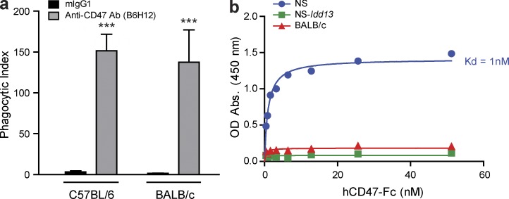
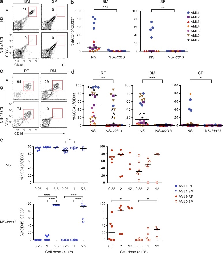
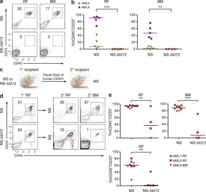
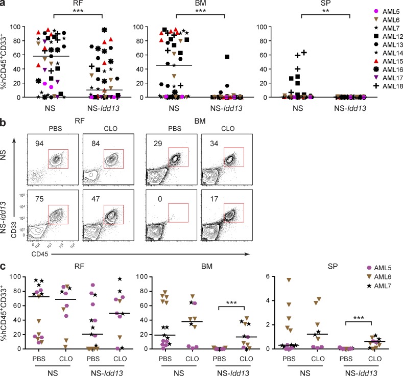
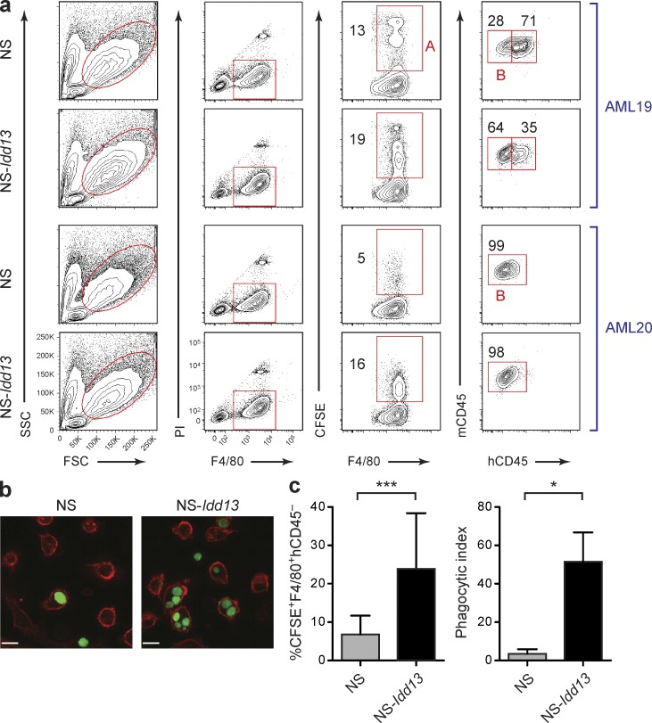
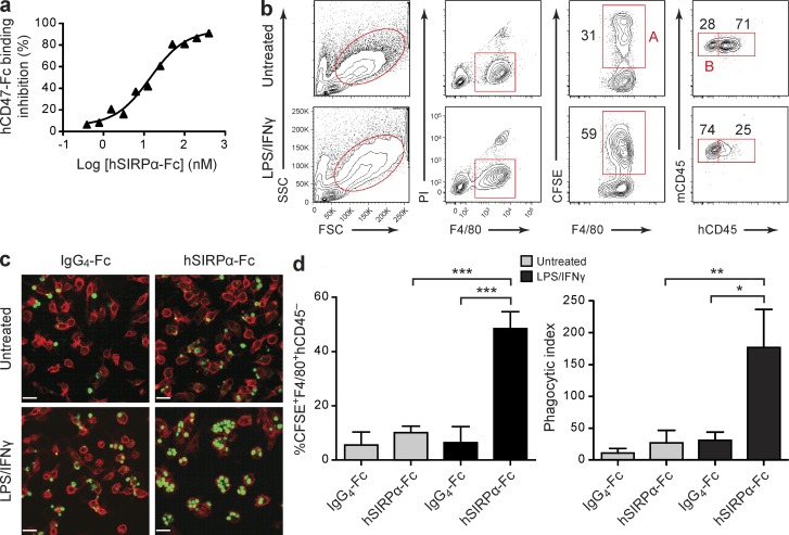
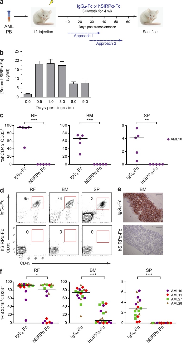
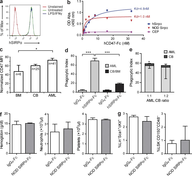
Although tumor surveillance by T and B lymphocytes is well studied, the role of innate immune cells, in particular macrophages, is less clear. Moreover, the existence of subclonal genetic and functional diversity in some human cancers such as leukemia underscores the importance of defining tumor surveillance mechanisms that effectively target the disease-sustaining cancer stem cells in addition to bulk cells. In this study, we report that leukemia stem cell function in xenotransplant models of acute myeloid leukemia (AML) depends on SIRPα-mediated inhibition of macrophages through engagement with its ligand CD47. We generated mice expressing SIRPα variants with differential ability to bind human CD47 and demonstrated that macrophage-mediated phagocytosis and clearance of AML stem cells depend on absent SIRPα signaling. We obtained independent confirmation of the genetic restriction observed in our mouse models by using SIRPα-Fc fusion protein to disrupt SIRPα-CD47 engagement. Treatment with SIRPα-Fc enhanced phagocytosis of AML cells by both mouse and human macrophages and impaired leukemic engraftment in mice. Importantly, SIRPα-Fc treatment did not significantly enhance phagocytosis of normal hematopoietic targets. These findings support the development of therapeutics that antagonize SIRPα signaling to enhance macrophage-mediated elimination of AML.


Figures

Figure 1.
Figure 1.
Anti-CD47 Ab induces phagocytosis of primary human AML cells by macrophages expressing a SIRPα variant that does not bind human CD47. (a) Phagocytosis of human AML cells (AML19) by untreated C57BL/6 or BALB/c macrophages with addition of 7 µg mouse IgG1 or anti-CD47 Ab (B6H12), as assessed by spinning disk confocal microscopy. The phagocytic index was determined as the number of engulfed AML cells per 100 macrophages, performed in triplicate. Bars indicate mean ± SD. ***, P < 0.001 (Student’s t test). (b) Analysis of binding of NS, NS-Idd13, or BALB/c macrophages to human CD47-Fc. NS-Idd13 and C57BL/6 macrophages express an identical SIRPα variant.
Figure 2.
Figure 2.
LSC engraftment in the NOD.SCID xenograft model depends on NOD-derived SIRPα. (a) Flow cytometric analysis of live cells in the BM (two femurs plus two tibias) and SP of NS and NS-Idd13 mice 8 wk after i.v. transplantation of human AML cells. Representative plots are from sample AML3. (b) Summary of human leukemic engraftment (CD45+CD33+) in murine BM and SP 8 wk after i.v. transplantation of AML cells from four patient samples (AML1–4) into NS (n = 14) and NS-Idd13 (n = 11) mice. (c) Flow cytometric analysis of live cells in the right femur (RF) and noninjected BM (left femur plus two tibias) of NS and NS-Idd13 mice 8 wk after transplantation of human AML cells into the RF. Representative plots are from sample AML7. (d) Summary of human leukemic engraftment in the RF, noninjected BM, and SP 8 wk after transplantation of AML cells from seven patient samples (AML1–7) into the RF of NS (n = 23) and NS-Idd13 (n = 21) mice. (e) Human leukemic engraftment in the RF and noninjected BM 8 wk after transplantation of AML cells from samples AML1 and AML9 at three different cell doses into the RF of NS and NS-Idd13 mice, as determined by flow cytometry. (a and c) The percentage of human CD45+CD33+ cells in the live cell gate is indicated. (b, d, and e) Each symbol represents one mouse. Bars indicate median values. *, P < 0.05; **, P < 0.01; and ***, P < 0.001 (b and d, Wald test; e, Tukey’s HSD).
Figure 3.
Figure 3.
LSC engraftment and serial transplantation ability depend on NOD-derived SIRPα. (a) Flow cytometric analysis of live cells in the right femur (RF) and noninjected BM of NS and NS-Idd13 mice 8 wk after transplantation of CD34+CD38 cells from sample AML8 into the RF. (b) Summary of human leukemic engraftment in the RF and noninjected BM 8 wk after transplantation of AML cells from two patient samples (AML6 and AML8) into the RF of NS (n = 10) and NS-Idd13 (n = 8) mice. (c) Protocol for serial transplantation of human AML cells harvested from primary NS and NS-Idd13 mice into secondary NS mice. Equal numbers of human CD45+ cells harvested from the injected RF or noninjected BM of primary engrafted NS or NS-Idd13 were injected into the RF of secondary NS mice. (d) Flow cytometric analysis of live cells harvested from the injected RF of primary NS and NS-Idd13 mice and the injected RF and noninjected BM of secondary NS recipient mice. Representative plots are from sample AML9-RF. (e) Summary of human leukemic engraftment in the injected RF, noninjected BM, and SP of secondary NS mice 10 wk after transplantation of AML cells from two patient samples harvested from primary NS (n = 8) and NS-Idd13 (n = 4) mice. (a and d) The percentage of human CD45+CD33+ cells in the live cell gate is indicated. (b and e) Each symbol represents one mouse. Bars indicate median values. **, P < 0.01; and ***, P < 0.001 (Wald test).
Figure 4.
Figure 4.
Macrophages but not NK cells limit AML engraftment in NS-Idd13 mice. (a) Summary of human leukemic engraftment (CD45+CD33+) in the right femur (RF), noninjected BM (left femur plus two tibias), and SP 8 wk after transplantation of 10 human AML samples into the RF of NS (n = 43) and NS-Idd13 (n = 45) mice treated with anti-CD122 Ab. (b) Flow cytometric analysis of live cells in the RF and noninjected BM of NS and NS-Idd13 mice 8 wk after transplantation of human AML cells into the RF. Mice were treated with PBS liposomes or CLO liposomes 48 h before transplantation and then once per week until sacrificed. The percentage of human CD45+CD33+ cells in the live gate is indicated. Representative plots are from sample AML7. (c) Summary of human leukemic engraftment in RF, noninjected BM, and SP 8 wk after transplantation of AML cells from three patient samples (AML5–7) into the RF of NS (n = 25) and NS-Idd13 (n = 26) mice. (a and c) Each symbol represents one mouse. Bars indicate median values. **, P < 0.01; and ***, P < 0.001 (Wald test).
Figure 5.
Figure 5.
Enhanced phagocytosis of AML cells by NS-Idd13 macrophages. (a) Quantification of phagocytosis of human AML cells by LPS/IFN-γ–treated NS and NS-Idd13 macrophages using flow cytometry. Phagocytosis of CFSE-labeled human primary AML targets was defined as the percentage of mouse macrophages (F4/80+) positive for CFSE (gate A) and negative for human CD45 (gate B). The percentage of mouse macrophages in each gate is indicated. Representative plots are from samples AML19 and AML20. (b) Visualization of engulfment of CFSE-labeled human AML cells by NS and NS-Idd13 LPS/IFN-γ–stimulated macrophages by spinning disk confocal microscopy. Bars, 25 µm. (c) Summary of human AML cell phagocytosis by NS and NS-Idd13 macrophages assessed by flow cytometry (left; n = 6 AML samples) and by spinning disk confocal microscopy (right; n = 3 AML samples). The phagocytic index was determined as the number of engulfed AML cells per 100 macrophages. Bars indicate mean ± SD. *, P < 0.05; and ***, P < 0.001 (Wald test).
Figure 6.
Figure 6.
hSIRPα-Fc fusion protein promotes phagocytosis of human AML cells by activated NS macrophages. (a) hSIRPα-Fc–mediated inhibition of human CD47 binding to SIRPα expressed on NOD macrophages. Data are expressed as percent reduction of hCD47 binding compared with NOD macrophages incubated with hCD47-Fc alone. Representative results from one of two experiments are shown. (b) Flow cytometric analysis of untreated or LPS/IFN-γ–stimulated NS macrophages incubated with CFSE-labeled AML cells from patient AML20 with addition of hSIRPα-Fc. Phagocytosis of AML cells was defined as the percentage of mouse macrophages (F4/80+) positive for CFSE (gate A) and negative for human CD45 (gate B). The percentage of mouse macrophages in each gate is indicated. (c) Visualization of engulfment of CFSE-labeled human AML cells by NS macrophages by spinning disk confocal microscopy. AML cells were coincubated with untreated or LPS/IFN-γ–stimulated macrophages with addition of IgG4-Fc or hSIRPα-Fc. Bars, 25 µm. (d) Summary of human AML cell phagocytosis by untreated or LPS/IFN-γ–stimulated NS macrophages assessed by flow cytometry (left; AML19, AML20, and AML23) and by spinning disk confocal microscopy (right; AML22–26). The phagocytic index was determined as the number of engulfed AML cells per 100 macrophages. Bars indicate mean ± SD. *, P < 0.05; **, P < 0.01; and ***, P < 0.001 (Wald test).
Figure 7.
Figure 7.
Disruption of SIRPα–CD47 interaction by hSIRPα-Fc impairs AML engraftment. (a) Overview of the experimental protocol. Mice were sacrificed 1–3 d after completion of hSIRPα-Fc treatment. (b) Pharmacokinetics of hSIRPα-Fc in NOD mice. Six groups of NOD mice (n = 4 mice/group) were injected intraperitoneally with a single dose of 50 µg hSIRPα-Fc. The mice were sacrificed at the indicated time points after injection, and serum hSIRPα-Fc protein concentration was determined by ELISA. Half-life for hSIRPα-Fc was calculated as 120 h. Bars indicate mean values ± SD. (c) Summary of human leukemic engraftment in the injected right femur (RF), noninjected BM (left femur plus two tibias), and SP of NS mice treated with IgG4-Fc control or hSIRPα-Fc for 4 wk starting 10 d after transplantation of cells from sample AML10 (Approach 1; n = 5 mice per treatment group). (d) Flow cytometric analysis of live cells in the injected RF, noninjected BM, and SP of NS mice treated with IgG4-Fc control or hSIRPα-Fc for 4 wk starting 28 d after transplantation of cells from patient AML11 (Approach 2). The percentage of human CD45+CD33+ cells in the live gate is indicated. (e) Murine BM sections stained for human CD45 from IgG4-Fc– and hSIRPα-Fc–treated mice. Bars, 50 µm. (f) Summary of human leukemic engraftment in the injected RF, noninjected BM, and SP of mice treated with IgG4-Fc control or hSIRPα-Fc for 4 wk starting 28 d after transplantation of cells from four AML samples (AML10, AML11, AML27, and AML28; Approach 2; n = 4 or 5 mice per treatment group). (c and f) Each symbol represents one mouse. Bars indicate median values. *, P < 0.05; **, P < 0.01; and ***, P < 0.001 (Wald test).
Figure 8.
Figure 8.
Disruption of SIRPα–CD47 interaction by hSIRPα-Fc enhances phagocytosis of AML but not normal hematopoietic targets. (a) SIRPα expression on human macrophages, untreated or stimulated with IFN-γ for 24 h and LPS for 1 h before flow cytometric analysis using anti–human SIRPα Ab. (b) Analysis of binding of NOR macrophages infected with lentiviral vectors expressing human SIRPα or NOD mouse SIRPα or empty vector (CEP), as indicated, to human CD47-Fc. (c) Flow cytometric analysis of CD47 expression on CD34+CD38 cells from normal human adult BM, CB, and primary AML patient samples. The number of samples analyzed is indicated. CD47 mean fluorescence intensity (MFI) for each sample was normalized for cell size and CD47 expression level on lymphocytes from the same sample. Error bars indicate SE. *, P < 0.05 (ANOVA). (d) Summary of phagocytosis of human AML cells (AML9, AML19, AML22, AML23, and AML26) or normal mononuclear cells from human CB (n = 2) or BM (n = 4) by LPS/IFN-γ–stimulated human CB-derived macrophages with addition of IgG4-Fc or hSIRPα-Fc as indicated. ***, P < 0.001 (ANOVA). (e) Phagocytosis of human AML (AML29) and CB mononuclear cells coincubated with LPS/IFN-γ–stimulated human CB-derived macrophages with addition of hSIRPα-Fc. *, P < 0.05 (Student’s t test). (d and e) The phagocytic index was determined by spinning disk confocal microscopy as the number of engulfed cells per 100 macrophages, with two to five replicates/sample. Bars indicate mean ± SD. (f) Peripheral blood hemoglobin concentration and neutrophil and platelet counts of NOD mice treated with NOD SIRPα-Fc (n = 4) or IgG4-Fc control (n = 3) for 4 wk. (g) Percentage of Sca1+cKit+ cells in the Lin population (LSK) and CD150+CD48 cells in the LSK fraction in the BM of NOD mice after 4 wk of treatment with NOD SIRPα-Fc or IgG4-Fc control (n = 3 mice/group). (f and g) Bars indicate mean ± SD. No statistically significant differences were observed (Student’s t test).

Similar articles

Cited by

References

    1. Anderson K., Lutz C., van Delft F.W., Bateman C.M., Guo Y., Colman S.M., Kempski H., Moorman A.V., Titley I., Swansbury J., et al. 2011. Genetic variegation of clonal architecture and propagating cells in leukaemia. Nature. 469:356–361 10.1038/nature09650 - DOI - PubMed
    1. Barazi H.O., Li Z., Cashel J.A., Krutzsch H.C., Annis D.S., Mosher D.F., Roberts D.D. 2002. Regulation of integrin function by CD47 ligands. Differential effects on alpha vbeta 3 and alpha 4beta1 integrin-mediated adhesion. J. Biol. Chem. 277:42859–42866 10.1074/jbc.M206849200 - DOI - PubMed
    1. Beatty G.L., Chiorean E.G., Fishman M.P., Saboury B., Teitelbaum U.R., Sun W., Huhn R.D., Song W., Li D., Sharp L.L., et al. 2011. CD40 agonists alter tumor stroma and show efficacy against pancreatic carcinoma in mice and humans. Science. 331:1612–1616 10.1126/science.1198443 - DOI - PMC - PubMed
    1. Bonnet D., Dick J.E. 1997. Human acute myeloid leukemia is organized as a hierarchy that originates from a primitive hematopoietic cell. Nat. Med. 3:730–737 10.1038/nm0797-730 - DOI - PubMed
    1. Brooke G., Holbrook J.D., Brown M.H., Barclay A.N. 2004. Human lymphocytes interact directly with CD47 through a novel member of the signal regulatory protein (SIRP) family. J. Immunol. 173:2562–2570 - PubMed

Publication types

MeSH terms