. 2012 Sep 3;198(5):865-80.
doi: 10.1083/jcb.201204107.

CYK4 inhibits Rac1-dependent PAK1 and ARHGEF7 effector pathways during cytokinesis

Affiliations

CYK4 inhibits Rac1-dependent PAK1 and ARHGEF7 effector pathways during cytokinesis

Ricardo Nunes Bastos et al. J Cell Biol. .

Abstract

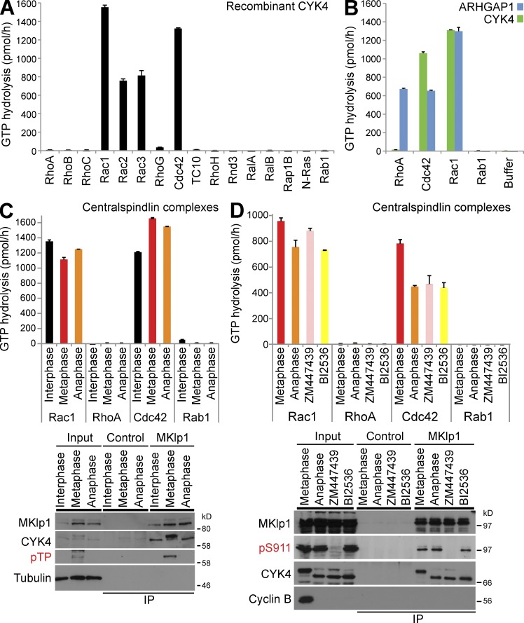
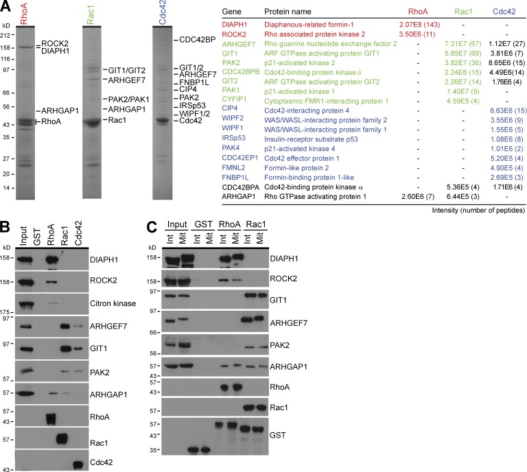
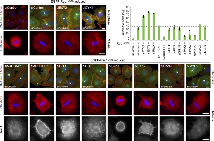
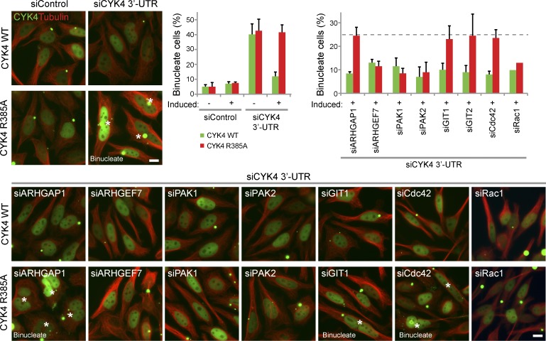
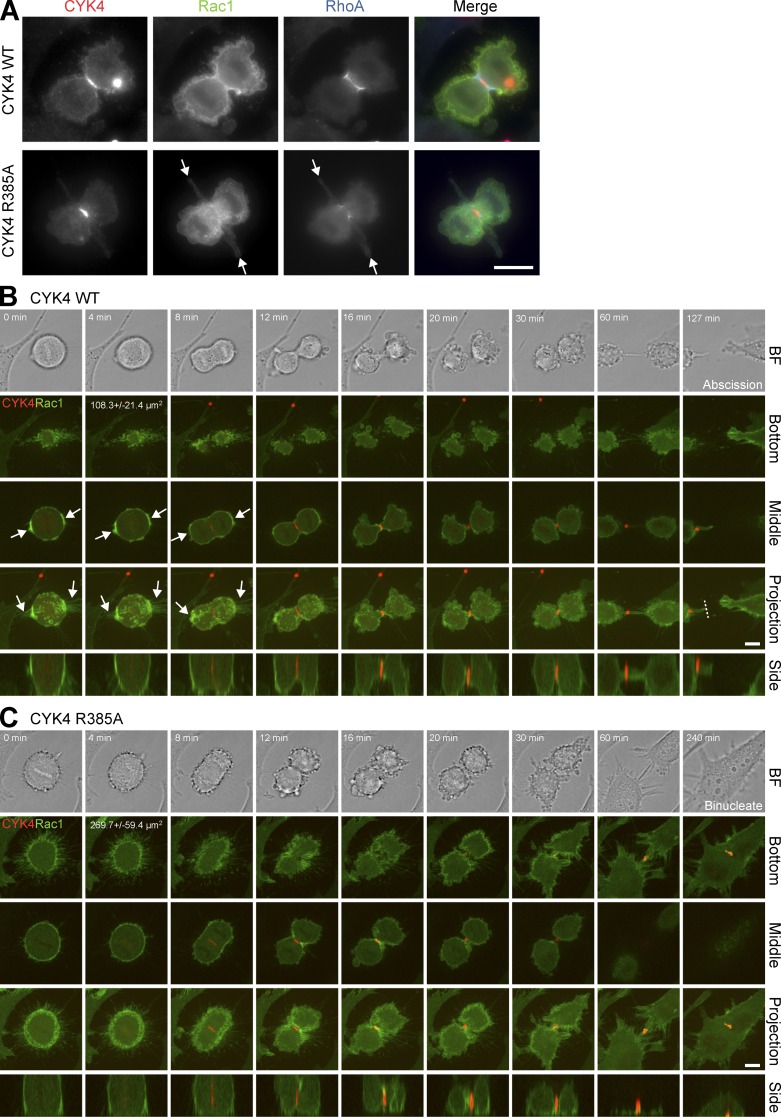
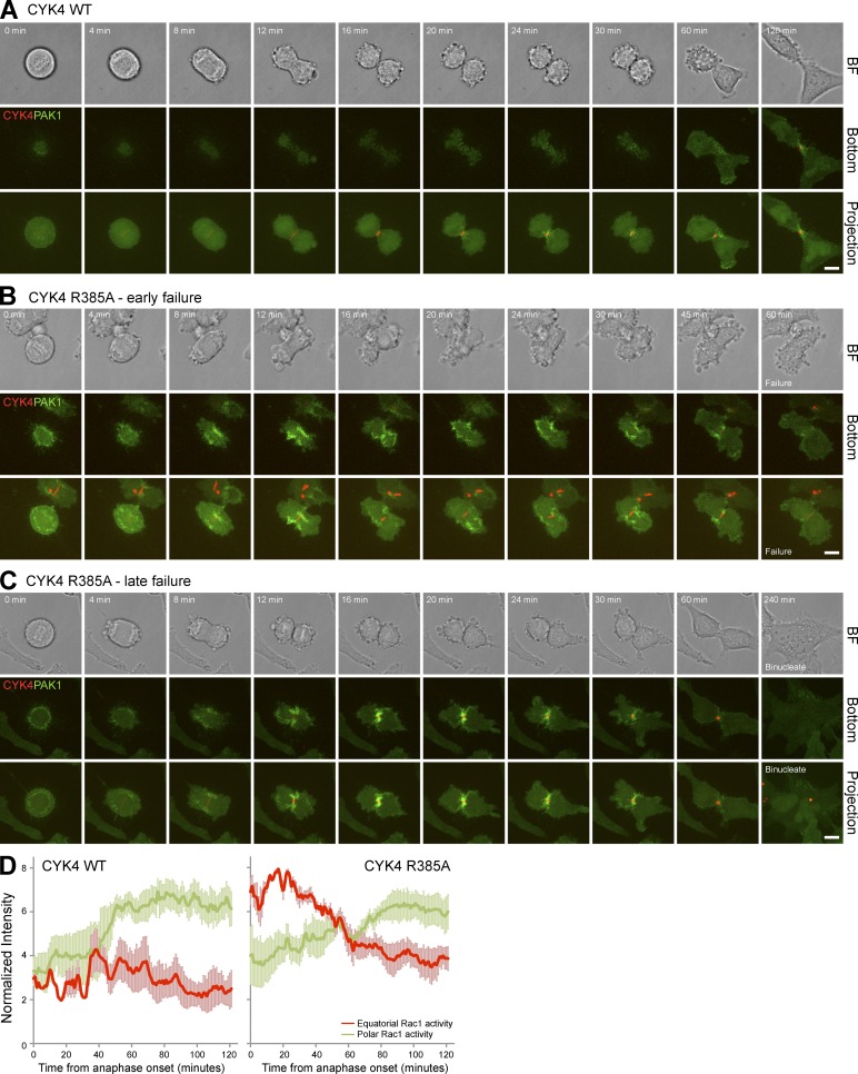
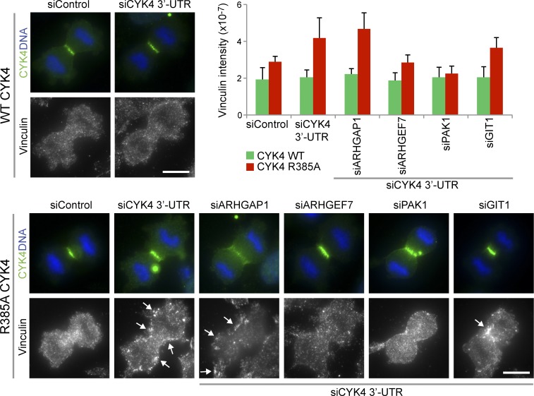
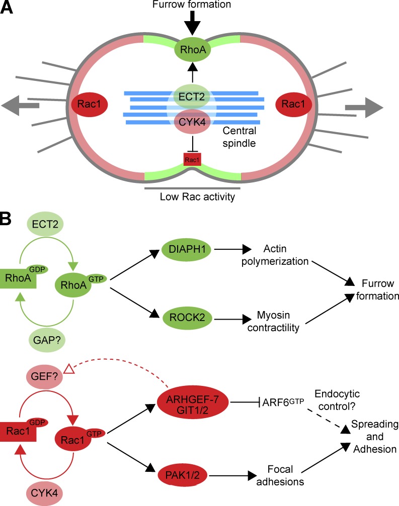
In mitosis, animal cells lose their adhesion to the surrounding surfaces and become rounded. During mitotic exit, they reestablish these adhesions and at the same time physically contract and divide. How these competing processes are spatially segregated at the cell cortex remains mysterious. To address this question, we define the specific effector pathways used by RhoA and Rac1 in mitotic cells. We demonstrate that the MKlp1-CYK4 centralspindlin complex is a guanosine triphosphatase-activating protein (GAP) for Rac1 and not RhoA and that CYK4 negatively regulated Rac1 activity at the cell equator in anaphase. Cells expressing a CYK4 GAP mutant had defects in cytokinesis and showed elevated staining for the cell adhesion marker vinculin. These defects could be rescued by depletion of ARHGEF7 and p21-activated kinase, Rac1-specific effector proteins required for cell adhesion. Based on these findings, we propose that CYK4 GAP activity is required during anaphase to inhibit Rac1-dependent effector pathways associated with control of cell spreading and adhesion.


Figures

Figure 1.
Figure 1.
Human CYK4 is a GAP for Rac and Cdc42 but not RhoA. (A) Recombinant human CYK4 was tested against a representative panel of Rho family GTPases, RalA, RalB, Rap1B, Ras, and Rab1. In brief, 100 pmol of each GTPase was incubated in the presence or absence of 0.5 pmol hexahistidine-tagged CYK4 as described in the Materials and methods. The amount of CYK4-dependent GTP hydrolysis in picomoles per hour was calculated and plotted as a bar graph. (B) Recombinant human ARHGAP1 and CYK4 were tested against RhoA, Rac1, Cdc42, Rab1, or a GAP storage buffer blank. The amount of GAP-dependent GTP hydrolysis in picomoles per hour was calculated and plotted as a bar graph. (C) Endogenous MKlp1–CYK4 centralspindlin complexes were immune precipitated using MKlp1 antibodies from synchronized populations of HeLa cells in interphase, metaphase, or anaphase states. Control isolations were performed using GFP antibodies. These complexes were then used for GAP assays with RhoA, Rac1, Cdc42, and Rab1 as a negative control. The amount of centralspindlin-dependent GTP hydrolysis in picomoles per hour was calculated and plotted as a bar graph. Aliquots of the total cell lysate (input) and isolated complexes were Western blotted for MKlp1, CYK4, phosphothreonine Cdk1-phosphorylated CYK4 (pTP), and tubulin. (D) Endogenous MKlp1–CYK4 centralspindlin complexes were immune precipitated using MKlp1 antibodies from synchronized populations of HeLa cells in metaphase or anaphase cells treated with ZM447439 (Aurora B inhibitor) or BI2536 (Plk1 inhibitor). Control isolations were performed using GFP antibodies. These complexes were then used for GAP assays with RhoA, Rac1, Cdc42, and Rab1 as a negative control. The amount of centralspindlin-dependent GTP hydrolysis in picomoles per hour was calculated and plotted as a bar graph. Aliquots of the total cell lysate (input) and isolated complexes were Western blotted for MKlp1, phosphoserine 911 Aurora-phosphorylated MKlp1 (pS911), CYK4, and tubulin. Error bars indicate the standard deviations. IP, immunoprecipitation.
Figure 2.
Figure 2.
Arginine finger mutations reduce CYK4 GAP activity toward Rac1. (A) A schematic of CYK4 shows the conserved coiled coil, C1 putative membrane-binding region, and GAP domain with arginine finger residue at amino acid 385. Recombinant human ARHGAP1, CYK4, CYK4R385A, CYK4S387A, and CYK4S387D were tested against RhoA, Rac1, Cdc42, and Rab1. The amount of GAP-dependent GTP hydrolysis in picomoles per hour was calculated and plotted as a bar graph. (B). HeLa cells were transfected with Myc-tagged CYK4, CYK4R385A, CYK4S387A, and CYK4S387D for 24 h, fixed, and then stained with antibodies to the Myc epitope, MKlp1, or tubulin. DNA was stained with DAPI. Bar, 10 µm. (C) HeLa cells stably expressing inducible Myc-tagged copies of CYK4, CYK4R385A, CYK4S387A, and CYK4S387D were treated for 36 h with an siRNA directed to the 3′-UTR of CYK4 to deplete the endogenous protein or a control siRNA. At the same time, the cells were treated with doxycycline to induce the Myc-tagged transgenes. MKlp1–CYK4 centralspindlin complexes were then immune precipitated using Myc antibodies. Control isolations were performed using GFP antibodies. These complexes were then used for GAP assays with RhoA and Rac1. The amount of centralspindlin-dependent GTP hydrolysis in picomoles per hour was calculated and plotted as a bar graph. Aliquots of the total cell lysate (input) and isolated complexes were Western blotted for Myc, CYK4, MKlp1, and tubulin. Arrowheads indicate endogenous CYK4, and arrows show Myc-tagged CYK4. Error bars indicate the standard deviations. IP, immunoprecipitation; WT, wild type.
Figure 3.
Figure 3.
Rac1 inactivation by CYK4 is required for cytokinesis. (A) HeLa cells stably expressing inducible Myc-tagged copies of CYK4 or CYK4R385A were treated for 36 h with an siRNA directed to the 3′-UTR of CYK4 to deplete the endogenous protein or a control siRNA. At the same time, the cells were treated with doxycycline to induce the Myc-tagged transgenes (induced) or left untreated (uninduced). The cells were fixed and then stained with antibodies to tubulin, the Myc epitope, or DAPI to detect DNA. The number of binucleate cells was counted for each condition and plotted as a bar graph. Error bars show the standard deviation from the means (n = 3). Asterisks mark binucleate cells that have failed cytokinesis. (B) HeLa cells stably expressing inducible EGFP-tagged copies of Rac1 or activated Rac1Q61L were treated for the time indicated with doxycycline to induce the EGFP-tagged transgenes. The cells were fixed and then stained with antibodies to tubulin or DAPI to detect DNA. Rac1 was directly visualized by EGFP fluorescence. The number of binucleate cells was counted for each condition and plotted as in the line graph. (C) Alternatively, the cells were fixed using TCA and stained for RhoA and with antibodies to EGFP to detect EGFP-Rac1. DAPI was used to detect DNA. siControl, nonsilencing control; WT, wild type. Bars, 10 µm.
Figure 4.
Figure 4.
RhoA and Rac1 couple to discrete effector pathways in mitotic cells. (A) Effector binding assays from mitotic HeLa cell lysate were performed using GST, RhoA, Rac1, and Cdc42 as described in the Materials and methods. Bound fractions were eluted using sample buffer and analyzed by SDS-PAGE and MS. Coomassie brilliant blue–stained gels of the RhoA, Rac1, and Cdc42 complexes are shown. Proteins identified by MS are listed in the table and marked by the side of the appropriate gel. Peptide number and intensity give a measure of the relative abundance. (B) Western blot analysis of the input and bound effector proteins was performed using the antibodies shown in the figure. (C) Effector binding assays from interphase (Int) and mitotic (Mit) HeLa cell lysate were performed using GST, RhoA, and Rac1 as described in the Materials and methods. Samples of the input material and bound fractions were analyzed by Western blotting using the antibodies shown in the figure.
Figure 5.
Figure 5.
Depletion of Rac1 effectors rescues the cytokinesis defect of cells expressing a hydrolysis-defective Rac1 mutant. HeLa cells expressing inducible EGFP-Rac1Q61L were transfected with siRNA duplexes targeting CYK4, ECT2, Rac1-specific effectors, Cdc42, a nonsilencing control (siControl), or Myh9, which was a protein binding nonspecifically to Sepharose beads in Rho GTPase pull-downs. Rac1Q61L expression was induced for 48 h, and the cells were fixed and then stained with phalloidin to detect actin or DAPI to detect DNA. Rac1 was directly visualized by EGFP fluorescence. Binucleate cells are marked by asterisks. The number of binucleate cells was counted for each condition and plotted as in the bar graph. Error bars indicate the standard deviation from the means (n = 3). The dotted line indicates the extent of cytokinesis failure. Bars, 10 µm.
Figure 6.
Figure 6.
Depletion of Rac1 effectors rescues the cytokinesis defect of cells expressing the CYK4 arginine finger mutant. HeLa cells expressing inducible EGFP-CYK4 or GAP activity–defective CYK4R385A were transfected with control or CYK4 3′-UTR siRNA duplexes in combination with siRNA duplexes targeting Rac1-specific effectors and Cdc42. CYK4 expression was induced for 60 h, and then, the cells were fixed and stained with antibodies to tubulin and DAPI to detect DNA. CYK4 was directly visualized by EGFP fluorescence. Binucleate cells are marked with asterisks. The number of binucleate cells was counted for each condition and plotted as in the bar graph. Error bars indicate the standard deviation from the means (n = 3). The dotted line indicates the extent of cytokinesis failure. siControl, nonsilencing control; WT, wild type. Bars, 10 µm.
Figure 7.
Figure 7.
Cell surface projections emerge from the cleavage furrow in CYK4 GAP-defective cells. (A) HeLa cells expressing EGFP-Rac1 and inducible mCherry-CYK4 or GAP activity–defective CYK4R385A were transfected with control or CYK4 3′-UTR siRNA duplexes. CYK4 expression was induced for 60 h, and then, the cells were TCA fixed and stained with antibodies to mCherry to detect CYK4, RhoA, or EGFP to detect Rac1. Arrows mark the cell surface projections seen emerging from the cleavage furrow region in CYK4R385A cells. (B and C) Alternatively, these CYK4 (B) or CYK4R385A (C) cells were used for time-lapse imaging. A bright-field (BF) image is shown at the time points indicated together with the bottom section where the cells touch the glass growth surface, a middle section through the cell equator, a maximum intensity projection along the z axis of all sections, or a side projection along the y axis. The dotted line indicates where abscission has occurred. The 4-min time point, bottom section, includes a quantitation of the area of the cell in contact with the surface (n = 20). Arrows mark Rac1 accumulation at the cell poles and associated retraction fibers. WT, wild type. Bars, 10 µm.
Figure 8.
Figure 8.
Increased Rac1 activity in the cleavage furrow of CYK4 GAP-defective cells. (A–C) HeLa cells expressing EGFP-PAK1 CRIB domain and inducible mCherry-CYK4 (A) or GAP activity–defective CYK4R385A (B and C) were transfected with CYK4 3′-UTR siRNA duplexes and then used for time-lapse imaging. A bright-field (BF) image is shown at the time points indicated together with the bottom section where the cells touch the glass growth surface and a maximum intensity projection along the z axis of all sections. Timings are relative to the onset of anaphase. Cells expressing only GAP activity–defective CYK4R385A showed either early (B) or late (C) stage failure of cytokinesis, and examples of both outcomes are therefore shown. CYK4 wild-type cells underwent abscission after 171.1 ± 29.8 min, whereas CYK4R385A cells showed early failure after 81.0 ± 21.8 min or late failure after 293.1 ± 53.6 min (n = 29). Bars, 10 µm. (D) Rac1 activity at the cell equator (red) and cell poles (green) was determined by measuring the intensity of the PAK1 CRIB domain probe every minute as cells exited mitosis using the Volocity 5 volume and quantitation tools. This was performed on cells expressing wild-type and R385A GAP mutant CYK4. Mean values are plotted in the graphs, and error bars indicate the standard deviation from the means (n = 5). WT, wild type.
Figure 9.
Figure 9.
Visualization of adhesion complexes using vinculin staining in CYK4 GAP-defective cells in anaphase. HeLa cells expressing inducible EGFP-CYK4 or GAP activity–defective CYK4R385A were transfected with control or CYK4 3′-UTR siRNA duplexes in combination with siRNA duplexes targeting Rac1-specific effectors. CYK4 expression was induced for 60 h, and the cells were fixed and then stained with antibodies to GFP to detect CYK4 and vinculin and DAPI to detect DNA. Arrows indicate especially bright vinculin spots. The vinculin staining intensity was measured using ImageJ and is plotted in the bar graph. Error bars indicate the standard deviation from the means (n = 7). siControl, nonsilencing control; WT, wild type. Bars, 10 µm.
Figure 10.
Figure 10.
A model for CYK4 regulation of Rac1 activity in anaphase. (A) At the onset of anaphase, cells adhere at the cell poles and contract in the equatorial region, driven by Rac1 and RhoA, respectively. The RhoA exchange factor ECT2 is enriched at the cell equator because of interactions with the MKlp1–CYK4 centralspindlin complex. This leads to local activation of RhoA in the furrow region (shaded pale green). Rac1 is inactivated in this region by the CYK4 GAP component of centralspindlin, creating a zone of low Rac1 activity. This does not occur at the cell poles, so Rac1 activity is higher in these regions (shaded pale red). Central spindle microtubules are shown in pale blue. (B) Regulatory and effector pathways for RhoA and Rac1 are summarized, together with the events they are known to control. The identity of the mitotic RhoA GAP remains to be assigned. GIT1/2 have been previously assigned as ARF6 GAPs and are therefore shown as inhibitors of ARF6 function. ARF6 has a complex function in cytokinesis together with Rab35 and modulates endocytic recycling (Chesneau et al., 2012).

Comment in

Similar articles

Cited by

References

    1. Barr F.A., Gruneberg U. 2007. Cytokinesis: placing and making the final cut. Cell. 131:847–860 10.1016/j.cell.2007.11.011 - DOI - PubMed
    1. Bassi Z.I., Verbrugghe K.J., Capalbo L., Gregory S., Montembault E., Glover D.M., D’Avino P.P. 2011. Sticky/Citron kinase maintains proper RhoA localization at the cleavage site during cytokinesis. J. Cell Biol. 195:595–603 10.1083/jcb.201105136 - DOI - PMC - PubMed
    1. Canman J.C., Lewellyn L., Laband K., Smerdon S.J., Desai A., Bowerman B., Oegema K. 2008. Inhibition of Rac by the GAP activity of centralspindlin is essential for cytokinesis. Science. 322:1543–1546 10.1126/science.1163086 - DOI - PMC - PubMed
    1. Castrillon D.H., Wasserman S.A. 1994. Diaphanous is required for cytokinesis in Drosophila and shares domains of similarity with the products of the limb deformity gene. Development. 120:3367–3377 - PubMed
    1. Chesneau L., Dambournet D., Machicoane M., Kouranti I., Fukuda M., Goud B., Echard A. 2012. An ARF6/Rab35 GTPase cascade for endocytic recycling and successful cytokinesis. Curr. Biol. 22:147–153 10.1016/j.cub.2011.11.058 - DOI - PubMed

Publication types

MeSH terms