

Age-related influence of vision and proprioception on Ia presynaptic inhibition in soleus muscle during upright stance
- PMID: 22946095
- PMCID: PMC3515837
- DOI: 10.1113/jphysiol.2012.228932
Age-related influence of vision and proprioception on Ia presynaptic inhibition in soleus muscle during upright stance
Abstract
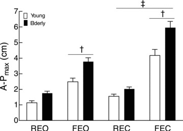
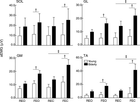
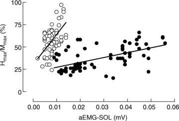
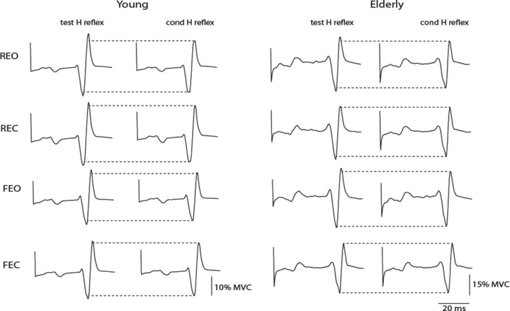
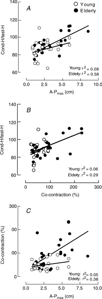
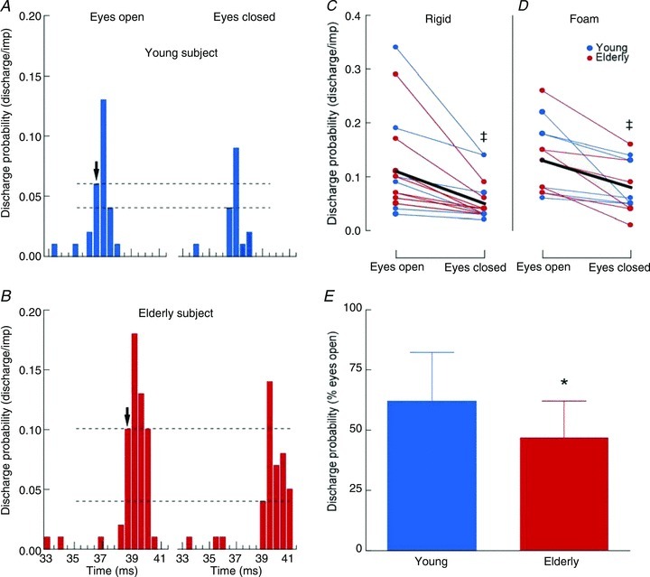
This study investigated the modulation of Ia afferent input in young and elderly adults during quiet upright stance in normal and modified visual and proprioceptive conditions. The surface EMG of leg muscles, recruitment curve of the soleus (SOL) Hoffmann (H) reflex and presynaptic inhibition of Ia afferents from SOL, assessed with the D1 inhibition and single motor unit methods, were recorded when young and elderly adults stood with eyes open or closed on two surfaces (rigid vs. foam) placed over a force platform. The results showed that elderly adults had a longer path length for the centre of pressure and larger antero-posterior body sway across balance conditions (P < 0.05). Muscle EMG activities were greater in elderly compared with young adults (P < 0.05), whereas the H(max) expressed as a percentage of the H(max) was lower (P = 0.048) in elderly (38 ± 16%) than young adults (58 ± 16%). The conditioned H reflex/test H reflex ratio (D1 inhibition method) increased with eye closure and when standing on foam (P < 0.05), with greater increases for elderly adults (P = 0.019). These changes were accompanied by a reduced peak motor unit discharge probability when standing on rigid and foam surfaces (P 0.001), with a greater effect for elderly adults (P = 0.026). Based on these latter results, the increased conditioned H reflex/test H reflex ratio in similar sensory conditions is likely to reflect occlusion at the level of presynaptic inhibitory interneurones. Together, these findings indicate that elderly adults exhibit greater modulation of Ia presynaptic inhibition than young adults with variation in the sensory conditions during upright standing.
Figures
Similar articles
-
Vision and proprioception do not influence the excitability of the corticomotoneuronal pathway during upright standing in young and elderly adults.Neuroscience. 2014 May 30;268:247-54. doi: 10.1016/j.neuroscience.2014.03.026. Epub 2014 Mar 22. Neuroscience. 2014. PMID: 24662846
-
Presynaptic inhibition of soleus Ia afferents does not vary with center of pressure displacements during upright standing.Neuroscience. 2015 Jul 9;298:63-73. doi: 10.1016/j.neuroscience.2015.04.010. Epub 2015 Apr 11. Neuroscience. 2015. PMID: 25869621
-
Input-output characteristics of soleus homonymous Ia afferents and corticospinal pathways during upright standing differ between young and elderly adults.Acta Physiol (Oxf). 2014 Mar;210(3):667-77. doi: 10.1111/apha.12233. Acta Physiol (Oxf). 2014. PMID: 24433254
-
Contributions to the understanding of gait control.Dan Med J. 2014 Apr;61(4):B4823. Dan Med J. 2014. PMID: 24814597 Review.
-
Soleus H-reflex and its relation to static postural control.Gait Posture. 2011 Feb;33(2):169-78. doi: 10.1016/j.gaitpost.2010.12.008. Epub 2011 Jan 5. Gait Posture. 2011. PMID: 21211976 Review.
Cited by
-
The Effect of Lightly Gripping a Cane on the Dynamic Balance Control.Open Biomed Eng J. 2015 Jul 23;9:146-50. doi: 10.2174/1874120701509010146. eCollection 2015. Open Biomed Eng J. 2015. PMID: 26312075 Free PMC article.
-
Cognitive demand does not influence the responsiveness of homonymous Ia afferents pathway during postural dual task in young and elderly adults.Eur J Appl Physiol. 2014 Feb;114(2):295-303. doi: 10.1007/s00421-013-2775-8. Epub 2013 Nov 19. Eur J Appl Physiol. 2014. PMID: 24248857
-
Spinal and corticospinal pathways are differently modulated when standing at the bottom and the top of a three-step staircase in young and older adults.Eur J Appl Physiol. 2017 Jun;117(6):1165-1174. doi: 10.1007/s00421-017-3603-3. Epub 2017 Apr 13. Eur J Appl Physiol. 2017. PMID: 28409395
-
Age-related reversal of spinal excitability during anticipatory postural control.Eur J Appl Physiol. 2018 Dec;118(12):2577-2585. doi: 10.1007/s00421-018-3982-0. Epub 2018 Sep 4. Eur J Appl Physiol. 2018. PMID: 30182185 Free PMC article.
-
Cortical Engagement Metrics During Reactive Balance Are Associated With Distinct Aspects of Balance Behavior in Older Adults.Front Aging Neurosci. 2021 Jul 14;13:684743. doi: 10.3389/fnagi.2021.684743. eCollection 2021. Front Aging Neurosci. 2021. PMID: 34335230 Free PMC article.
References
-
- Baudry S, Lecoeuvre G, Duchateau J. Age-related changes in the behavior of the muscle-tendon unit of the gastrocnemius medialis during upright stance. J Appl Physiol. 2012;112:296–304. - PubMed
-
- Benjuya N, Melzer I, Kaplanski J. Aging-induced shifts from a reliance on sensory input to muscle cocontraction during balanced standing. J Gerontol A Biol Sci Med Sci. 2004;59:166–171. - PubMed
-
- Billot M, Simoneau EM, Van Hoecke J, Martin A. Age-related relative increases in electromyography activity and torque according to the maximal capacity during upright standing. Eur J Appl Physiol. 2010;109:669–680. - PubMed
-
- Burke D, Adams RW, Skuse NF. The effects of voluntary contraction on the H reflex of human limb muscles. Brain. 1989;112:417–433. - PubMed
-
- Capaday C, Lavoie BA, Comeau F. Differential effects of a flexor nerve input on the human soleus H-reflex during standing versus walking. Can J Physiol Pharmacol. 1995;73:436–449. - PubMed
Publication types
MeSH terms
LinkOut - more resources
Full Text Sources
Medical