. 2012 Sep 4:13:451.
doi: 10.1186/1471-2164-13-451.

Using BAC transgenesis in zebrafish to identify regulatory sequences of the amyloid precursor protein gene in humans

Affiliations

Using BAC transgenesis in zebrafish to identify regulatory sequences of the amyloid precursor protein gene in humans

Leighcraft A Shakes et al. BMC Genomics. .

Abstract

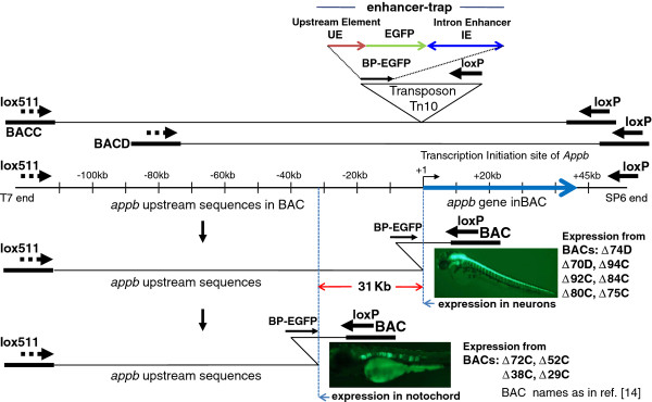
Background: Non-coding DNA in and around the human Amyloid Precursor Protein (APP) gene that is central to Alzheimer's disease (AD) shares little sequence similarity with that of appb in zebrafish. Identifying DNA domains regulating expression of the gene in such situations becomes a challenge. Taking advantage of the zebrafish system that allows rapid functional analyses of gene regulatory sequences, we previously showed that two discontinuous DNA domains in zebrafish appb are important for expression of the gene in neurons: an enhancer in intron 1 and sequences 28-31 kb upstream of the gene. Here we identify the putative transcription factor binding sites responsible for this distal cis-acting regulation, and use that information to identify a regulatory region of the human APP gene.

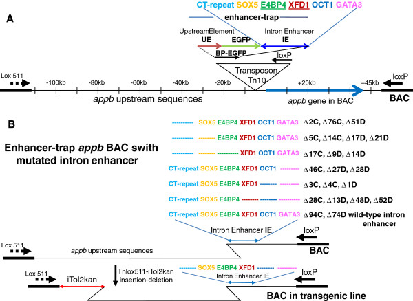
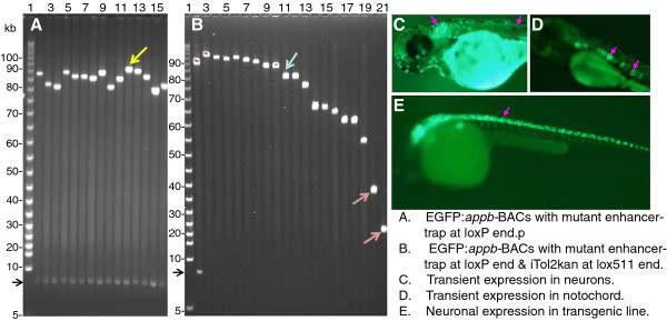
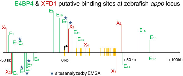
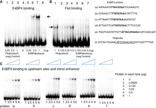
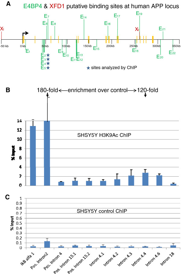
Results: Functional analyses of intron 1 enhancer mutations in enhancer-trap BACs expressed as transgenes in zebrafish identified putative binding sites of two known transcription factor proteins, E4BP4/ NFIL3 and Forkhead, to be required for expression of appb. A cluster of three E4BP4 sites at -31 kb is also shown to be essential for neuron-specific expression, suggesting that the dependence of expression on upstream sequences is mediated by these E4BP4 sites. E4BP4/ NFIL3 and XFD1 sites in the intron enhancer and E4BP4/ NFIL3 sites at -31 kb specifically and efficiently bind the corresponding zebrafish proteins in vitro. These sites are statistically over-represented in both the zebrafish appb and the human APP genes, although their locations are different. Remarkably, a cluster of four E4BP4 sites in intron 4 of human APP exists in actively transcribing chromatin in a human neuroblastoma cell-line, SHSY5Y, expressing APP as shown using chromatin immunoprecipitation (ChIP) experiments. Thus although the two genes share little sequence conservation, they appear to share the same regulatory logic and are regulated by a similar set of transcription factors.

Conclusion: The results suggest that the clock-regulated and immune system modulator transcription factor E4BP4/ NFIL3 likely regulates the expression of both appb in zebrafish and APP in humans. It suggests potential human APP gene regulatory pathways, not on the basis of comparing DNA primary sequences with zebrafish appb but on the model of conservation of transcription factors.


Figures

Figure 1
Figure 1
Scanning the appb genomic region of zebrafish by enhancer trapping using BACs represented schematically (summary of results from reference[14]). The two BACs C and D used in this study overlap one another and contain different lengths of sequences upstream of appb gene. They are shown schematically as the top two lines. The inverted triangle represents enhancer-trap in Tn10 transposon, which is comprised of 0.35 kb of DNA immediately upstream of appb (UE), followed by EGFP gene with basal promoter, and ~1 kb intron 1 enhancer (IE). The entire enhancer-trap cassette is named BP-EGFP. The appb gene region in the BACs, with the thick blue arrow to represent the total length of exons and introns of the gene, is drawn to scale and shown below it. Insertion of the enhancer-trap into the appb BAC DNA and subsequent Cre recombination between the transposed loxP and the BAC-end loxP deletes the BAC DNA from that end and simultaneously inserts the enhancer-trap (shown as BP-EGFP). The enhancer-trap is in front of loxP in the transposon and is retained in the BAC after Cre-mediated loxP-loxP deletion. This end-truncation is represented by the bent line to illustrate the location of that transposon-end retained in the BAC after the loxP-Cre deletion. The earlier study [14] found that appb BACs that had the enhancer-trap located close to the appb transcription start site expressed EGFP fluorescence in neurons (e.g. BACs Δ74D, Δ70D, Δ94C, Δ92C Δ84C, Δ80C, Δ75C), while appb BACs that had the enhancer-trap inserted further upstream beyond -31 kb of the appb gene expressed EGFP fluorescence in the notochord (e.g. BACs Δ72C, Δ52C, Δ38C, Δ29C). The vertical blue dotted lines, separated by ~31 kb, mark these locations on the appb BACs. The names of BACs are indicated adjacent to the pictures of EGFP expression in zebrafish neurons or the notochord.
Figure 2
Figure 2
Panel A: Schematic representation of bio-informatically predicted transcription factor binding sites in the intron 1 enhancer (IE) within the enhancer-trap (taken from Additional file 2: Figure S2 of reference [14]), highlighted by the colored letters. Panel B: Mutated enhancer-trap BACs used in this study: Mutations and deletions, represented schematically with dotted lines of same color in place of the letters, were engineered into the intron enhancer (IE) in the small enhancer-trap transposon plasmid at the sites marked by CT-repeat, SOX5, E4BP4, XFD1, OCT1, and GATA3. Each mutated enhancer-trap transposon was inserted into appb BACs C and D to generate the enhancer-trap BACs indicated on the side of the corresponding mutation. For example the blue dashed line in the first row indicates a deletion of the CT-repeat sequence in enhancer-trap (shown as dotted blue line), and the BACs containing this mutation are Δ2C, Δ76C and Δ51D. Because SOX5 and E4BP4 sites overlapped, only point mutations were introduced into the SOX5 site to obtain the plasmid with wild type E4BP4 and point mutation in SOX5 and deletion of CT-repeat, shown in second row Panel B. Enhancer-trap BACs with these mutations are Δ5C Δ14C, Δ17D Δ21D. Row 7 shows the wild type enhancer-trap with Δ94C, and Δ74D as representatives. The results of expressions of these BACs are summarized in Table 1. The last enhancer-trap BAC in Panel B (row 8) has enhancer-trap deleted for CT-repeat, OCT1 and GATA3. This BAC is also deleted from the lox511 end of BAC with the Tnlox511-iTol2kan to make the germline transgenic zebrafish shown in Figure 3, Panel E. The bent lines on both ends of the BAC represent end-truncations by the transposons, and illustrate the location of the particular transposon end preserved after either the loxP-loxP or lox511-lox511 deletions mediated by Cre protein, respectively.
Figure 3
Figure 3
Panel A: FIGE analysis of enhancer-trap BACs with mutated intron 1 enhancer from different libraries. Panel B: FIGE analysis of clone DNA from the library generated by inserting Tnlox511-iTol2kan at the lox511 end of the enhancer-trap BAC in lane 12, Panel A (marked by yellow arrowhead). Panel C: EGFP fluorescence from transient expression in neurons (marked by the pink arrowheads) of zebrafish injected with mutated but functional intron enhancer-trap BAC with intact upstream DNA, Panel D: EGFP expression in notochord (indicated with pink arrowheads) from injecting enhancer trap BAC with mutated but functional intron enhancer and with 31 kb upstream DNA deleted (such as clone in lane 21, panel B, red arrowhead) taken from the same library as the BAC used for Panel C. Panel E: EGFP fluorescence in neurons (marked by the pink arrowhead) from a F2 transgenic zebrafish line obtained from the enhancer-trap BAC shown in lane 11 of Panel B (marked by blue arrowhead). The mutated but functional intron enhancer used was deleted for GATA3, OCT1 and the CT-repeat element (clone shown schematically in row 8, Panel B of Figure 2). Additional examples of germline transgenic fish with slightly smaller enhancer-trap BAC transgenes, but containing the upstream ~31 kb sequence, are shown in Figure 6 panels A and B of reference [23]. The BAC vector DNA band from Not I digestion is shown by the black arrowhead to the left of panels A and B. Lane 2, Panel B, contains the same DNA as lane 12, Panel A. Lanes 3–21 in panel B do not have this band because insertion of Tnlox511-iTol2kan at the lox511 end of BAC DNA and subsequent lox511-lox511 deletion eliminates the Not I site at that end [23].
Figure 4
Figure 4
Location of E4BP4 (E, in green), and XFD1 (X, in red), sites in non-coding DNA within zebrafish appb and surrounding 50 kb DNA. The green and red vertical lines above or below the horizontal line indicate sites in the forward and reverse strand of DNA, respectively. The short yellow vertical bars indicate exons of appb. Stars mark the E4BP4 and XFD1 sites shown to specifically bind the DNA-binding domains of zebrafish E4BP4 or Forkhead proteins respectively, by EMSA. The bent arrow indicates transcription start site of appb gene.
Figure 5
Figure 5
The DNA binding domain common to members of zebrafish E4BP4 or Forkhead (Fkd) families were each expressed in E. coli, partially purified under non-denaturing conditions, and tested for specific binding to their recognition sequences identified in and around the zebrafish appb gene. Panel A: E4BP4 protein, indicated below each lane in μg, added to Fkd probe (f), lanes 1–3, or the E4BP4 probe (e), lanes 4–8. Panel B: Fkd protein, indicated below each lane in μg, added to Fkd probe (f), lanes 1–4, or E4BP4 probe (e), lanes 5–7. Panel C: E4BP4 protein binding to probes (a through f) spanning upstream E4BP4 sites (a-d), E4BP4 site in intron 1 enhancer (e), and Fkd site in intron enhancer (f). E4BP4 protein used in each lane (μg) is indicated on right. Probes (b-d) correspond to cluster of three E4BP4 sites at −31 kb.
Figure 6
Figure 6
Panel A: Location of E4BP4 (E, green) and XFD1 (X, red) sites in non-coding DNA within the human APP gene and surrounding 50 kb DNA. The green and red vertical lines above or below the horizontal line indicate sites in the forward and reverse strand of DNA, respectively. The short yellow vertical bars indicate exons of APP. The first three exons are very close to one another near the transcription start site. Stars indicate the E4BP4 sites marked by H3K9Ac in chromatin immunoprecipitation (ChIP) assays in a human cell-line SHSY5Y expressing APP gene. The bent arrow indicates transcription start site of APP gene. The SHSY5Y cells were analyzed by ChIP using anti-H3K9Ac antibodies as described in Methods. Y-axis represents the amount of material present in anti-H3K9Ac (Panel B) or non-specific IgG (Panel C) immunoprecipitates compared to input chromatin used for the assay. Note the different scales for the two panels. Results shown are the average of three independent ChIP experiments, each of which was assayed in duplicates. Error bars represent standard deviation between experiments. Amplification from IkBα served as a positive control. The fold enrichment, over control IgG, of H3K9Ac activity at the APP intron 2 site (positive control) and the intron 4 sites are 180-fold versus 120-fold, respectively, as indicated at the top of Panel 6B.

Similar articles

Cited by

References

    1. Dillen K, Annaert W. A two decade contribution of molecular cell biology to the centennial of Alzheimer’s disease: are we progressing toward therapy? Int Rev Cytol. 2006;254:215–300. - PubMed
    1. Perneczky R, Tsolakidou A, Arnold A, Diehl-Schmid J, Grimmer T, Förstl H, Kurz A, Alexopoulos P. CSF soluble amyloid precursor proteins in the diagnosis of incipient Alzheimer disease. Neurology. 2011;77:35–38. doi: 10.1212/WNL.0b013e318221ad47. - DOI - PubMed
    1. Theuns J, Brouwers N, Engelborghs S, Sleegers K, Bogaerts V, Corsmit E, De Pooter T, van Duijn CM, De Deyn PP, Van Broeckhoven C. Promoter mutations that increase amyloid precursor-protein expression are associated with Alzheimer disease. Am J Hum Genet. 2006;78:936–946. doi: 10.1086/504044. - DOI - PMC - PubMed
    1. Salbaum JM, Weidemann A, Lemaire HG, Masters CL, Beyreuther K. The promoter of Alzheimer’s disease amyloid A4 precursor gene. EMBO J. 1988;7:2807–2813. - PMC - PubMed
    1. Yoshikai SI, Sasaki H, Dohura K, Fururya H, Sakaki Y. Genomic organization of the human amyloid beta-protein precursor gene. Gene. 1990;87:257–263. doi: 10.1016/0378-1119(90)90310-N. - DOI - PubMed

Publication types

MeSH terms

Substances