

Induction of pulmonary mucosal immune responses with a protein vaccine targeted to the DEC-205/CD205 receptor
- PMID: 22947140
- PMCID: PMC3461253
- DOI: 10.1016/j.vaccine.2012.08.051
Induction of pulmonary mucosal immune responses with a protein vaccine targeted to the DEC-205/CD205 receptor
Abstract
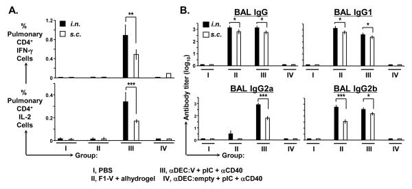
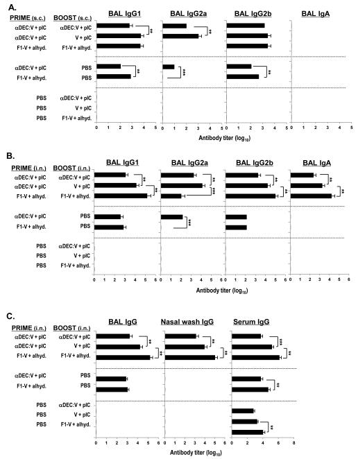
It is of great interest to develop a pneumonic plague vaccine that would induce combined humoral and cellular immunity in the lung. Here we investigate a novel approach based on targeting of dendritic cells using the DEC-205/CD205 receptor (DEC) via the intranasal route as way to improve mucosal cellular immunity to the vaccine. Intranasal administration of Yersinia pestis LcrV (V) protein fused to anti-DEC antibody together with poly IC as an adjuvant induced high frequencies of IFN-γ secreting CD4(+) T cells in the airway and lung as well as pulmonary IgG and IgA antibodies. Anti-DEC:LcrV was more efficient to induce IFN-γ/TNF-α/IL-2 secreting polyfunctional CD4(+) T cells when compared to non-targeted soluble protein vaccine. In addition, the intranasal route of immunization with anti-DEC:LcrV was associated with improved survival upon pulmonary challenge with the virulent CO92 Y. pestis. Taken together, these data indicate that targeting dendritic cells via the mucosal route is a potential new avenue for the development of a mucosal vaccine against pneumonic plague.
Copyright © 2012 Elsevier Ltd. All rights reserved.
Figures
Similar articles
-
Broad T cell immunity to the LcrV virulence protein is induced by targeted delivery to DEC-205/CD205-positive mouse dendritic cells.Eur J Immunol. 2008 Jan;38(1):20-9. doi: 10.1002/eji.200737799. Eur J Immunol. 2008. PMID: 18081041 Free PMC article.
-
Targeting of LcrV virulence protein from Yersinia pestis to dendritic cells protects mice against pneumonic plague.Eur J Immunol. 2010 Oct;40(10):2791-6. doi: 10.1002/eji.201040511. Eur J Immunol. 2010. PMID: 20812236
-
A Recombinant Attenuated Yersinia pseudotuberculosis Vaccine Delivering a Y. pestis YopENt138-LcrV Fusion Elicits Broad Protection against Plague and Yersiniosis in Mice.Infect Immun. 2019 Sep 19;87(10):e00296-19. doi: 10.1128/IAI.00296-19. Print 2019 Oct. Infect Immun. 2019. PMID: 31331960 Free PMC article.
-
Protecting against plague: towards a next-generation vaccine.Clin Exp Immunol. 2013 Apr;172(1):1-8. doi: 10.1111/cei.12044. Clin Exp Immunol. 2013. PMID: 23480179 Free PMC article. Review.
-
Dendritic cell-targeted protein vaccines: a novel approach to induce T-cell immunity.J Intern Med. 2012 Feb;271(2):183-92. doi: 10.1111/j.1365-2796.2011.02496.x. Epub 2012 Jan 4. J Intern Med. 2012. PMID: 22126373 Free PMC article. Review.
Cited by
-
In vivo targeting of protein antigens to dendritic cells using anti-DEC-205 single chain antibody improves HIV Gag specific CD4+ T cell responses protecting from airway challenge with recombinant vaccinia-gag virus.Immun Inflamm Dis. 2019 Jun;7(2):55-67. doi: 10.1002/iid3.151. Epub 2017 Mar 13. Immun Inflamm Dis. 2019. PMID: 28474788 Free PMC article.
-
A Replication-Defective Human Type 5 Adenovirus-Based Trivalent Vaccine Confers Complete Protection against Plague in Mice and Nonhuman Primates.Clin Vaccine Immunol. 2016 Jul 5;23(7):586-600. doi: 10.1128/CVI.00150-16. Print 2016 Jul. Clin Vaccine Immunol. 2016. PMID: 27170642 Free PMC article.
-
Applications of Antibody-Based Antigen Delivery Targeted to Dendritic Cells In Vivo.Antibodies (Basel). 2022 Jan 25;11(1):8. doi: 10.3390/antib11010008. Antibodies (Basel). 2022. PMID: 35225867 Free PMC article. Review.
-
A dendritic cell targeted vaccine induces long-term HIV-specific immunity within the gastrointestinal tract.Mucosal Immunol. 2016 Sep;9(5):1340-52. doi: 10.1038/mi.2015.133. Epub 2016 Jan 6. Mucosal Immunol. 2016. PMID: 26732678 Free PMC article.
-
Human XCR1+ dendritic cells derived in vitro from CD34+ progenitors closely resemble blood dendritic cells, including their adjuvant responsiveness, contrary to monocyte-derived dendritic cells.J Immunol. 2014 Aug 15;193(4):1622-35. doi: 10.4049/jimmunol.1401243. Epub 2014 Jul 9. J Immunol. 2014. PMID: 25009205 Free PMC article.
References
-
- Reed DS, Martinez MJ. Respiratory immunity is an important component of protection elicited by subunit vaccination against pneumonic plague. Vaccine. 2006 Mar 20;24(13):2283–9. - PubMed
-
- Eyles JE, Williamson ED, Spiers ID, Alpar HO. Protection studies following bronchopulmonary and intramuscular immunisation with yersinia pestis F1 and V subunit vaccines coencapsulated in biodegradable microspheres: a comparison of efficacy. Vaccine. 2000 Aug 1;18(28):3266–71. - PubMed
-
- Baca-Estrada ME, Foldvari MM, Snider MM, Harding KK, Kournikakis BB, Babiuk LA, et al. Intranasal immunization with liposome-formulated Yersinia pestis vaccine enhances mucosal immune responses. Vaccine. 2000 Apr 28;18(21):2203–11. - PubMed
Publication types
MeSH terms
Substances
Grants and funding
LinkOut - more resources
Full Text Sources
Other Literature Sources
Medical
Research Materials
Miscellaneous
