. 2012 Dec;16(12):2978-89.
doi: 10.1111/j.1582-4934.2012.01625.x.

Rho-kinase mediates diphosphorylation of myosin regulatory light chain in cultured uterine, but not vascular smooth muscle cells

Affiliations

Rho-kinase mediates diphosphorylation of myosin regulatory light chain in cultured uterine, but not vascular smooth muscle cells

Hector N Aguilar et al. J Cell Mol Med. 2012 Dec.

Abstract

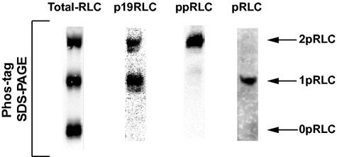
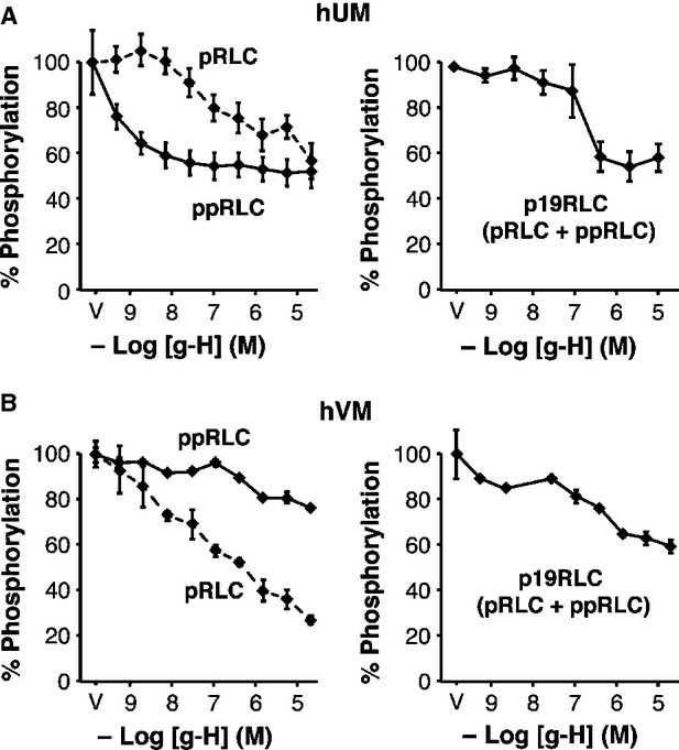
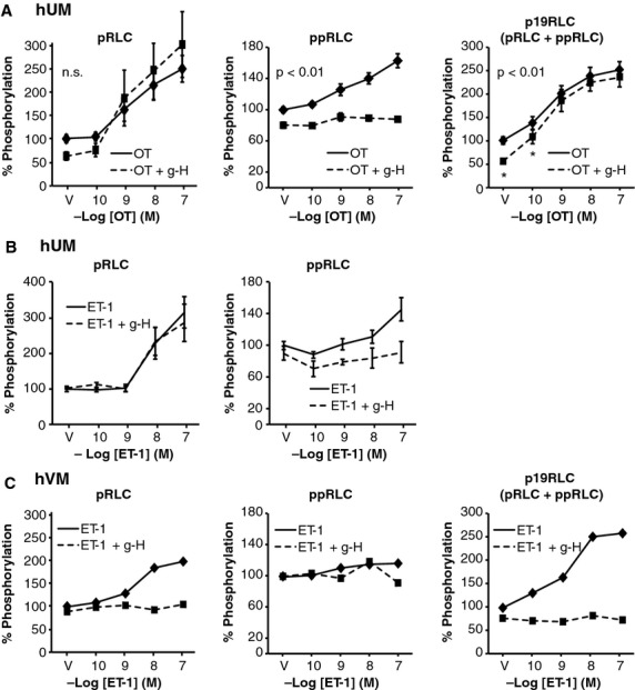
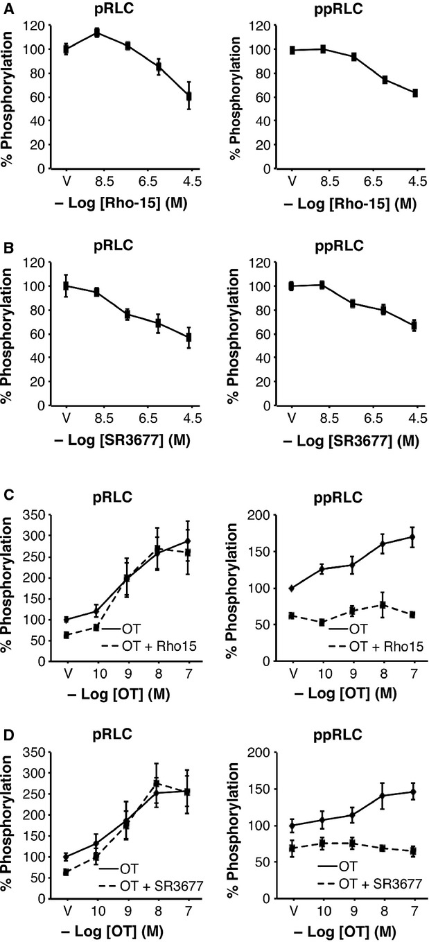
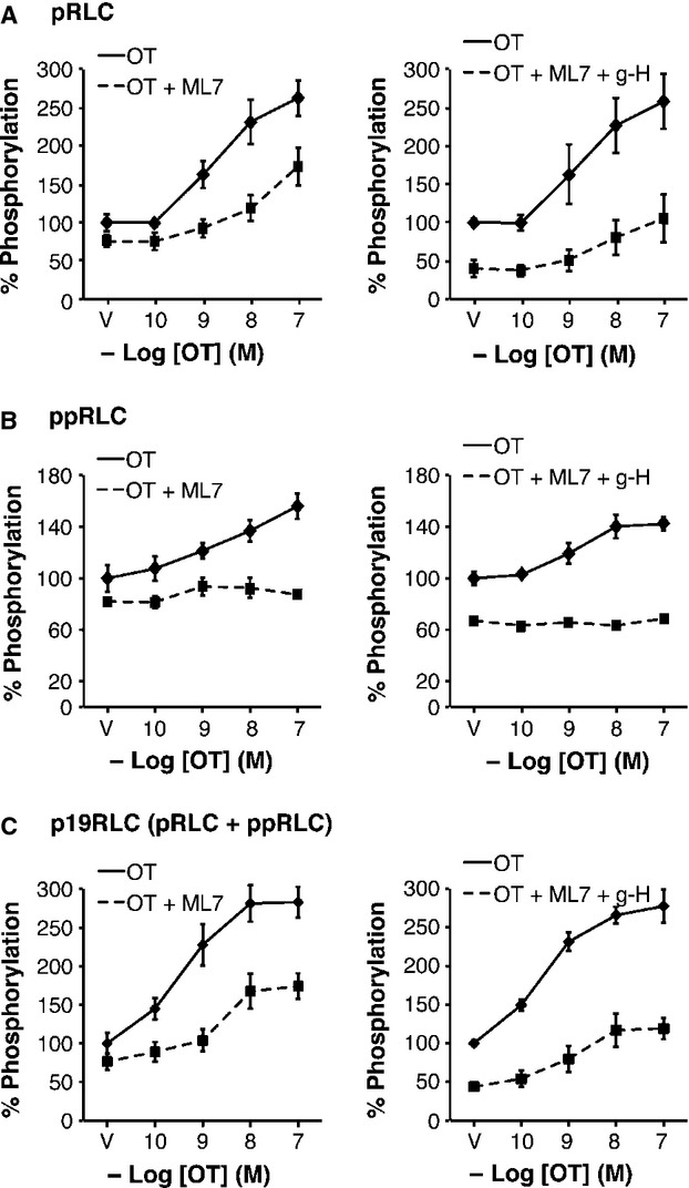
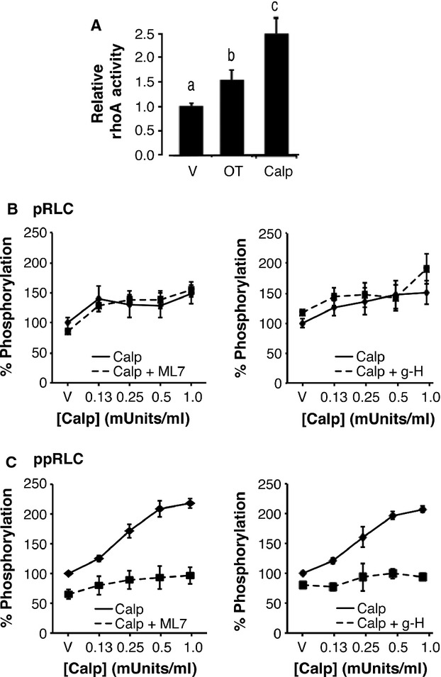
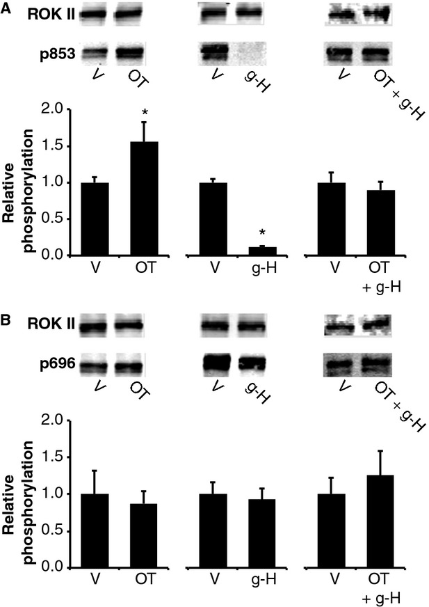
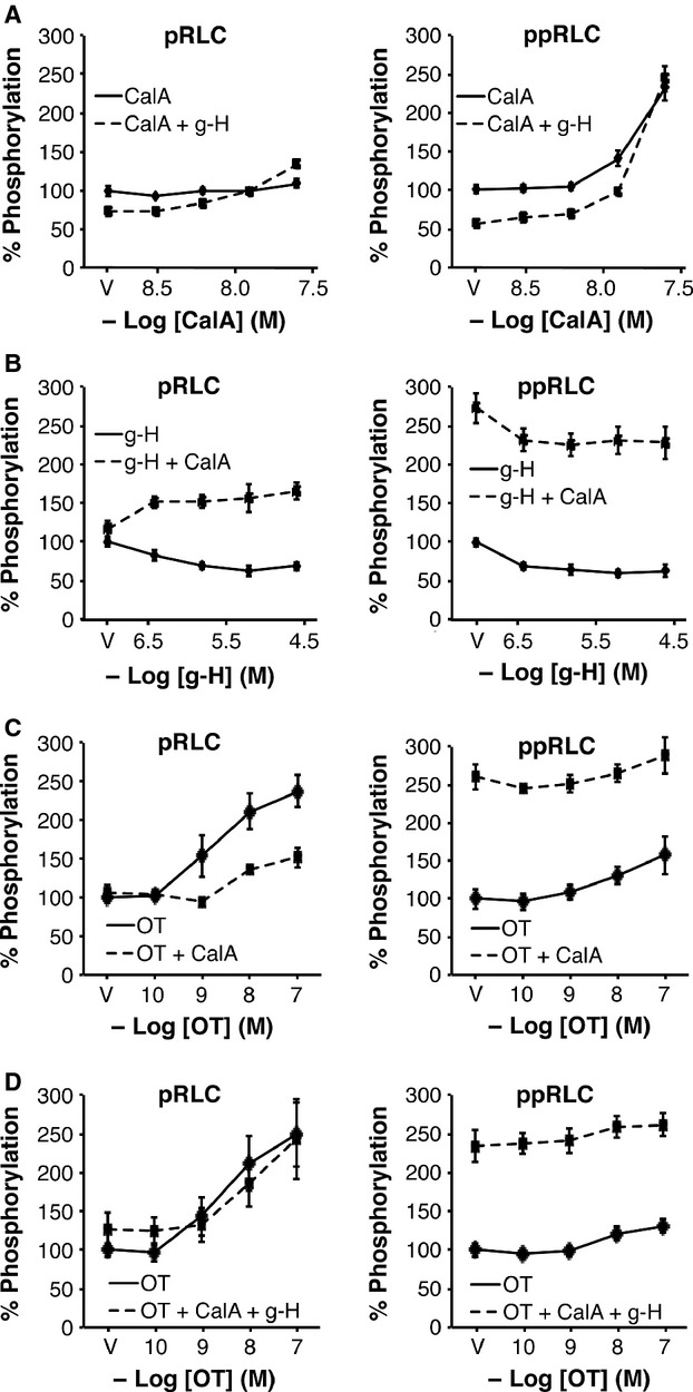
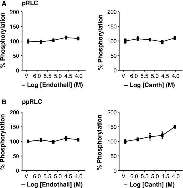
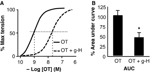
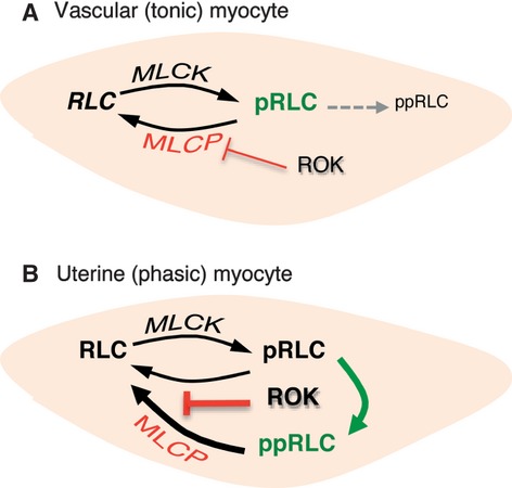
Phosphorylation of myosin regulatory light chain (RLC) triggers contraction in smooth muscle myocytes. Dephosphorylation of phosphorylated RLC (pRLC) is mediated by myosin RLC phosphatase (MLCP), which is negatively regulated by rho-associated kinase (ROK). We have compared basal and stimulated concentrations of pRLC in myocytes from human coronary artery (hVM), which has a tonic contractile pattern to myocytes from human uterus (hUM), which has a phasic contractile pattern. Our studies reveal fundamental differences between hVM and hUM regarding the mechanisms regulating phosphorylation RLC. Whereas hVM responded to stimulation by phosphorylation of RLC at S19, hUM responded by forming diphosphorylated RLC (at T18 and S19; ppRLC), which, compared to pRLC, causes two to threefold greater activation of myosin ATPase that provides energy to power the contraction. Importantly, the conversion of pRLC to ppRLC is mediated by ROK. In hUM, MLCP has high activity for ppRLC and this is inhibited by ROK through phosphorylation of the substrate targeting subunit (MYPT1) at T853. Inhibitors of ROK significantly reduce contractility in both hVM and hUM. We demonstrated that inhibition of ppRLC in phasic myocytes (hUM) is 100-fold more sensitive to ROK inhibitors than is pRLC in tonic myocytes (hVM). We speculate that these differences in phosphorylation of RLC might reflect evolution of different contractile patterns to perform distinct physiological functions. Furthermore, our data suggest that low concentrations of ROK inhibitors might inhibit uterine contractions with minimal effects on vascular tone, thus posing a novel strategy for prevention or treatment of conditions such as preterm birth.


Figures

Fig 1
Fig 1
Demonstration of phospho-regulatory light chain (RLC) antibody specificity in lysates of human uterine lysates. Western blots were produced by separation of proteins in lysates from hUM using phos-tag SDS-PAGE. Single lanes were loaded with ∽25 μg of protein/lane from the same OT-stimulated cells. From left to right: Abs directed against the C-terminus of total-RLC, total phospho-S19-RLC (p19RLC, which includes pRLC + ppRLC), diphosphorylated-T18,S19- (ppRLC) and mono-S19-phosphorylated RLC. The lower, middle and upper bands in these blots correspond to non-, mono- and di-phosphorylated RLC, denoted as 0pRLC, 1pRLC and 2pRLC.
Fig 2
Fig 2
Effects of rho-associated kinase (ROK) inhibition on phosphorylation of regulatory light chain (RLC) in resting hUM and hVM. In-cell Western (ICW) assays using specific Ab measured monophospho-S19-RLC (pRLC, dashed lines), diphospho-T18/S19-RLC (ppRLC, solid lines) or total-phospho-S19-RLC [p19RLC (pRLC + ppRLC)]. The first data point in all graphs corresponds to the vehicle control (V, 100%). The specific ROK inhibitor glycyl-H-1152 (g-H) caused a concentration-dependent decrease in phosphorylated RLC in human uterine myocytes (hUM) (A) and human vascular myocytes (hVM) (B). For hUM, n = 4. As for all ICW data, results are reported as means ± SEM of% changes compared to vehicle-treated controls. For hVM, there were three independent experiments using different passages of human coronary artery myocytes from passages two to five. For hUM, the effect of g-H on ppRLC was significantly greater (P < 0.01) than on pRLC, determined by two-way anova. In contrast, for hVM, the effect of g-H was greater on pRLC compared to ppRLC.
Fig 3
Fig 3
Effects of rho-associated kinase (ROK) inhibition on phosphorylation of regulatory light chain (RLC) in stimulated hUM and hVM. (A) OT caused a significant dose-dependent increase in pRLC and ppRLC in hUM. The ROK inhibitor g-H (1.0 μM) reduced basal concentrations of both pRLC and ppRLC. However, g-H had no significant effect on the pRLC response to OT, but abolished the ppRLC response. These findings were confirmed using the total p19RLC antibody (right panel). The asterisks denote that suppression of the p19RLC signal was significant (two-way anova; P ≤ 0.01), which confirms that the g-H induced suppression was due to reduction in S19 phosphorylation. (B) The responses of hUM to ET-1 in the absence or presence of g-H were similar to the responses to OT. (C) In hVM, treatment with ET-1 caused a brisk response in pRLC, but no significant response in ppRLC. Again, the p19RLC antibody confirmed that the phosphorylation in hVM was at S19.
Fig 4
Fig 4
Effects of isoform-selective inhibitors of rho-associated kinase (ROK) 1 and ROK 2 on resting and stimulated phosphorylation of regulatory light chain (RLC) in hUM. Both the ROK 1-selective inhibitor (Rho-15) (A) and the ROK 2-selective inhibitor (SR3677) (B) suppressed basal pRLC and ppRLC in hUM (n = 4; P < 0.01 by one-way anova), similarly to the less selective inhibitor g-H as noted in Figure2A. Neither Rho-15 (25 μM) (C) nor SR3677 (25 μM) (D) affected the OT-induced responses in pRLC, but both abolished the responses in ppRLC (n = 4; P < 0.01 by two-way anova), in a pattern identical to g-H (Fig.3).
Fig 5
Fig 5
Effects of inhibition of MLCK alone and combined with inhibition of rho-associated kinase (ROK) on OT-stimulated phosphorylation of regulatory light chain (RLC) in hUM. (A) Inhibition of MLCK using ML7 (25 μM) caused significant reduction in the concentrations of pRLC in response to OT (n = 4; P < 0.01 by two-way anova). Addition of g-H (1 μM) further reduced the basal pRLC level, but the response to OT was not altered compared to ML7 alone (n = 4; P < 0.01 by two-way anova). (B) Treatment with ML7 with or without g-H abolished the OT-stimulated response in ppRLC. (C) The responses assessed using the total p19RLC antibody confirmed the findings in panels A and B, again indicating that the primary effect of inhibition of MLCK is in decreasing phosphorylation at S19.
Fig 6
Fig 6
Effects of activation of rhoA on phosphorylation of regulatory light chain (RLC) in hUM. (A) Activation of rhoA, assessed by measuring GTP-bound rhoA, was significantly increased by OT (100 nM) and by the rhoA activator Calpeptin (Calp; 0.5 units/ml). These data have been normalized to rhoA peptide content assessed using Western blot. (n = 4; histograms with different superscripts are significantly different from each other using one-way anova and post hoc Tukey test). (B) Treatment with Calp caused an increase in pRLC that was not significantly affected by inhibition of MLCK using ML7 (25 μM) or inhibition of ROK using g-H (1 μM). (C) In contrast, the Calp-stimulated responses in ppRLC were significantly decreased with either ML7 or g-H (1 μM) (n = 4–8; P < 0.01 by two-way anova).
Fig 7
Fig 7
Effects of OT, Calpeptin and g-H on phosphorylation of MYPT1 in hUM. (A) Phosphorylation of MYPT1 at T853 was significantly increased by OT and Calp and was significantly decreased by the ROK inhibitor g-H (n = 4–6; P < 0.05 compared to corresponding vehicle controls (V) using unpaired t-test, as indicated by *). Representative Western blots are indicated above the histograms. The data were normalized to ROK 2, which was used as the loading control. (B) There was no effect of OT, Calpeptin or g-H on phosphorylation of MYPT1 at T696.
Fig 8
Fig 8
Effects of inhibition of phosphoprotein phosphatase type1 inhibition with or without ROK inhibition on basal and OT-stimulated phosphorylation of regulatory light chain (RLC) in hUM. (A) Inhibition of PP1 using Calyculin A (CalA) alone had no significant effect on basal levels of pRLC. In the presence of the ROK inhibitor g-H (1 μM), there was a slight, but significant increase in pRLC (n = 8, P < 0.01 by two-way anova). However, CalA caused a significant increase in ppRLC. Treatment with g-H suppressed basal ppRLC, but the response to increasing CalA was maintained (n = 4–8; P < 0.01 by two-way anova). (B) Treatment with g-H caused a concentration-dependent suppression of pRLC and ppRLC. However, in the presence of constant CalA (25 nM), increasing g-H caused a significant increase in pRLC. Conversely, in the absence of g-H, CalA caused a large increase in ppRLC concentrations (the vehicle (V) point in the right panel) and increasing g-H resulted in a significant suppression of this ppRLC. (C) CalA (25 nM) caused significant suppression of the OT-induced response in pRLC. CalA again cause a massive increase in ppRLC with only a small although significant increase in response to OT. (D) In the presence of inhibition of both PP1 and ROK, there was no significant change in the OT-induced increase in pRLC in response to OT. Again, treatment with CalA markedly enhanced ppRLC and OT was unable to stimulate further significant increase. (n = 4–8; all analyses used two-way anova).
Fig 9
Fig 9
Effects of inhibition of phosphoprotein phosphatase type 2 on phosphorylation of regulatory light chain (RLC) in hUM. Inhibition of PP2 using endothall or Cantharidic acid (Canth) had no significant effect on concentrations of pRLC (A) or ppRLC (B) with the exception that the maximal concentration of Canth resulted in a small increase in ppRLC (n = 4; P < 0.05 by one-way anova).
Fig 10
Fig 10
Sub-cellular localization of diphosphorylated regulatory light chain (RLC) in hUM. Immunofluorescent staining of hUM. (A–C) Filamentous actin (F-actin) stained with rhodamine-phalloidin (red). (D–F) Identical fields corresponding to panels A–C showing staining with a secondary Ab conjugated to Alexa-Fluor 488 (green). In panel D, the primary Ab has been omitted (secondary Ab only). In panels E and F, the anti-ppRLC Ab was used. (G) and (H) Merged images after combining panels B and E, or C and F respectively. Panels B, E and G correspond to vehicle (V) treatment, and panels C, F and H to cells stimulated with OT (100 nM). In all panels, nuclei are stained with DAPI (blue). Images are shown at 400× magnification. White bars represent 25 micrometres.
Fig 11
Fig 11
Effects of inhibition of ROK on activity of human uterine strips ex vivo. Concentration-response curves of uterine strips to OT. The calculated EC50 was 1.3 nM, but in the presence of the ROK inhibitor g-H (1 μM) the EC50 was increased to 27.1 nM. There was a corresponding significant decrease (to 46 ± 15% as indicated by *) in the area under the response curve (n = 4; unpaired t-test).
Fig 12
Fig 12
Proposed distinguishing mechanisms of phosphorylation of regulatory light chain (RLC) in tonic and phasic smooth muscle. (A) Myocytes from smooth muscle with tonic patterns of contractile activity (hVM) initiate contractions by simple phosphorylation of RLC at S19. This is predominantly regulated by Ca2+-dependent activation of MLCK. Return to a lower contractile state can be accomplished by increasing activity of MLCP, which is negatively regulated by ROK. (B) For myocytes obtained from smooth muscle with a phasic pattern of contractility (hUM), contraction is also initiated by MLCK-mediated increase in pRLC, but this is augmented by ROK-mediated phosphorylation of pRLC at T18 to form ppRLC, which enhances energy production from myosin ATPase. In addition, increasing ROK activity strongly inhibits the phosphatase activity of MLCP that is predominantly directed towards ppRLC.

References

    1. Kamm KE, Stull JT. The function of myosin and myosin light chain kinase phosphorylation in smooth muscle. Annu Rev Pharmacol Toxicol. 1985;25:593–620. - PubMed
    1. Longbottom ER, Luckas MJ, Kupittayanant S, et al. The effects of inhibiting myosin light chain kinase on contraction and calcium signalling in human and rat myometrium. Pflugers Arch. 2000;440:315–21. - PubMed
    1. Takashima S. Phosphorylation of myosin regulatory light chain by myosin light chain kinase, and muscle contraction. Circ J. 2009;73:208–13. - PubMed
    1. Ikebe M, Inagaki M, Kanamaru K, et al. Phosphorylation of smooth muscle myosin light chain kinase by Ca2+-activated, phospholipid-dependent protein kinase. J Biol Chem. 1985;260:4547–50. - PubMed
    1. Ikebe M, Koretz J, Hartshorne DJ. Effects of phosphorylation of light chain residues threonine 18 and serine 19 on the properties and conformation of smooth muscle myosin. J Biol Chem. 1988;263:6432–7. - PubMed

Publication types