. 2012 Dec;16(12):2990-3000.
doi: 10.1111/j.1582-4934.2012.01626.x.

Characterization of the tissue-level Ca2+ signals in spontaneously contracting human myometrium

Affiliations

Characterization of the tissue-level Ca2+ signals in spontaneously contracting human myometrium

Gilles Bru-Mercier et al. J Cell Mol Med. 2012 Dec.

Abstract

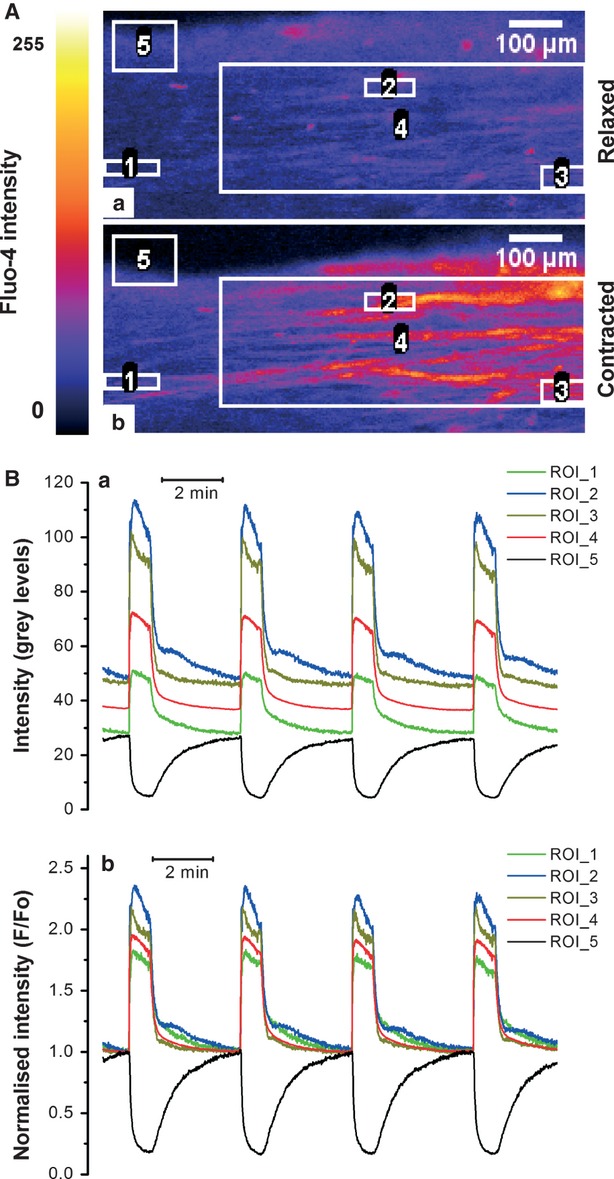
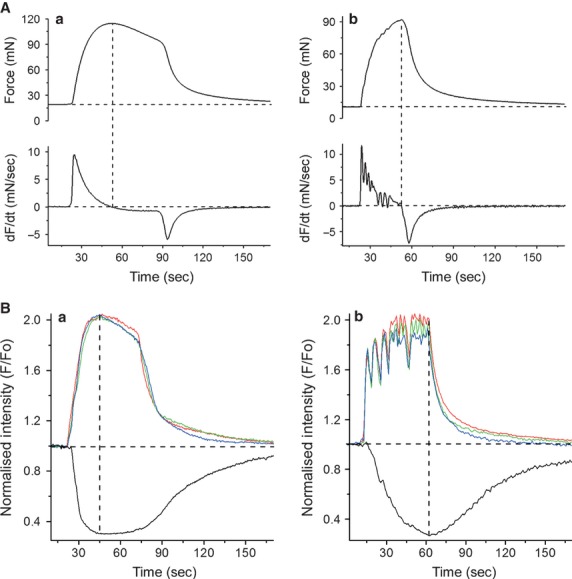
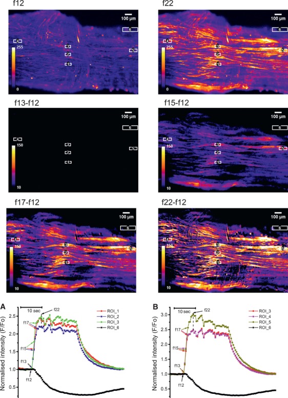
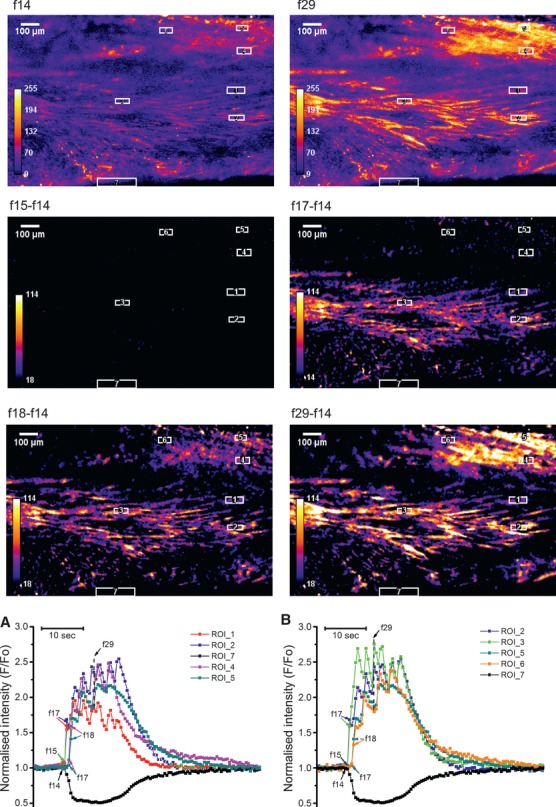
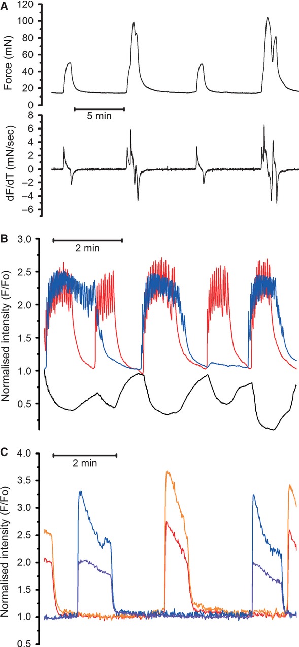
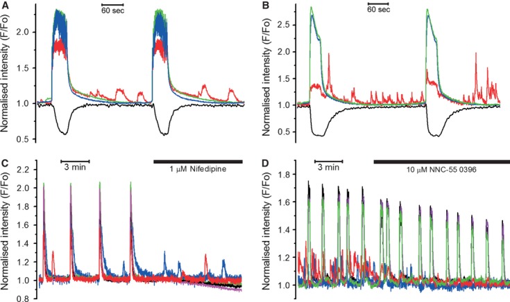
In the labouring uterus, millions of myocytes forming the complex geometrical structure of myometrium contract in synchrony to increase intrauterine pressure, dilate the cervix and eventually expel the foetus through the birth canal. The mechanisms underlying the precise coordination of contractions in human myometrium are not completely understood. In the present study, we have characterized the spatio-temporal properties of tissue-level [Ca(2+)](i) transients in thin slices of intact human myometrium. We found that the waveform of [Ca(2+)](i) transients and isotonic contractions recorded from thin slices was similar to the waveform of isometric contractions recorded from the larger strips in traditional organ bath experiments, suggesting that the spatio-temporal information obtained from thin slices is representative of the whole tissue. By comparing the time course of [Ca(2+)](i) transients in individual cells to that recorded from the bundles of myocytes we found that the majority of myocytes produce rapidly propagating long-lasting [Ca(2+)](i) transients accompanied by contractions. We also found a small number of cells showing desynchronized [Ca(2+)](i) oscillations that did not trigger contractions. The [Ca(2+)](i) oscillations in these cells were insensitive to nifedipine, but readily inhibited by the T-type Ca(2+) channel inhibitor NNC55-0396. In conclusion, our data suggest that the spread of [Ca(2+)](i) signals in human myometrium is achieved via propagation of long-lasting action potentials. The propagation was fast when action potentials propagated along bundles of myocytes and slower when propagating between the bundles of uterine myocytes.


Figures

Fig 1
Fig 1
Confocal imaging of spontaneous [Ca2+]i transients and contraction in thin slices of human myometrium. (A) Raw fluorescence images of Fluo-4 loaded myometrial slice in the relaxed and fully contracted state show the arrangement of regions of interest (ROIs) for extraction of the intensity versus time data: single-cell ROIs (ROI 1-3), multi-cell ROI (ROI 4) and a contraction-monitoring ROI (ROI 5) are shown. (B) Intensity versus time traces extracted from the ROI. (Ba) Raw data; (Bb) Normalized intensity showing repetitive increases in [Ca2+]i (red, blue, green and olive traces) and the slice edge displacement indicative of contraction (black trace).
Fig 2
Fig 2
Comparison of the waveforms of isometric contractions recorded in organ bath and [Ca2+]i transients and isotonic twitches recorded from the thin slices of myometrium. (A) Isometric force (upper trace) and its first derivative (lower trace) recorded from strips exhibiting the plateau-type (Aa) or burst-type (Ab) activity. The main difference between these two types was the position of the peak force: it occurred earlier in strips with plateau-type activity and was followed by a slow decay of force before the rapid relaxation at the end of the contraction-relaxation cycle. In the burst-type strips, contractile force continued to rise until the end of the contraction-relaxation cycle. The rising phase of contraction had a ‘rugged’ appearance, more evident in the first derivative trace. (B) [Ca2+]i transients (red, blue and green traces from multi-cell ROIs positioned at opposite ends of the viewing field) and isotonic contractions (black traces) recorded from two different slices, one exhibiting the plateau-type (Ba) and another the burst-type (Bb) activity. In both cases, the ROIs were positioned over cell groups belonging to a single bundle of myocytes present in each slice. The burst-type [Ca2+]i transient had a ‘rugged’ appearance that resulted from a fusion of many individual increments in [Ca2+]i presumably corresponding to repetitive spikes in the burst-type action potential, whereas the plateau-type [Ca2+]i transient has a smooth appearance. Vertical dashed lines in all panels indicate the peaks of contraction.
Fig 3
Fig 3
Synchronous activation of myocytes in myometrial slice containing a single bundle of myocytes. Frames in top row show a Fluo-4 loaded slice at rest (f12) and at peak of [Ca2+]i transient (f22). The ‘resting fluorescence’ frame (f12) was subtracted from subsequent frames during the rising phase of [Ca2+]i transients to yield the ‘delta F’ images where cells are visible only when they rise [Ca2+]i above the resting level. The ROI were positioned along the longitudinal axis (ROI 4, 3 and 5) or perpendicular to it (ROI 1-3). Graph in (a) shows [Ca2+]i traces extracted from ROIs positioned in longitudinal direction. Graph in (b) shows [Ca2+]i traces extracted from ROIs positioned perpendicular to the longitudinal axis. Black traces correspond to a contraction-monitoring ROI 6. Arrows next to frame numbers in both graphs indicate data points corresponding to the corresponding frames shown above. Note that [Ca2+]i transient spread between cells in both sets of ROIs without measurable delay indicating synchronous activation of cells in both directions.
Fig 4
Fig 4
Propagation of [Ca2+]i transient in myometrial slice containing two bundles of myocytes, one in the middle, another in the top right corner. Data presentation is similar to that in Figure3 with the exception that two sets of three ROIs were used in this experiment: ROI 1-2 and ROI 4-5 to measure propagation perpendicular to the longitudinal axis in each bundle and ROI 2-3 and ROI5-6 to measure propagation in axial direction. In this experiment, there was a 1 sec. time delay between the onset of [Ca2+]i transients in neighbouring bundles but cells within each bundle were activated simultaneously. Typical of seven experiments on slices from seven different samples.
Fig 5
Fig 5
Desynchronized [Ca2+]i transients elicit contractions of alternating amplitude. (A) Example of alternating isometric force (upper trace) and its first derivative (lover trace) recorded from a strip in an organ bath experiment (typical of 23 recordings from 23 different samples). (B) Multi-cell [Ca2+]i transients showing no synchronization (typical of five other slices from five different samples). Red and blue traces correspond to ROI positioned over two different bundles. Note that larger contractions occurred when [Ca2+]i transients in different bundles coincided (marked by asterisks). (C) Single-cell ROIs positioned over two different cells in one bundle (red and orange traces) and two more cells in another bundle (blue and violet traces) in a slice with desynchronized bundles. Note that the [Ca2+]i transients in individual cells within each bundle remained synchronized.
Fig 6
Fig 6
Oscillating cells in myometrial slices (observed in 18 of 27 slices). Unsynchronized, low-amplitude [Ca2+]i oscillations (red trace) in a burst-type (A) and plateau-type (B) slices. Note that only tissue-level [Ca2+]i transients (blue and green traces in both graphs) were capable of triggering contractions (black traces). (C) Nifedipine inhibited tissue-level [Ca2+]i transients, but not [Ca2+]i oscillations in desynchronized cells. (D) [Ca2+]i oscillations were inhibited by NNC-55 0396, a specific T-type Ca2+ channel inhibitor.

References

    1. Bolton TB. Cholinergic Mechanisms in Smooth-Muscle. Br Med Bull. 1979;35:275–83. - PubMed
    1. Kuriyama H, Kitamura K, Itoh T, et al. Physiological features of visceral smooth muscle cells, with special reference to receptors and ion channels. Physiol Rev. 1998;78:811–920. - PubMed
    1. Wikland M, Lindblom B. Relationship between electrical and mechanical activity of the isolated term-pregnant human myometrium. Eur J Obstet Gynecol Reprod Biol. 1985;20:337–46. - PubMed
    1. Nakao K, Inoue Y, Okabe K, et al. Oxytocin enhances action potentials in pregnant human myometrium - A study with microelectrodes. Am J Obstet Gynecol. 1997;177:222–8. - PubMed
    1. Kawarabayashi T, Izumi H, Ikeda M, et al. Modification by Magnesium of the Excitatory Effect of Oxytocin in Electrical and Mechanical Activities of Pregnant Human Myometrium. Obst Gynecol. 1990;76:183–8. - PubMed

Publication types