

Enamel pits in hamster molars, formed by a single high fluoride dose, are associated with a perturbation of transitional stage ameloblasts
- PMID: 22947666
- PMCID: PMC3591722
- DOI: 10.1159/000341802
Enamel pits in hamster molars, formed by a single high fluoride dose, are associated with a perturbation of transitional stage ameloblasts
Abstract
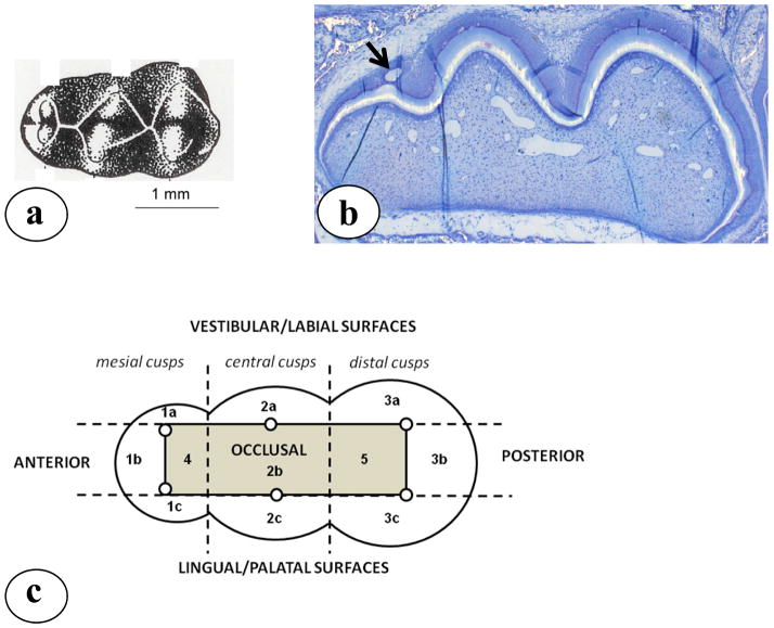
Excessive intake of fluoride (F) by young children results in the formation of enamel subsurface porosities and pits, called enamel fluorosis. In this study, we used a single high dose of F administered to hamster pups to determine the stage of ameloblasts most affected by F and whether pit formation was related to F-related sub-ameloblastic cyst formation. Hamster pups received a single subcutaneous injection of either 20 mg or 40 mg NaF/kg body weight, were sacrificed 24 h later, and the number of cysts formed in the first molars were counted. Other pups were sacrificed 8 days after F injection, when the first molars had just erupted, to score for enamel defects. All F-injected pups formed enamel defects in the upper half of the cusps in a dose-dependent way. After injection of 20 mg NaF/kg, an average of 2.5 white spots per molar was found but no pits. At 40 mg NaF/kg, almost 4.5 spots per molar were counted as well as 2 pits per molar. The defects in erupted enamel were located in the upper half of the cusps, sites where cysts had formed at the transition stage of ameloblast differentiation. These results suggest that transitional ameloblasts, located between secretory- and maturation-stage ameloblasts, are most sensitive to the effects of a single high dose of F. F-induced cysts formed earlier at the pre-secretory stage were not correlated to either white spots or enamel pits, suggesting that damaged ameloblasts overlying a F-induced cyst regenerate and continue to form enamel.
Copyright © 2012 S. Karger AG, Basel.
Figures


References
-
- Angmar-Mansson B, Whitford GM. Plasma fluoride levels and enamel fluorosis in the rat. Caries Res. 1982;16:334–339. - PubMed
-
- Angmar-Mansson B, Whitford GM. Enamel fluorosis related to plasma F levels in the rat. Caries Res. 1984;18:25–32. - PubMed
-
- DenBesten PK, Crenshaw MA, Wilson MH. Changes in the fluoride-induced modulation of maturation stage ameloblasts of rats. J Dent Res. 1985;64:1365–1370. - PubMed
-
- DenBesten PK. Biological mechanisms of dental fluorosis relevant to the use of fluoride supplements. Community Dent Oral Epidemiol. 1999;27:41–47. - PubMed
