. 2012 Aug 8;103(3):453-463.
doi: 10.1016/j.bpj.2012.06.043.

Binding of β-amyloid (1-42) peptide to negatively charged phospholipid membranes in the liquid-ordered state: modeling and experimental studies

Affiliations

Binding of β-amyloid (1-42) peptide to negatively charged phospholipid membranes in the liquid-ordered state: modeling and experimental studies

Hasna Ahyayauch et al. Biophys J. .

Abstract

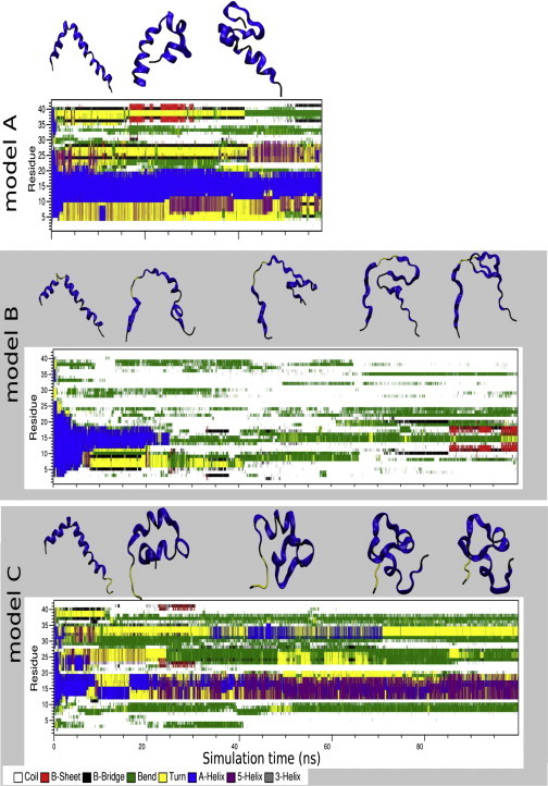
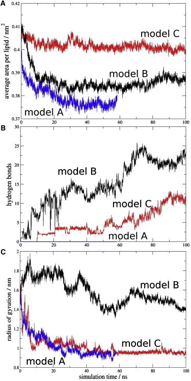
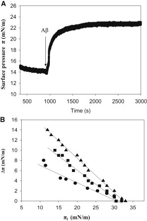
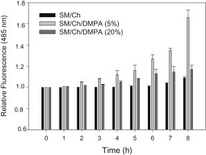
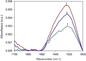
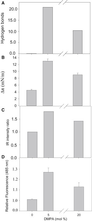
To explore the initial stages of amyloid β peptide (Aβ42) deposition on membranes, we have studied the interaction of Aβ42 in the monomeric form with lipid monolayers and with bilayers in either the liquid-disordered or the liquid-ordered (L(o)) state, containing negatively charged phospholipids. Molecular dynamics (MD) simulations of the system have been performed, as well as experimental measurements. For bilayers in the L(o) state, in the absence of the negatively charged lipids, interaction is weak and it cannot be detected by isothermal calorimetry. However, in the presence of phosphatidic acid, or of cardiolipin, interaction is detected by different methods and in all cases interaction is strongest with lower (2.5-5 mol%) than higher (10-20 mol%) proportions of negatively charged phospholipids. Liquid-disordered bilayers consistently allowed a higher Aβ42 binding than L(o) ones. Thioflavin T assays and infrared spectroscopy confirmed a higher proportion of β-sheet formation under conditions when higher peptide binding was measured. The experimental results were supported by MD simulations. We used 100 ns MD to examine interactions between Aβ42 and three different 512 lipid bilayers consisting of palmitoylsphingomyelin, dimyristoyl phosphatidic acid, and cholesterol in three different proportions. MD pictures are different for the low- and high-charge bilayers, in the former case the peptide is bound through many contact points to the bilayer, whereas for the bilayer containing 20 mol% anionic phospholipid only a small fragment of the peptide appears to be bound. The MD results indicate that the binding and fibril formation on the membrane surface depends on the composition of the bilayer, and is the result of a subtle balance of many inter- and intramolecular interactions between the Aβ42 and membrane.


Figures

Figure 1
Figure 1
Time evolution of the Aβ42 peptide secondary structure in models A (SM/Chol/DMPA = 50:50:0), B (47.5:47.5:5.0), and C (40:40:20). The secondary structures were calculated by the DSSP program (50). The snapshot structures of Aβ42 were taken from trajectories at 20-ns intervals.
Figure 2
Figure 2
Time evolution of the MD simulations in the model A (SM/Chol/DMPA = 50:50:0), B (47,5:47.5:5.0), and C (40:40:20). For the sake of clarity, the data were averaged every 20 ps. (A) Average area per lipid molecule. (B) Hydrogen bonds between Aβ42 and membrane phospholipids. The proton donor-acceptor distance threshold was 0.35 nm and the angle was 30°. (C) Radius of gyration of the Aβ42 peptide in all models.
Figure 3
Figure 3
Hydrogen bond occurrence between the residues of Aβ42 and membrane lipids in the models B and C. Distance and angle threshold between the hydrogen bond donor and acceptor was 0.35 nm and 30°, respectively. Relative occurrences for multivalent interactions are summed to individual residues.
Figure 4
Figure 4
Changes in surface pressure of lipid monolayers, upon insertion of Aβ42 monomers at varying initial pressures. (A) A representative experiment, obtained with SM/Chol/DMPA (40/40/20) at 15 mN/m. (B) Equilibrium values. ●, SM/Chol (1:1), ▴, SM/Chol/DMPA (47.5/47.5/5), ■, SM/Chol/DMPA (40/40/20). Average values mean ± SE (n = 3). Aβ42 stock solution was 50 μM. Aβ42 final concentration in the trough was 1.22 μM.
Figure 5
Figure 5
Time course of ThT fluorescence in the presence of Aβ42 in mixtures with LUV of varying lipid compositions. Lipid/protein mol ratio was 200:1, at 5 μM Aβ42. Average values mean ± SE (n = 3).
Figure 6
Figure 6
Difference IR spectra of Aβ42 in mixtures with multilamellar vesicles (MLV) of varying lipid compositions. Spectra of free peptide were subtracted from spectra of MLV-peptide mixtures. MLV composition was: 1, SM/Chol (1/1); 2, SM/Chol/DMPA (47.5/47.5/5); 3, SM/Chol/DMPA (40/40/20). Lipid/protein mol ratio was 200:1 at 80 μM Aβ42.
Figure 7
Figure 7
A comparison of data concerning Aβ42 binding to bilayers composed of SM/Chol (1:1 mol ratio), to which increasing amounts of anionic phospholipid (phosphatidic acid) had been added. (A) MD calculations of average H-bonds established between peptide and membrane for the last 20 ns. Data taken from Fig. 2B. (B) Langmuir balance. Increase in surface pressure of lipid monolayers (16 mN/m) of the compositions given previously due to the presence of Aβ42. Data are taken from Fig. 4B. (C) Relative change in intensity of the IR band centered at 1622 cm−1, assigned to antiparallel β-sheet vibration. Data taken from Fig. 6. (D) Changes in ThT fluorescence after 6 h incubation with Aβ42 and LUVs. Data taken from Fig. 5.

References

    1. Ross C.A., Poirier M.A. Protein aggregation and neurodegenerative disease. Nat. Med. 2004;10(Suppl):S10–S17. - PubMed
    1. Selkoe D.J. Alzheimer's disease: genes, proteins, and therapy. Physiol. Rev. 2001;81:741–766. - PubMed
    1. Kojro E., Gimpl G., Fahrenholz F. Low cholesterol stimulates the nonamyloidogenic pathway by its effect on the alpha -secretase ADAM 10. Proc. Natl. Acad. Sci. USA. 2001;98:5815–5820. - PMC - PubMed
    1. Ehehalt R., Keller P., Simons K. Amyloidogenic processing of the Alzheimer β-amyloid precursor protein depends on lipid rafts. J. Cell Biol. 2003;160:113–123. - PMC - PubMed
    1. Haass C., Lemere C.A., Selkoe D.J. The Swedish mutation causes early-onset Alzheimer's disease by β-secretase cleavage within the secretory pathway. Nat. Med. 1995;1:1291–1296. - PubMed

Publication types