. 2012 Aug 22;103(4):702-10.
doi: 10.1016/j.bpj.2012.06.045.

Two-step mechanism of membrane disruption by Aβ through membrane fragmentation and pore formation

Affiliations

Two-step mechanism of membrane disruption by Aβ through membrane fragmentation and pore formation

Michele F M Sciacca et al. Biophys J. .

Abstract

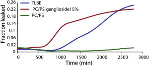
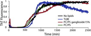
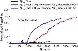
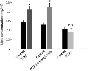
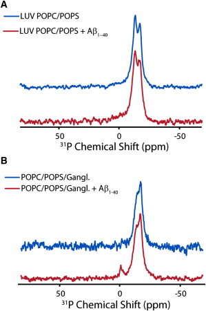
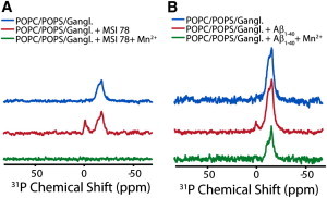
Disruption of cell membranes by Aβ is believed to be one of the key components of Aβ toxicity. However, the mechanism by which this occurs is not fully understood. Here, we demonstrate that membrane disruption by Aβ occurs by a two-step process, with the initial formation of ion-selective pores followed by nonspecific fragmentation of the lipid membrane during amyloid fiber formation. Immediately after the addition of freshly dissolved Aβ(1-40), defects form on the membrane that share many of the properties of Aβ channels originally reported from single-channel electrical recording, such as cation selectivity and the ability to be blockaded by zinc. By contrast, subsequent amyloid fiber formation on the surface of the membrane fragments the membrane in a way that is not cation selective and cannot be stopped by zinc ions. Moreover, we observed that the presence of ganglioside enhances both the initial pore formation and the fiber-dependent membrane fragmentation process. Whereas pore formation by freshly dissolved Aβ(1-40) is weakly observed in the absence of gangliosides, fiber-dependent membrane fragmentation can only be observed in their presence. These results provide insights into the toxicity of Aβ and may aid in the design of specific compounds to alleviate the neurodegeneration of Alzheimer's disease.


Figures

Figure 1
Figure 1
Membrane disruption induced by Aβ1-40, as measured by the 6-carboxyfluorescein dye leakage assay. The graph illustrates the release of 6-carboxyfluorescein induced by 10 μM Aβ1-40 from 0.2 mg/ml POPC/POPS LUVs 7:3 (green line), 0.2 mg/ml POPC/POPS/ganglioside LUVs 5.5/3/1.5 (red line), and 0.2 mg/ml TLBE LUVs (blue line). Dye leakage occurs only after a lag period and is detected only in ganglioside-containing membranes. Experiments were performed at room temperature in 10 mM phosphate buffer, 100 mM NaCl, pH 7.4. Results are the average of three measurements.
Figure 2
Figure 2
Kinetics of Aβ1-40 amyloid formation measured by ThT fluorescent emission. The graph shows 10 μM Aβ1-40 in buffer in the absence of membranes (black line) and in the presence of 0.2 mg/ml POPC/POPS 7:3 LUVs (green line), 0.2 mg/ml POPC/POPS/ganglioside 5.5/3/1.5 LUVs (red line), and 0.2 mg/ml TLBE LUVs (blue line). Experiments were performed at room temperature in 10 mM phosphate buffer, 100 mM NaCl, pH 7.4. Results are the average of three measurements.
Figure 3
Figure 3
Ca+2 influx into LUVs after the addition of freshly dissolved Aβ1-40. The maximum values of Ca+2 ion influx detected by encapsulated Fura-2 after 30 min of incubation with freshly dissolved Aβ1-40 from 0.2 mg/ml TLBE LUVs (dark gray bars) or 0.2 mg/ml POPC/POPS 7:3 LUVs (light gray bars) are shown. In contrast to the 6-carboxyfluorescein dye release assay, Ca+2 influx occurs shortly after the addition of Aβ1-40 and occurs weakly in the absence of gangliosides. The antimicrobial peptide MSI-78 was used as a reference for total disruption of the membrane (black bar).
Figure 4
Figure 4
Zn2+ inhibits the pore activity of freshly dissolved Aβ1-40. The graph shows the influx of Ca+2 (red line) and Zn2+ (blue line) ions induced by Aβ1-40 on 0.2 mg/ml TLBE LUVs. Freshly dissolved Aβ1-40 was added to each sample at time zero, and Ca+2 or Zn+2 was added at 600 s as indicated by the dashed line. Fura-2 is sensitive to both Ca+2 and Zn2+ ions, as indicated by the control membrane disruptive MSI-78 peptide (Fig. S4).
Figure 5
Figure 5
Zn2+ ions cannot block the fiber-dependent step of membrane disruption. The figure indicates the influx of Ca+2 (red line) and Zn2+ (blue line) ions induced by adding freshly dissolved Aβ1-40 to 0.2 mg/ml TLBE LUVs incubated with preformed fibers. Freshly dissolved Aβ1-40 was added to each sample at time zero, and Ca+2 or Zn+2 was added at 600 s as indicated by the dashed line. No Ca+2 influx was detected after the addition of preformed Aβ1-40 fibers (black line). The influx of both Ca+2 and Zn2+ ions (red and blue lines, respectively) were detected by seeding Aβ1-40 fiber formation with preformed fiber. This finding suggests that the fiber-dependent step of membrane disruption is not correlated with pore formation. LUVs were made from TLBE lipids.
Figure 6
Figure 6
Membrane fragmentation induced by prolonged incubation with Aβ1-40. Lipid concentrations in the supernatant after centrifugation from brain extract LUVs (dark gray bar) and POPC/POPS/gangliosides 5.5/3/1.5 LUVs (gray bar) after incubation with Aβ1-40 for 48 h are shown. The failure of lipid vesicles to sediment is an indication of their disruption to smaller micelle-like structures. No significant lipids were detected in the supernatant of samples containing POPC/POPS 7:3 (light gray bar), in agreement with the 6-carboxyfluorescein dye leakage assay (Fig. 1). Results are the average of three independent measures, and error bars represent the standard deviation.
Figure 7
Figure 7
31P solid-state NMR of LUVs incubated with Aβ1-40. 31P chemical shift spectra of large unilamellar vesicles composed of 7:3 POPC/POPS (A) and 5.5:3:1.5 POPC/POPS/ganglioside (B) before (blue line) and after the addition of Aβ1-40 (red line) are illustrated. The small peak near 0 ppm in the ganglioside-containing spectra indicates the formation of small, rapidly tumbling lipid structures indicative of membrane fragmentation. The absence of a corresponding peak for samples without ganglioside is an indication that membrane fragmentation does not occur. All spectra were obtained at 37°C and referenced with respect to 85% H3PO4 at 0.0 ppm.
Figure 8
Figure 8
Absence of membrane defects after fiber formation as detected by paramagnetic quenching. (A and B) 31P chemical shift spectra of POPC/POPS/ganglioside LUVs before (top), after the addition of MSI-78 (A) and Aβ1-40 (B) (middle), and after the addition of 500 μM Mn+2 (bottom). Mn+2 completely quenches the peaks originating from both the isotropic and lamellar phases in the MSI-78 sample, but only partially quenches the lamellar phase in the Aβ1-40 sample, indicating the absence of membrane defects after fiber formation is complete. Aβ1-40 was allowed to incubate on the membrane for 4 days before acquisition. All spectra were collected at 37°C and referenced with respect to 85% H3PO4 at 0.0 ppm.

References

    1. Hardy J.A., Higgins G.A. Alzheimer’s disease: the amyloid cascade hypothesis. Science. 1992;256:184–185. - PubMed
    1. Benilova I., Karran E., De Strooper B. The toxic Aβ oligomer and Alzheimer’s disease: an emperor in need of clothes. Nat. Neurosci. 2012;15:349–357. - PubMed
    1. Small D.H. Dysregulation of calcium homeostasis in Alzheimer’s disease. Neurochem. Res. 2009;34:1824–1829. - PubMed
    1. Mattson M.P., Cheng B., Rydel R.E. β-Amyloid peptides destabilize calcium homeostasis and render human cortical neurons vulnerable to excitotoxicity. J. Neurosci. 1992;12:376–389. - PMC - PubMed
    1. Pollard H.B., Arispe N., Rojas E. Ion channel hypothesis for Alzheimer amyloid peptide neurotoxicity. Cell. Mol. Neurobiol. 1995;15:513–526. - PMC - PubMed

Publication types

MeSH terms