. 2012 Oct 19;287(43):36059-70.
doi: 10.1074/jbc.M112.360487. Epub 2012 Sep 4.

Subtype-specific modulation of acid-sensing ion channel (ASIC) function by 2-guanidine-4-methylquinazoline

Affiliations

Subtype-specific modulation of acid-sensing ion channel (ASIC) function by 2-guanidine-4-methylquinazoline

Omar Alijevic et al. J Biol Chem. .

Abstract

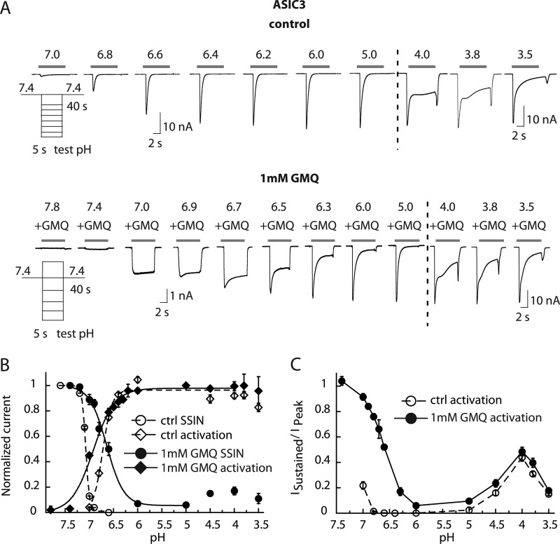
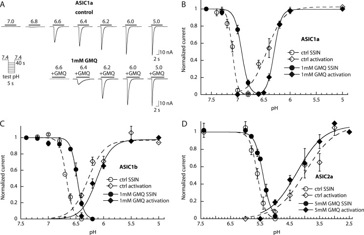
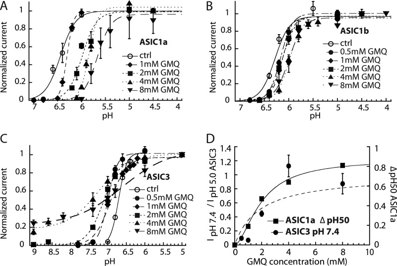
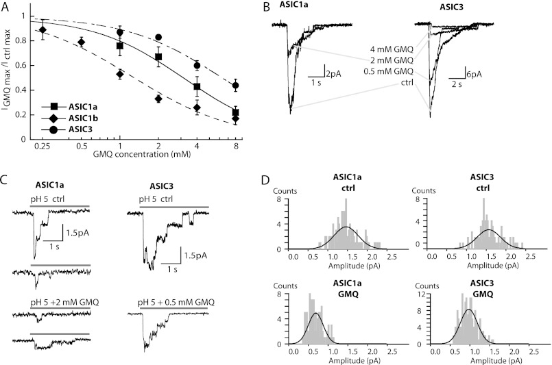
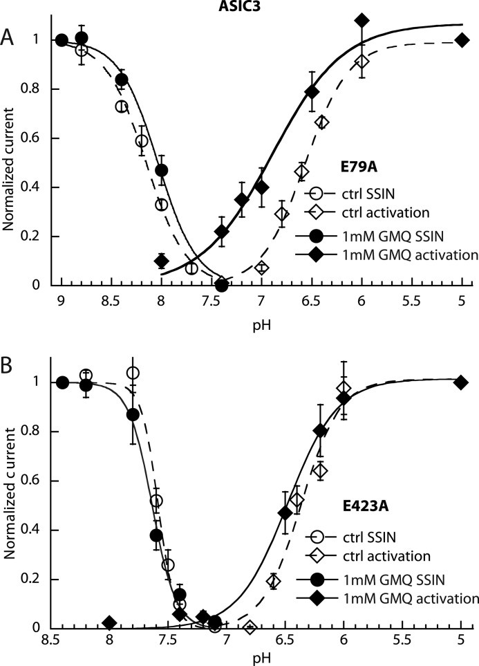
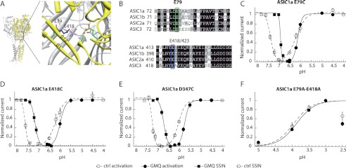
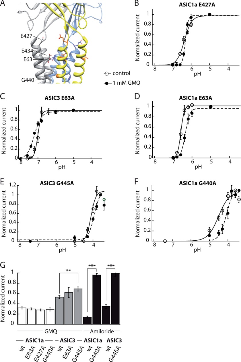
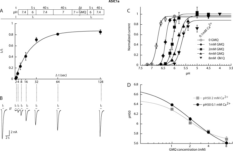
Acid-sensing ion channels (ASICs) are neuronal Na(+)-selective channels that are transiently activated by extracellular acidification. ASICs are involved in fear and anxiety, learning, neurodegeneration after ischemic stroke, and pain sensation. The small molecule 2-guanidine-4-methylquinazoline (GMQ) was recently shown to open ASIC3 at physiological pH. We have investigated the mechanisms underlying this effect and the possibility that GMQ may alter the function of other ASICs besides ASIC3. GMQ shifts the pH dependence of activation to more acidic pH in ASIC1a and ASIC1b, whereas in ASIC3 this shift goes in the opposite direction and is accompanied by a decrease in its steepness. GMQ also induces an acidic shift of the pH dependence of inactivation of ASIC1a, -1b, -2a, and -3. As a consequence, the activation and inactivation curves of ASIC3 but not other ASICs overlap in the presence of GMQ at pH 7.4, thereby creating a window current. At concentrations >1 mM, GMQ decreases maximal peak currents by reducing the unitary current amplitude. Mutation of residue Glu-79 in the palm domain of ASIC3, previously shown to be critical for channel opening by GMQ, disrupted the GMQ effects on inactivation but not activation. This suggests that this residue is involved in the consequences of GMQ binding rather than in the binding interaction itself. This study describes the mechanisms underlying the effects of a novel class of ligands that modulate the function of all ASICs as well as activate ASIC3 at physiological pH.


Figures

FIGURE 1.
FIGURE 1.
GMQ changes the pH dependence of ASIC3 gating and impairs inactivation. Currents were measured by whole-cell patch clamp at a holding potential of −60 mV from CHO cells stably expressing ASIC3. A, current traces of ASIC3 in the absence (top) or presence (bottom) of 1 mm GMQ. Traces obtained at pH 4.0–3.5 (right panel) are of a different experiment. B, ASIC3 was activated by switching from a conditioning pH of 7.4 to various acidic test pH values for 5 s once every 40 s. For the activation curve, the normalized current response is plotted as a function of the stimulation pH for control condition (open symbols) or in the presence of 1 mm GMQ in the acidic test solutions (filled symbols) (n = 4–11). For SSIN, a conditioning pH in the range of pH 7.6–6.5 was applied for 60 s, and the fraction of non-inactivated channels was measured by applying pH 6 for 5 s. This protocol was repeated with increasingly acidic conditioning pH values. In the SSIN curve, normalized currents are plotted as a function of the conditioning pH for the control condition (open symbols) and in the presence of 1 mm GMQ in the conditioning solution (filled symbols) (n = 4–5). For fit parameters, see Table 1. C, pH dependence of the sustained ASIC3 current fraction in the absence or presence of 1 mm GMQ. The ratio of sustained/peak current amplitude is plotted (n = 4–8). Error bars represent S.E.
FIGURE 2.
FIGURE 2.
GMQ shifts the pH dependence of ASIC1a, -1b, and -2a. The currents were measured by whole-cell voltage clamp to −60 mV from CHO cells stably expressing the ASIC homomultimers (ASIC1a, -1b, and -2a). A, current traces of representative activation curves of ASIC1a in the absence (top) or presence (bottom) of 1 mm GMQ. Activation and SSIN curves were measured as described in the text and in the legend to Fig. 1 and are plotted in B for ASIC1a (n = 5–6), C for ASIC1b (n = 3–12), and D for ASIC2a (n = 3–6). Open symbols correspond to data obtained in control conditions, and filled symbols correspond to data obtained with 1 mm GMQ (5 mm GMQ for ASIC2a) in the acidic test solutions for activation curves and conditioning solutions for SSIN curves, respectively. Error bars represent S.E.
FIGURE 3.
FIGURE 3.
Concentration dependence of the effects of GMQ on ASIC gating. Currents were measured by whole-cell voltage clamp to −60 mV from CHO cells stably expressing the ASIC homomultimers (ASIC1a, -1b, and -3). A–C, activation curves obtained in the presence of the indicated GMQ concentrations in the stimulation solution for ASIC1a (A), ASIC1b (B), and ASIC3 (C). The lines represent fits to the Hill equation (see “Experimental Procedures”). D, the amplitude of the GMQ-induced ASIC3 current at pH 7.4 normalized to the pH 5-induced current amplitude and the shift in pH50 of ASIC1a are plotted as a function of the GMQ concentration. Fit parameters are for ASIC3: EC50, 1.83 ± 0. 97 mm (n = 4–6). For ASIC1a, the EC50 of the GMQ-induced shift in pH50, obtained from the fit shown as a solid line, was 1.97 ± 0.29 mm (n = 4–6). Error bars represent S.E.
FIGURE 4.
FIGURE 4.
Pore block by GMQ. A, the maximal peak current amplitude of ASIC1a (measured at pH 4.5), ASIC1b (pH 4.5), and ASIC3 (pH 5.0) is plotted as a function of GMQ concentration (n = 3–6). Data are from whole-cell voltage clamp to −60 mV. IC50 values determined from the fit are indicated in the text. B–D, data are from excised outside-out patches voltage-clamped to −60 mV from CHO cells except where noted. B, current traces of ASIC1a (left panel; from Xenopus oocytes) and ASIC3 (right panel) from patches containing a large number of channels measured at pH 5.0 in the absence or presence of different concentrations of GMQ. C, current traces of ASIC1a (left panel) and ASIC3 (right panel) from patches with small numbers of channels measured at pH 5.0 in the absence (upper panels) or presence (lower panels) of 2 mm GMQ (ASIC1a) or 0.5 mm GMQ (ASIC3). In these experiments, GMQ perfusion was started briefly (3 s) before switching to the acidic solutions. D, amplitude histograms. Single-channel amplitudes were determined from Gaussian fits of amplitude histograms derived from different patches (see “Experimental Procedures”). Error bars represent S.E.
FIGURE 5.
FIGURE 5.
Partial suppression of GMQ action on ASIC3 by palm mutations. Currents were measured by whole-cell voltage clamp to −60 mV from CHO cells transfected with the mutant ASIC3 channels. SSIN curves and activation curves are shown for the mutants ASIC3-E79A (A) and ASIC3-E423A (B) in the absence (open symbols) or presence of 1 mm GMQ (filled symbols). Fit parameters are shown in supplemental Table S1 (n = 4–5). Error bars represent S.E.
FIGURE 6.
FIGURE 6.
Mutations in the lower pore domain of ASIC1a impair the gating effect of GMQ only partially. A, structural model, based on the chicken ASIC1 structure, illustrating the location of residues Glu-79 in ASIC1a (green) and Glu-418 in ASIC1a (blue). Parts of the upper two β-strands of the palm in the subunit shown in yellow are removed to show the residues of interest. B, alignment of the β-strands β1 and β12 of the lower palm domains of ASIC1a, -1b, -2a, and -3. The proposed GMQ binding site residues in ASIC3, Glu-79 and Glu-423, corresponding to Glu-79 and Glu-418 in ASIC1a, are highlighted. C–F, currents were measured by two-electrode voltage clamp at −60 mV from Xenopus oocytes expressing the ASIC1a constructs. Activation and SSIN curves are shown for different ASIC1a mutants in the absence (open symbols) or presence of 1 mm GMQ (filled symbols). Data are shown for ASIC1a-E79C (C), ASIC1a-E418C (D), and ASIC1a-D347C (E). F, activation curves of the double mutant ASIC1a-E79A/E418A. The current amplitude decreased at pH 2.5; these values were not used for the fit. The pH50 and pHIn50 values, obtained from the fits to individual experiments, are indicated in supplemental Table S2 (n = 2–10). Error bars represent S.E.
FIGURE 7.
FIGURE 7.
Mutation of the conserved amiloride binding site in ASIC3 impairs pore block by GMQ. A, structural model of human ASIC1a, based on the chicken ASIC1 structure, illustrating the location of potential GMQ binding residues in the outer pore domain that were mutated. B–G, the currents were measured by whole-cell voltage clamp to −60 mV from CHO cells expressing the indicated mutants of ASIC1a or ASIC3. B–E, activation curves obtained in the absence (open symbols) or presence of 1 mm GMQ (filled symbols). B, ASIC1a-E427A; C, ASIC3-E63A; D, ASIC1a-E63A; E, ASIC3-G445A; F, ASIC1a-G440A. G, bar graph presenting the inhibition of ASIC current amplitudes by 4 mm GMQ or 100 μm amiloride at acidic pH expressed as the current (in the presence of 4 mm GMQ or 100 μm amiloride)/control current (n = 3–9). **, different from WT, p < 0.01; ***, different from WT, p < 0.001. Error bars represent S.E.
FIGURE 8.
FIGURE 8.
Properties of GMQ binding for gating effects in ASIC1a. The currents were measured by whole-cell voltage clamp to −60 mV from CHO cells stably expressing ASIC1a. A, upper panel, schematic view of the experimental recovery protocol; lower panel, the recovered current fraction (I2/I1 in the scheme) is plotted as a function of the duration of the exposure to 1 mm GMQ, pH 7.0 (n = 5). B, current traces of a representative recovery experiment. C, activation curves for ASIC1a obtained in the presence of the indicated GMQ concentrations with 0.1 mm Ca2+ in the stimulation solution. The lines represent fits to the Hill equation. D, pH50 values of experiments of A (0.1 mm Ca2+ condition) and Fig. 3A (2 mm Ca2+ condition) are plotted as a function of the GMQ concentration. Because of the logarithmic axis, pH50 values in the absence of GMQ are not shown. They were 6.75 ± 0.02 (0.1 mm Ca2+) and 6.49 ± 0.02 (2 mm Ca2+). The EC50 values of the GMQ-induced shift in pH50, obtained from the fits shown as solid lines, were 2.16 ± 0.17 mm (n = 8–6) for 2 mm Ca2+ (gray squares) and 1.90 ± 0.36 mm (n = 5–6) for 0.1 mm Ca2+ (filled circles). Error bars represent S.E.

Similar articles

Cited by

References

    1. Kellenberger S., Schild L. (2002) Epithelial sodium channel/degenerin family of ion channels: a variety of functions for a shared structure. Physiol. Rev. 82, 735–767 - PubMed
    1. Wemmie J. A., Price M. P., Welsh M. J. (2006) Acid-sensing ion channels: advances, questions and therapeutic opportunities. Trends Neurosci. 29, 578–586 - PubMed
    1. Xiong Z. G., Zhu X. M., Chu X. P., Minami M., Hey J., Wei W. L., MacDonald J. F., Wemmie J. A., Price M. P., Welsh M. J., Simon R. P. (2004) Neuroprotection in ischemia: blocking calcium-permeable acid-sensing ion channels. Cell 118, 687–698 - PubMed
    1. Ziemann A. E., Allen J. E., Dahdaleh N. S., Drebot I. I., Coryell M. W., Wunsch A. M., Lynch C. M., Faraci F. M., Howard M. A., 3rd, Welsh M. J., Wemmie J. A. (2009) The amygdala is a chemosensor that detects carbon dioxide and acidosis to elicit fear behavior. Cell 139, 1012–1021 - PMC - PubMed
    1. Wemmie J. A., Chen J., Askwith C. C., Hruska-Hageman A. M., Price M. P., Nolan B. C., Yoder P. G., Lamani E., Hoshi T., Freeman J. H., Jr., Welsh M. J. (2002) The acid-activated ion channel ASIC contributes to synaptic plasticity, learning, and memory. Neuron 34, 463–477 - PubMed

Publication types