. 2012 Oct 26;287(44):37406-19.
doi: 10.1074/jbc.M112.360982. Epub 2012 Sep 4.

Toll-like receptor 2 (TLR2), transforming growth factor-β, hyaluronan (HA), and receptor for HA-mediated motility (RHAMM) are required for surfactant protein A-stimulated macrophage chemotaxis

Affiliations

Toll-like receptor 2 (TLR2), transforming growth factor-β, hyaluronan (HA), and receptor for HA-mediated motility (RHAMM) are required for surfactant protein A-stimulated macrophage chemotaxis

Joseph P Foley et al. J Biol Chem. .

Abstract

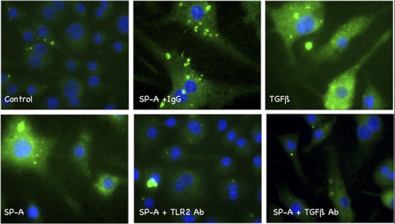
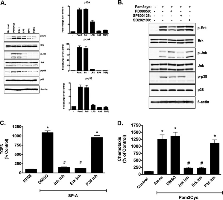
The innate immune system protects the host from bacterial and viral invasion. Surfactant protein A (SPA), a lung-specific collectin, stimulates macrophage chemotaxis. However, the mechanisms regulating this function are unknown. Hyaluronan (HA) and its receptors RHAMM (receptor for HA-mediated motility, CD168) and CD44 also regulate cell migration and inflammation. We therefore examined the role of HA, RHAMM, and CD44 in SPA-stimulated macrophage chemotaxis. Using antibody blockade and murine macrophages, SPA-stimulated macrophage chemotaxis was dependent on TLR2 but not the other SPA receptors examined. Anti-TLR2 blocked SPA-induced production of TGFβ. In turn, TGFβ1-stimulated chemotaxis was inhibited by HA-binding peptide and anti-RHAMM antibody but not anti-TLR2 antibody. Macrophages from TLR2(-/-) mice failed to migrate in response to SPA but responded normally to TGFβ1 and HA, effects that were blocked by anti-RHAMM antibody. Macrophages from WT and CD44(-/-) mice had similar responses to SPA, whereas those from RHAMM(-/-) mice had decreased chemotaxis to SPA, TGFβ1, and HA. In primary macrophages, SPA-stimulated TGFβ production was dependent on TLR2, JNK, and ERK but not p38. Pam3Cys, a specific TLR2 agonist, stimulated phosphorylation of JNK, ERK, and p38, but only JNK and ERK inhibition blocked Pam3Cys-stimulated chemotaxis. We have uncovered a novel pathway for SPA-stimulated macrophage chemotaxis where SPA stimulation via TLR2 drives JNK- and ERK-dependent TGFβ production. TGFβ1, in turn, stimulates macrophage chemotaxis in a RHAMM and HA-dependent manner. These findings are highly relevant to the regulation of innate immune responses by SPA with key roles for specific components of the extracellular matrix.


Figures

FIGURE 1.
FIGURE 1.
SPA-stimulated macrophage chemotaxis requires TLR-2, RHAMM, and HA. A, as assessed in a modified Boyden chemotaxis chamber assay, SPA stimulated macrophage chemotaxis in a dose-dependent manner with maximum stimulation at 100 μg/ml (*, p < 0.05 versus DM). Defined medium (DMEM) was used as a negative control. B, SPA (100 μg/ml)-stimulated chemotaxis was tested in the presence of blocking antibodies against SIRPα, calreticulin/CD91, TLR4, and TLR2 with nonimmune rabbit, rat, and chicken IgGs as species-specific controls. SPA alone significantly stimulated macrophage chemotaxis (*, p < 0.05 versus DMEM). SPA-stimulated chemotaxis was only inhibited by anti-TLR2 antibody (#, p < 0.05 versus SPA alone, SPA + IgG). C, SPA-stimulated chemotaxis (100 μg/ml) in the presence of HABPep, anti-RHAMM, and three CD44 antibodies, IM-7, KM81, or CD44v3, with nonimmune IgG and HABPep incubated with an excess of HA used as controls. SPA alone significantly stimulated macrophage chemotaxis (*, p < 0.05 versus DMEM). SPA-stimulated chemotaxis was significantly inhibited to base line in the presence of HABPep or anti-RHAMM antibody (#, p < 0.05 versus SPA alone/IgG/HA + HABPep), but not with any CD44 antibody tested. All data are normalized as percent of control (DM) presented as mean ± S.E. for three independent separate experiments, each with samples run at least in triplicate.
FIGURE 2.
FIGURE 2.
SPA-stimulated increase in HA and chemotaxis is dependent on TGFβ, and SPA-stimulated TGFβ production is dependent on TLR2. A, media were assayed for HA content by an ELISA-like assay after incubation with SPA (100 μg/ml) or TGFβ1 (10 ng/ml) for 1, 6, 12, and 24 h and normalized to the protein content of the cells in each culture. Data are presented as nanograms of HA per mg of protein ± S.E. for three independent experiments with samples run in triplicate within each experiment. Macrophages cultured in DMEM had no increase in HA content of the medium over time. SPA or TGFβ1 treatment produced similar time-dependent increases in HA production that were significant at 12 and 24 h (*, p < 0.05 versus DMEM). B, SPA stimulated HA production in the presence of TGFβ antibody or rabbit IgG (each 50 μg/ml). SPA alone or with IgG resulted in a significant increase in media HA concentration (*, p < 0.05 versus DMEM). SPA stimulation of HA was significantly inhibited to base line in the presence of a pan-specific TGFβ antibody (#, p < 0.05 versus SPA/SPA+IgG). C, SPA stimulated chemotaxis in the presence of anti-TGFβ, anti-RHAMM, and anti-TLR2 antibodies. SPA alone significantly stimulated macrophage chemotaxis (*, p < 0.05 versus DMEM). Anti-TGFβ antibody inhibited SPA-stimulated chemotaxis to levels similar to that achieved by anti-RHAMM and anti-TLR-2 antibodies (#, p < 0.05 versus SPA/SPA + IgG). Data are representative of three independent experiments with at least four replicates within each experiment. D, media were assayed for TGFβ content after incubation with SPA in the presence of either anti-TLR2 or anti-RHAMM antibodies. Data are presented as mean ± S.E. of percent of DM control. SPA + IgG produced significant induction of TGFβ (*, p < 0.05 versus DMEM). SPA induction of TGFβ was significantly inhibited to base line in the presence of anti-TLR2 antibody (#, p < 0.05 versus SPA/SPA + IgG). However, anti-RHAMM antibody failed to block SPA stimulation of TGFβ secretion. Data are representative of three experiments with at least three replicates within each experiment. Collectively, these data show that SPA stimulates TGFβ production via interaction with TLR2, and TGFβ stimulates macrophage chemotaxis in a RHAMM-dependent manner. E, genetic confirmation of TLR2 requirement for SPA-stimulated TGFβ production. BMDM from WT, RHAMM−/−, and TLR2−/− mice were exposed to SPA, and active TGFβ content of the media was measured by ELISA. SPA stimulated TGFβ production in both WT and RHAMM−/− mice, a response that was significantly blunted in TLR2−/− macrophages. Data are presented as mean ± S.E. and are representative of two independent experiments with samples run in triplicate within each experiment.
FIGURE 3.
FIGURE 3.
Effect of SPA on macrophages obtained from CAGA-GFP mice. BMDM were obtained from CAGA-GFP mice expressing a transgene consisting of 12 repeats of the PAI1 promoter linked to GFP as a reporter (39). The PAI1 promoter is highly TGFβ-sensitive, and GFP expression correlates with active TGFβ expression and signaling. Cells were plated and made quiescent overnight in 1% FCS. Three hours after stimulation, they were observed under direct fluorescence microscopy and imaged at ×400 magnification. Quiescent cells (Control) showed little to no GFP. Stimulation with active TGFβ1, a positive control, showed robust GFP staining. Similar increases in GFP staining were observed with SPA, and both anti-TLR2 and anti-TGFβ antibodies blocked this response. Nonimmune IgG had little to no effect on the GFP signal stimulated by SPA. Data shown are representative of two independent experiments with samples examined in triplicate within each experiment.
FIGURE 4.
FIGURE 4.
TGFβ1 stimulation of chemotaxis is dependent on RHAMM and HA. A, TGFβ1-stimulated macrophage chemotaxis was examined in the presence of anti-TGFβ, anti-TLR2, and anti-RHAMM antibodies. TGFβ1 alone significantly stimulated macrophage chemotaxis (*, p < 0.05 versus DMEM). This effect was significantly inhibited to base line in the presence of anti-TGFβ and anti-RHAMM antibodies (#, p < 0.05 versus TGFβ1 or TGFβ1 + ΙgG) but not by anti-TLR2 antibody. B, we next determined the chemotactic response to HA6 and the receptors involved in this response. HA6 (4 mm) stimulated a 4–5-fold response in macrophage chemotaxis (*, p < 0.05 versus DMEM). Only anti-RHAMM antibody and HABPep blocked this response to base line (#, p < 0.05 versus HA6 alone). Anti-TLR2, -TLR4, and -TGFβ and three antibodies to CD44 (IM7, KM81, and CD44v3) had no effect. C, to compare the effects of HMW and low molecular weight HA, we used HA6 (6-mer HA, 100 μg/ml) and HA900 (900-kDa HA, 100 μg/ml) as chemoattractants. HA6 stimulated macrophage chemotaxis 6-fold (*, p < 0.05 versus DMEM and HA900), whereas HA900 had no effect. Interestingly, HA900 inhibited HA6-stimulated chemotaxis when the two HA products were combined (#, p < 0.05 versus HA6). D, HA of various molecular sizes, each at 4 mm except HA900 (100 μg/ml), were tested in the modified Boyden chamber assay. A molecular size-dependent increase in HA-stimulated chemotaxis was observed with maximal chemotaxis observed with 34-mer HA (*, p < 0.05 versus DMEM; #, p < 0.01 versus DMEM; n.s. means not significant versus DMEM). HA900 had no effect on chemotaxis. All chemotaxis data are presented as mean ± S.E. normalized as percent of control (DM) for three separate experiments, with at least four replicates within each experiment.
FIGURE 5.
FIGURE 5.
Chemotactic responses in TLR2−/−, CD44−/−, and RHAMM−/− macrophages. A, BMDM were obtained from WT and TLR2−/− mice, and chemotaxis to SPA, TGFβ, and HA6 was determined. Chemotaxis is represented by cells per high power field in three independent experiments with four replicates per group in each experiment. Data are presented as mean ± S.E. In concordance with the data obtained with the RAW264.7 murine macrophage cell line, WT macrophages had 5–6-fold chemotactic responses to SPA (100 μg/ml), TGFβ (10 ng/ml), and HA6 (4 mm) (*, p < 0.05 versus DM). Each of these responses was blocked to base line by anti-RHAMM antibody (#, p < 0.05 versus SPA, TGFβ, or HA6) but not by normal IgG. Complete medium (CM, DMEM + 10% FCS) was used as a positive control. TLR2−/− macrophages did not respond to SPA (^, p < 0.05 versus CM, TGFβ, and HA) but had robust 4-fold responses to TGFβ and HA6, both of which were inhibited to base line by anti-RHAMM antibody (#, p < 0.05 versus TGFβ or HA alone or with normal IgG). B, BMDM were obtained from WT and CD44−/− mice, and chemotactic responses to SPA and TGFβ were examined. Both sets of macrophages showed equivalent chemotactic responses to SPA and TGFβ (*, p < 0.05 versus DM), and these responses were inhibited to base line by anti-RHAMM antibody (#, p < 0.05 versus SPA or TGFβ alone or with IgG). C, WT and CD44−/− macrophages also had similar 4–6-fold chemotactic responses to HA6 (*, p < 0.05 versus RPMI). D, BMDM were obtained from WT and RHAMM−/− mice, and chemotaxis to SPA, TGFβ and HA6 was determined. RHAMM WT macrophages demonstrated significant chemotactic responses to all three stimulants. RHAMM−/− macrophages showed significantly lower chemotactic responses to SPA and no response to TGFβ and HA6 (#, p < 0.05 versus WT macrophages). Collectively, these data confirm that CD44 is dispensable, and RHAMM is a key mediator in the chemotactic responses to SPA, TGFβ, and HA.
FIGURE 6.
FIGURE 6.
Cytoskeletal changes in response to SPA, TGFβ, and HA6 in WT and TLR2−/− mice. BMDM from WT and TLR2−/− mice were plated and made quiescent overnight. Three hours after stimulation with SPA, TGFβ, or HA6, the cytoskeleton was stained using FITC-phalloidin (green) and nuclei with DAPI (blue). A, WT macrophages that were mostly round in shape at base line showed increased formation of filopodia and lamellipodia when treated with SPA with or without normal IgG. Treatment with anti-RHAMM antibody (R36) inhibited SPA-stimulated cytoskeletal changes. B, both WT and TLR2−/− macrophages exposed to TGFβ or HA6 showed similar robust cytoskeletal changes with formation of lamellipodia and filopodia as seen in WT macrophages exposed to SPA, and these responses were completely inhibited when cells were also exposed to R36.
FIGURE 7.
FIGURE 7.
Pam3Cys, a TLR2 ligand, stimulates TGFβ production and chemotaxis that requires RHAMM. A, dose response to Pam3Cys-stimulated chemotaxis. RAW264.7 cells, examined in the chemotaxis assay, showed a dose-dependent increase in chemotaxis to Pam3Cys with the optimal dose being 5 μm. This dose of Pam3Cys was used for all subsequent experiments. B, Pam3Cys stimulated chemotaxis to the same degree as 10 ng/ml TGFβ1. Chemotaxis to Pam3Cys was inhibited by antibodies to TGFβ and RHAMM (R36), as well as HABPep, but not by scrambled peptide or normal rabbit IgG used as controls. (*, p < 0.05 versus Pam3Cys + TGFβ and Pam3Cys + R36; #, p < 0.05 versus Pam3Cys alone.) C, Pam3Cys stimulated the production of both latent and active TGFβ with maximal effect at even the lowest concentrations studied (*, p < 0.05 versus DMEM without Pam3Cys).
FIGURE 8.
FIGURE 8.
Pam3Cys-stimulated TGFβ production and chemotaxis requires ERK and JNK but not p38. We examined the intracellular signaling events, specifically the MAPKs ERK, JNK, and p38, stimulated by TLR2 ligation, TLR4 ligation, HA6, and TGFβ1, and we used pharmacologic inhibitors to test the contribution of each pathway to Pam3Cys-stimulated TGFβ production and chemotaxis. A, in RAW264.7 cells, Pam3Cys, a TLR2/1 ligand (2nd lane), and FSL1, a TLR2/6 ligand (3rd lane), stimulated the phosphorylation of ERK, JNK, and p38. LPS (25 μg/ml, 4th lane), a TLR4 ligand, only stimulated ERK phosphorylation. HA6 (4 mm, 5th lane) and TGFβ1 (10 ng/ml, 6th lane) had no significant effects on any MAPK and remained similar to that of DMEM control cells (No treat, 1st lane). Data are presented as mean ± S.E. of densitometry from three independent experiments. B, using BMDM from WT mice, we examined the effects of Pam3Cys and blockers of ERK, JNK, and p38 on the phosphorylation of these proteins. Pam3Cys treatment resulted in a robust phosphorylation of ERK, JNK, and p38 and all three blockers (ERK, PD98059; JNK, SP600125; and p38, SB202190; each at 10 μm concentration) blocked the relevant pathway confirming the specificity of each blocker in these primary cells. C, in WT BMDM, SPA (100 μg/ml) stimulated a 10-fold increase in TGFβ production (*, p < 0.05 DMSO versus RPMI). Pharmacologic inhibition of JNK and ERK, but not p38, inhibited Pam3Cys-stimulated TGFβ production to base line (#, p < 0.05 versus SPA + DMSO). D, also in WT BMDM, pharmacologic inhibition of JNK and ERK, but not p38, inhibited Pam3Cys-stimulated chemotaxis (*, p < 0.05 versus control; #, p < 0.05 versus Pam3Cys + DMSO). Data are presented as mean ± S.E. from three experiments with samples run in triplicate within each experiment. Collectively, these data confirm that TLR2 stimulation by SPA or Pam3Cys results in JNK- and ERK-dependent TGFβ production and subsequent chemotaxis and confirm that universal ligation of TLR2 has the same effects as SPA.
FIGURE 9.
FIGURE 9.
Schematic describing the pathway for TLR2-mediated TGFβ production and chemotaxis. The studies presented here show that SPA or Pam3Cys ligation of TLR2 increases TGFβ production in a JNK/ERK-dependent manner. In turn, TGFβ acts to recruit macrophages to the site of infection/injury, an event that is mediated by TGFβ receptors, but requires RHAMM and HA. When TGFβ concentrations are high enough at the site of infection/injury, there is a general suppression of inflammation. Modulation of RHAMM and HA may provide novel therapeutic targets to modulate innate immune responses.

Similar articles

Cited by

References

    1. Waters P., Vaid M., Kishore U., Madan T. (2009) Lung surfactant proteins A and D as pattern recognition proteins. Adv. Exp. Med. Biol. 653, 74–97 - PubMed
    1. Wright J. R. (2005) Immunoregulatory functions of surfactant proteins. Nat. Rev. Immunol. 5, 58–68 - PubMed
    1. Chroneos Z. C., Sever-Chroneos Z., Shepherd V. L. (2010) Pulmonary surfactant. An immunological perspective. Cell Physiol. Biochem. 25, 13–26 - PMC - PubMed
    1. McCormack F. X., Whitsett J. A. (2002) The pulmonary collectins, SP-A and SP-D, orchestrate innate immunity in the lung. J. Clin. Invest. 109, 707–712 - PMC - PubMed
    1. Wright J. R., Youmans D. C. (1993) Pulmonary surfactant protein A stimulates chemotaxis of alveolar macrophage. Am. J. Physiol. 264, L338–L344 - PubMed

Publication types

MeSH terms

Substances