. 2012 Dec;8(12):1724-40.
doi: 10.4161/auto.21857. Epub 2012 Sep 4.

MAPK15/ERK8 stimulates autophagy by interacting with LC3 and GABARAP proteins

Affiliations

MAPK15/ERK8 stimulates autophagy by interacting with LC3 and GABARAP proteins

David Colecchia et al. Autophagy. 2012 Dec.

Abstract

Macroautophagy (hereafter referred to as autophagy) is an evolutionarily conserved catabolic process necessary for normal recycling of cellular constituents and for appropriate response to cellular stress. Although several genes belonging to the core molecular machinery involved in autophagosome formation have been discovered, relatively little is known about the nature of signaling networks controlling autophagy upon intracellular or extracellular stimuli. We discovered ATG8-like proteins (MAP1LC3B, GABARAP and GABARAPL1) as novel interactors of MAPK15/ERK8, a MAP kinase involved in cell proliferation and transformation. Based on the role of these proteins in the autophagic process, we demonstrated that MAPK15 is indeed localized to autophagic compartments and increased, in a kinase-dependent fashion, ATG8-like proteins lipidation, autophagosome formation and SQSTM1 degradation, while decreasing LC3B inhibitory phosphorylation. Interestingly, we also identified a conserved LC3-interacting region (LIR) in MAPK15 responsible for its interaction with ATG8-like proteins, for its localization to autophagic structures and, consequently, for stimulation of the formation of these compartments. Furthermore, we reveal that MAPK15 activity was induced in response to serum and amino-acid starvation and that this stimulus, in turn, required endogenous MAPK15 expression to induce the autophagic process. Altogether, these results suggested a new function for MAPK15 as a regulator of autophagy, acting through interaction with ATG8 family proteins. Also, based on the key role of this process in several human diseases, these results supported the use of this MAP kinase as a potential novel therapeutic target.


Figures

None
Figure 1. MAPK15 interacted, in vitro and in vivo, with GABARAP, GABARAPL1 and LC3B. (A) Two positive clones, identified by two-hybrid screening, encoding GABARAP and GABARAPL1 were tested to be devoid of autoactivation. Yeast cells were cotransformed with the pACT2 vector containing the Gabarap (upper row) and Gabarapl1 (lower row) cDNA, respectively, with pGBKT7 alone (left) or with pGBKT7-MAPK15_C-term (right), and streaked on selective medium. (B) Bacterially expressed GST-GABARAP (lanes 3–4), GST-GABARAPL1 (lanes 5–6) and GST-LC3B (lanes 7–8) or GST alone (lanes 1–2), immobilized on glutathione-Sepharose Beads 4B, were incubated, for affinity precipitation (AP), with total lysates (TL) of 293T cells transiently transfected with a control vector or with HA-MAPK15, then analyzed by western blot (WB). (C) HeLa cells were cotransfected with a control vector or with GFP-LC3B, GFP-GABARAPL1, GFP-GABARAP in presence or absence of MAPK15–6XHis. Lysates (1 mg) were incubated with NiNTA-Sepharose Beads 4B, subjected to in vivo affinity precipitation, and then analyzed by WB. The total and MAPK15-bound amounts of GFP-tagged ATG8-like protein were quantified by NIH ImageJ software. (D) HeLa cells lysates (5 mg) were immunoprecipitated with control IgG or anti-MAPK15 antibody and analyzed by WB. LC3B was detected with anti-LC3B antibody (Sigma Aldrich).
None
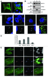
Figure 2. MAPK15 localized to autophagic structures. (A) HeLa cells were transfected with HA-MAPK15 cDNA. Cells were permeabilized with 0.2% Triton X-100. HA-MAPK15 proteins were immuno-labeled with anti-MAPK15 antibody and revealed with AlexaFluor488-conjugated secondary antibody. Nuclei were stained with DAPI. (B) HeLa cells stably expressing HA-MAPK15 were subjected to fractionation using the Subcellular Protein Fractionation Kit (Thermo Scientific). Lysates were subjected to SDS-PAGE followed by WB with anti-MAPK15 (top), anti-NFKBIA (middle top), anti-Calnexin (middle bottom), and anti-JUN (bottom) antibodies. C, cytoplasmic fraction; M, membrane fraction; N, nuclear fraction. (C) A clone of HeLa cells stably expressing HA-MAPK15 were permeabilized with 100 μg/ml digitonin. Cells were stained with appropriate antibodies (anti-MAPK15, anti-LC3B MBL, anti-GABARAP, anti-SQSTM1 and anti-LAMP1) and revealed with AlexaFluor488- and AlexaFluor555-conjugated secondary antibodies. Nuclei were stained with DAPI. The region enclosed in the white square has been enlarged in the smaller panel for better appreciation of the colocalizations. Arrows indicate colocalization areas. Similar results were obtained in at least 3 independent clones. (D) Colocalization rate of MAPK15 and LC3, GABARAP, SQSTM1 and LAMP1 respectively was obtained by analyzing at least 400 cells/sample from three different experiments (n = 3). (E) HeLa cells stably expressing HA-MAPK15 were transfected with pCEFL-GFP-LC3B. Twenty-four hours later, cells were permeabilized with 100 μg/ml digitonin. Cells were stained with anti-SQSTM1 and anti-MAPK15 antibodies and revealed with AlexaFluor555- and AlexaFluor647-conjugated secondary antibodies. Nuclei were stained with DAPI. The region enclosed in the white square has been enlarged in the lower panels for better appreciation of the colocalizations. Arrows indicate colocalization areas.
None
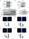
Figure 3. MAPK15 induced autophagy. (A) HeLa cells were transfected with control vector or HA-MAPK15. Two hours (h) before harvesting, cells were starved (Starv) or treated with rapamycin (Rap, 200 nM), where indicated. Lysates were analyzed by WB, with indicated antibodies. LC3B was detected with anti-LC3B antibody (Nanotools). The LC3B-II and MAPK1 amounts were quantified by NIH ImageJ software. (B) HeLa cells were transfected with control vector or HA-MAPK15. One hour before harvesting, cells were treated with E64d (10 μg/ml), where indicated. Lysates were analyzed by WB, with indicated antibodies. LC3B was detected with anti-LC3B antibody (Nanotools). The LC3B-II and MAPK1 amounts were quantified by NIH ImageJ software. (C) HeLa T-Rex MAPK15 cells were treated with doxycycline (Doxy, 1 μM) for different periods. Two hrs before harvesting, cells were starved, where indicated. Lysates were analyzed by WB, with indicated antibodies. LC3B was detected with anti-LC3B antibody (Nanotools). The LC3B-II and MAPK1 amounts were quantified by NIH ImageJ software. (D) HeLa T-Rex MAPK15 cells were treated with Doxy for 16 h, where indicated. Cells were fixed and then permeabilized with 100 μg/ml digitonin. Cells were stained with anti-LC3B antibody (MBL) and revealed with AlexaFluor488-conjugated antibody. Nuclei were stained with DAPI. Lower panel of each figure indicates the amount of LC3B dots per cell quantified by Volocity software. Measures were obtained by analyzing at least 400 cells/sample from five different experiments (n = 5). Measures were subjected to one-way ANOVA test. Asterisks were attributed for the following significance value: **p < 0.01. (E) Same as in (D), but, 4 h before fixing, cells were treated with Bafilomycin A1 (Baf, 100 nM). (F) HeLa cells were transfected with control vector or HA-MAPK15. Before harvesting, cells were starved (Starv) for the indicated periods. Lysates were analyzed by WB, with indicated antibodies. (G) HeLa cells were transfected with nonsilencing siRNA or with MAPK15-specific siRNA. After 72 h, cells were fixed and permeabilized with 100 μg/ml digitonin. Cells were stained with anti-LC3B (MBL) antibody and revealed with AlexaFluor488-conjugated secondary antibody. Nuclei were stained with DAPI. Lower panel of each figure indicate the amount of LC3B dots per cell quantified by Volocity software. Measures were obtained by analyzing at least 400 cells/sample from three different experiments (n = 3). Measures were subjected to one-way ANOVA test. Asterisks were attributed for the following significance value: ***p < 0.001. (H) Same as in (G), but, cells were starved for 1 h before fixing.
None
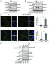
Figure 4. MAPK15 induced SQSTM1 degradation and reduced LC3B inhibitory phosphorylation. (A) HeLa cells were transfected with control vector or HA-MAPK15. Four hrs before harvesting, cells were treated with Rap or CQ, where indicated. Lysates were analyzed by WB, with indicated antibodies. The SQSTM1 and MAPK1 amounts were quantified by NIH ImageJ software. (B) HeLa T-Rex MAPK15 cells were treated with Doxy for 24 and 32 h, where indicated. Four hrs before harvesting, cells were starved or treated with Rap, where indicated. Lysates were analyzed by WB, with indicated antibodies. The SQSTM1 and MAPK1 amounts were quantified by NIH ImageJ software. (C) HeLa T-Rex MAPK15 cells were treated with Doxy for 16 h or starved as indicated. Cells were fixed and then permeabilized with 100 μg/ml digitonin. Cells were stained with anti-SQSTM1 and anti-MAPK15 antibodies and revealed with AlexaFluor488-conjugated and AlexaFluor555-conjugated secondary antibodies, respectively. Right panels, indicate the amount of SQSTM1 dots per cell quantified by Volocity software. Measures were obtained by analyzing at least 400 cells/sample from four different experiments (n = 4). Measures were subjected to one-way ANOVA test. Asterisks were attributed for the following significance value: ***p < 0.001. (D) HeLa cells were transfected with control vector or HA-MAPK15. One hour before harvesting, cells were treated with Rap, where indicated. Lysates were analyzed by WB, with indicated antibodies. Total LC3B was detected with anti-LC3B antibody (Sigma Aldrich). The phospho-LC3B, LC3B-I and MAPK1 amounts were quantified by NIH ImageJ software.
None
Figure 5. MAPK15 bound ATG8-like protein through region 300–373 aa. (A) Scheme depicts MAPK15 full length and its deletion mutants. (B) Bacterially expressed GST-GABARAP (lanes 3–7), GST-GABARAPL1 (lanes 8–12), GST-LC3B (lanes 13–17) or GST alone (lanes 1–2) immobilized on glutathione-Sepharose Beads 4B, were incubated with lysates of 293T cells transiently transfected with a control vector or with full length HA-MAPK15_WT or with HA-MAPK15 deletion mutants, subjected to affinity precipitation and then analyzed by WB. (C) Colocalization rate of LC3 with MAPK15 or its deletion mutants in HeLa transfected cells was obtained by analyzing at least 400 cells/sample from three different experiments (n = 3). Measures were subjected to one-way ANOVA test. Asterisks were attributed for the following significance value: ***p < 0.001. (D) The graph indicates the amount of LC3B dots per cell in HeLa cells transfected with MAPK15 or its deletion mutants, quantified by Volocity software. Measures were obtained by analyzing at least 400 cells/sample from three different experiments (n = 3). Measures were subjected to one-way ANOVA test. Asterisks were attributed for the following significance value: **p < 0.01.
None
Figure 6. MAPK15 bound ATG8-like protein through a specific LIR-containing region. (A) Alignment of a portion of MAPK15 protein sequence across the species showing the position of the conserved LIR motif. (B) GST, GST-MAPK15_300–373_WT or GST-MAPK15_300–373_AXXA were incubated for 1 h at 4°C with 6xHis-GABARAP or 6xHis-LC3B. Proteins were then affinity-purified by glutathione-Sepharose Beads 4B, extensively washed and loaded on SDS-PAGE gels. Total proteins were stained with Coomassie stain. The total and MAPK15-bound amounts of 6xHis-tagged ATG8-like protein were quantified by NIH ImageJ software. (C) HeLa cells were cotransfected with a control vector or with GFP-LC3B, MAPK15_WT-V5–6xHis or MAPK15_AXXA-V5–6xHis. Lysates (1 mg) were incubated with NiNTA-Sepharose Beads 4B, subjected to in vivo affinity precipitation, and then analyzed by WB. The total and MAPK15-bound amounts of GFP-tagged ATG8-like protein were quantified by NIH ImageJ software. (D) Colocalization rate of LC3 with MAPK15_WT or MAPK15_AXXA in HeLa transfected cells was obtained by analyzing at least 400 cells/sample from three different experiments (n = 3). Measures were subjected to one-way ANOVA test. Asterisks were attributed for the following significance value: ***p < 0.001. (E) HeLa cells were transfected with control vector or HA-MAPK15 (WT) or its mutants. Lysates were analyzed by WB, with indicated antibodies. LC3B was detected with anti-LC3B antibody (Nanotools). The LC3B-II, SQSTM1 and MAPK1 amounts were quantified by NIH ImageJ software. (F) The graph indicates the amount of LC3B dots per cell in HeLa cells transfected with MAPK15_WT or MAPK15_AXXA, quantified by Volocity software. Measures were obtained by analyzing at least 400 cells/sample from three different experiments (n = 3). Measures were subjected to one-way ANOVA test. Asterisks were attributed for the following significance value: **p < 0.01.
None
Figure 7. The catalytic activity of MAPK15 was required for its ability to control autophagy. (A) HeLa cells were transfected with control vector or HA-MAPK15_WT or HA-MAPK15_KD. Lysates were analyzed by WB, with indicated antibodies. LC3B was detected with anti-LC3B antibody (Nanotools). Right panel, steady-state autophagy. Left panel, autophagic flux. (B) The graph indicates the amount of LC3B dots per cell in HeLa cells transfected with MAPK15_WT or MAPK15_KD, quantified by Volocity software. Measures were obtained by analyzing at least 400 cells/sample from three different experiments (n = 3). Measures were subjected to one-way ANOVA test. Asterisks were attributed for the following significance value: **p < 0.01. (C) For affinity precipitation (AP), bacterially expressed GST-LC3B or GST alone, immobilized on glutathione-Sepharose Beads 4B were incubated with lysates of 293T cells transiently transfected with a control vector or with HA-MAPK15_WT or HA-MAPK15_KD, then analyzed by western blot. (D) Colocalization rate of LC3 and MAPK15 in HeLa cells stably expressing MAPK15_WT or MAPK15_KD was obtained by analyzing at least 400 cells/sample from three different experiments (n = 3). (E) HeLa T-Rex MAPK15 cells were treated with Doxy for 2 h, then Ro-318220 (1 μM) was added to culture medium for the following 6 h, where indicated. Cells were fixed and then permeabilized with 100 μg/ml digitonin. Cells were stained with anti-LC3B (MBL) antibody and revealed with AlexaFluor488-conjugated antibody. Nuclei were stained with DAPI. The LC3B-positive dots per cell were quantified using Volocity software (right panel). Measures were obtained by analyzing at least 400 cells/sample from three different experiments (n = 3). Measures were subjected to one-way ANOVA test. Asterisks were attributed for the following significance values: *p < 0.05, **p < 0.01. (F) HeLa cells were treated with Ro-318220 (1 μM) for 6 h and/or then starved for 1 h, where indicated. Cells were fixed and permeabilized with 100 μg/ml digitonin, then stained with anti-LC3B antibody (MBL) and revealed with AlexaFluor488-conjugated secondary antibody. Nuclei were stained with DAPI. The number of LC3B positive dots per cell was quantified using the Volocity software (right panel). Measures were obtained by analyzing at least 400 cells/sample from three different experiments (n = 3). Measures were subjected to one-way ANOVA test. Asterisks were attributed for the following significance values: *p < 0.05, **p < 0.01. At the concentration used, the Ro-318220 compound did not affect cell viability. (G) HeLa cells were treated as in (E). Lysates were analyzed by WB, with indicated antibodies. LC3B was detected with anti-LC3B antibody (Nanotools). (H) HeLa cells were treated as in (F). Lysates were analyzed by WB, with indicated antibodies. LC3B was detected with anti-LC3B antibody (Nanotools).
None
Figure 8. MAPK15 in the control of the autophagic process. A fraction of the MAPK15 protein is localized to autophagic structures by binding to mammalian ATG8 proteins, through a conserved LIR. When localized to these compartments, in basal conditions as well as upon starvation, MAPK15 enzymatic activity is able to control the rate of autophagy by phosphorylating downstream substrates, possibly additional ATG proteins, ultimately mediating its effect on this process.

Similar articles

Cited by

  • Disturbed Flow Induces Autophagy, but Impairs Autophagic Flux to Perturb Mitochondrial Homeostasis.
    Li R, Jen N, Wu L, Lee J, Fang K, Quigley K, Lee K, Wang S, Zhou B, Vergnes L, Chen YR, Li Z, Reue K, Ann DK, Hsiai TK. Li R, et al. Antioxid Redox Signal. 2015 Nov 20;23(15):1207-19. doi: 10.1089/ars.2014.5896. Epub 2015 Jun 29. Antioxid Redox Signal. 2015. PMID: 26120766 Free PMC article.
  • Autophagy processes are dependent on EGF receptor signaling.
    De Iuliis V, Marino A, Caruso M, Capodifoglio S, Flati V, Marynuk A, Marricareda V, Ursi S, Lanuti P, Talora C, Conti P, Martinotti S, Toniato E. De Iuliis V, et al. Oncotarget. 2018 Jul 13;9(54):30289-30303. doi: 10.18632/oncotarget.25708. eCollection 2018 Jul 13. Oncotarget. 2018. PMID: 30100990 Free PMC article.
  • MAPK15 protects from oxidative stress-dependent cellular senescence by inducing the mitophagic process.
    Franci L, Tubita A, Bertolino FM, Palma A, Cannino G, Settembre C, Rasola A, Rovida E, Chiariello M. Franci L, et al. Aging Cell. 2022 Jul;21(7):e13620. doi: 10.1111/acel.13620. Epub 2022 Jun 1. Aging Cell. 2022. PMID: 35642724 Free PMC article.
  • Atypical MAP kinases - new insights and directions from amoeba.
    Hadwiger JA, Aranda RG, Fatima S. Hadwiger JA, et al. J Cell Sci. 2023 Oct 15;136(20):jcs261447. doi: 10.1242/jcs.261447. Epub 2023 Oct 16. J Cell Sci. 2023. PMID: 37850857 Free PMC article. Review.
  • Guidelines for the use and interpretation of assays for monitoring autophagy (3rd edition).
    Klionsky DJ, Abdelmohsen K, Abe A, Abedin MJ, Abeliovich H, Acevedo Arozena A, Adachi H, Adams CM, Adams PD, Adeli K, Adhihetty PJ, Adler SG, Agam G, Agarwal R, Aghi MK, Agnello M, Agostinis P, Aguilar PV, Aguirre-Ghiso J, Airoldi EM, Ait-Si-Ali S, Akematsu T, Akporiaye ET, Al-Rubeai M, Albaiceta GM, Albanese C, Albani D, Albert ML, Aldudo J, Algül H, Alirezaei M, Alloza I, Almasan A, Almonte-Beceril M, Alnemri ES, Alonso C, Altan-Bonnet N, Altieri DC, Alvarez S, Alvarez-Erviti L, Alves S, Amadoro G, Amano A, Amantini C, Ambrosio S, Amelio I, Amer AO, Amessou M, Amon A, An Z, Anania FA, Andersen SU, Andley UP, Andreadi CK, Andrieu-Abadie N, Anel A, Ann DK, Anoopkumar-Dukie S, Antonioli M, Aoki H, Apostolova N, Aquila S, Aquilano K, Araki K, Arama E, Aranda A, Araya J, Arcaro A, Arias E, Arimoto H, Ariosa AR, Armstrong JL, Arnould T, Arsov I, Asanuma K, Askanas V, Asselin E, Atarashi R, Atherton SS, Atkin JD, Attardi LD, Auberger P, Auburger G, Aurelian L, Autelli R, Avagliano L, Avantaggiati ML, Avrahami L, Awale S, Azad N, Bachetti T, Backer JM, Bae DH, Bae JS, Bae ON, Bae SH, Baehrecke EH, Baek SH, Baghdiguian S, Bagniewska-Zadworna A, Bai H, Bai J, Bai XY, Bailly Y, Balaji KN, … See abstract for full author list ➔ Klionsky DJ, et al. Autophagy. 2016;12(1):1-222. doi: 10.1080/15548627.2015.1100356. Autophagy. 2016. PMID: 26799652 Free PMC article. No abstract available.

References

    1. Klionsky DJ, Emr SD. Autophagy as a regulated pathway of cellular degradation. Science. 2000;290:1717–21. doi: 10.1126/science.290.5497.1717. - DOI - PMC - PubMed
    1. Tooze SA, Yoshimori T. The origin of the autophagosomal membrane. Nat Cell Biol. 2010;12:831–5. doi: 10.1038/ncb0910-831. - DOI - PubMed
    1. Seglen PO, Bohley P. Autophagy and other vacuolar protein degradation mechanisms. Experientia. 1992;48:158–72. doi: 10.1007/BF01923509. - DOI - PubMed
    1. Ohsumi Y. Molecular dissection of autophagy: two ubiquitin-like systems. Nat Rev Mol Cell Biol. 2001;2:211–6. doi: 10.1038/35056522. - DOI - PubMed
    1. Klionsky D, Abdalla FC, Abeliovich H, Abraham RT, Acevedo-Arozena A, Adeli K, et al. Guidelines for the use and interpretation of assays for monitoring autophagy. Autophagy. 2012;8:445–544. doi: 10.4161/auto.19496. - DOI - PMC - PubMed

Publication types

MeSH terms