Clinical Trial
. 2012 Dec;32(12):2171-80.
doi: 10.1038/jcbfm.2012.123. Epub 2012 Sep 5.

CADASIL mutations and shRNA silencing of NOTCH3 affect actin organization in cultured vascular smooth muscle cells

Affiliations
Clinical Trial

CADASIL mutations and shRNA silencing of NOTCH3 affect actin organization in cultured vascular smooth muscle cells

Saara Tikka et al. J Cereb Blood Flow Metab. 2012 Dec.

Abstract

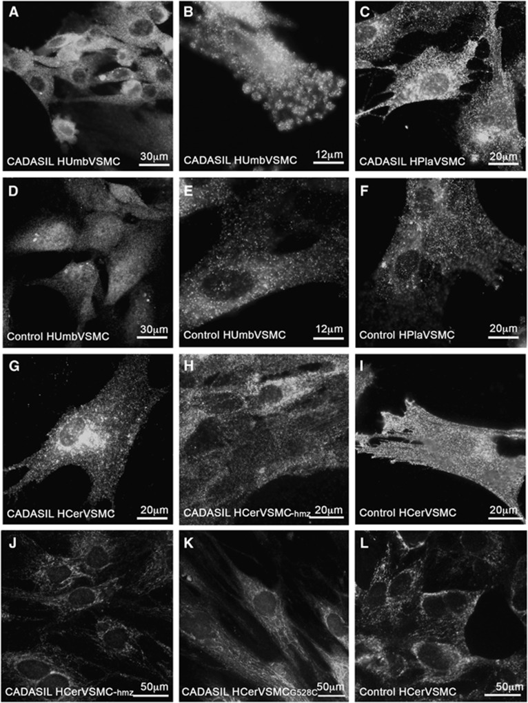
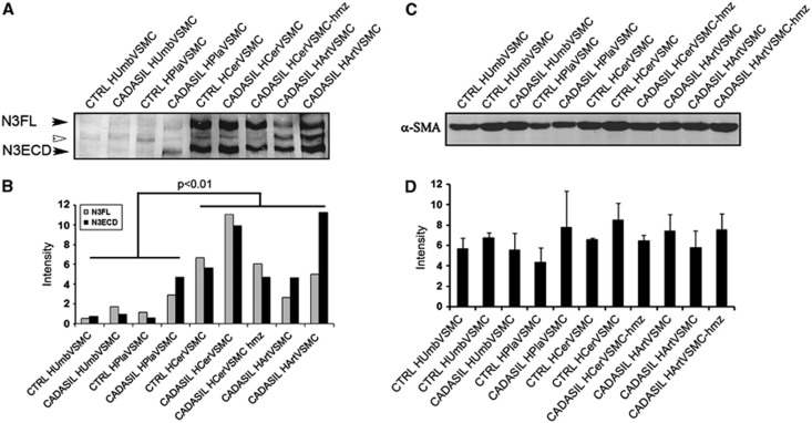
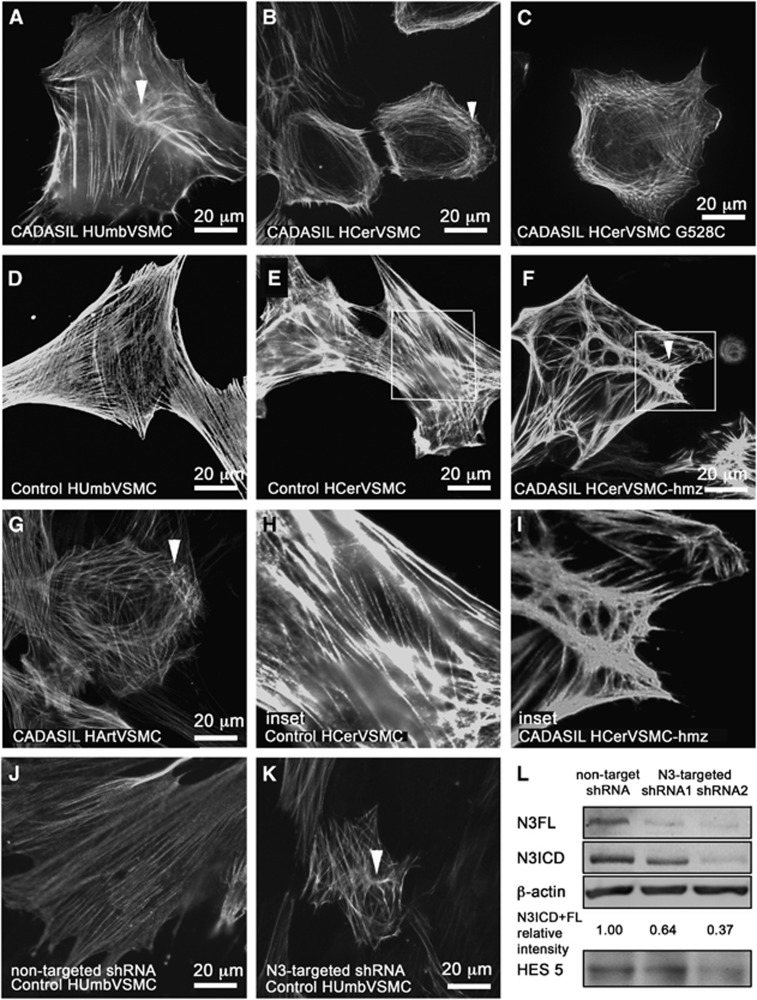
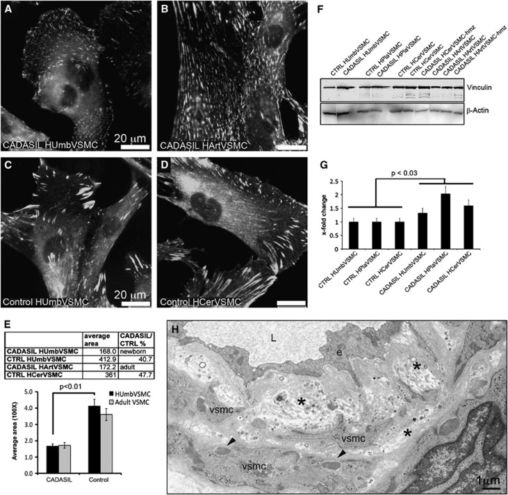
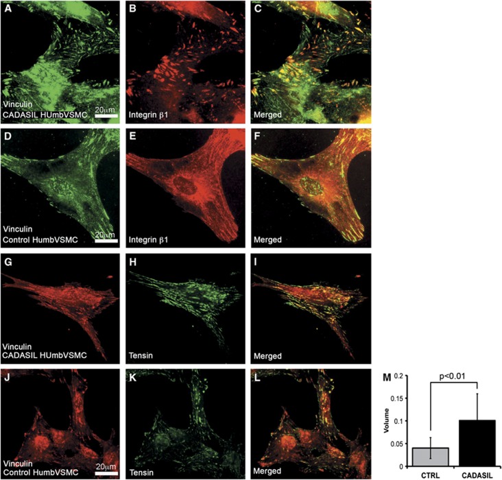
Cerebral autosomal dominant arteriopathy with subcortical infarcts and leukoencephalopathy (CADASIL) is the most common hereditary vascular dementia caused by mutations in NOTCH3 gene. Pathology is manifested in small- and middle-sized arteries throughout the body, though primarily in cerebral white matter. Hemodynamics is altered in CADASIL and NOTCH3 is suggested to regulate actin filament polymerization and thereby vascular tone. We analyzed NOTCH3 expression and morphology of actin cytoskeleton in genetically genuine cultured human CADASIL vascular smooth muscle cells (VSMCs) (including a cell line homozygous for p.Arg133Cys mutation) derived from different organs, and in control VSMCs with short hairpin RNA (shRNA)-silenced NOTCH3. NOTCH3 protein level was higher in VSMCs derived from adult than newborn arteries in both CADASIL and control VSMCs. CADASIL VSMCs showed altered actin cytoskeleton including increased branching and node formation, and more numerous and smaller adhesion sites than control VSMCs. Alterations in actin cytoskeleton in shRNA-silenced VSMCs were similar as in CADASIL VSMCs. Severity of the alterations in actin filaments corresponded to NOTCH3 expression level being most severe in VSMCs derived from adult cerebral arteries. These observations suggest that hypomorphic NOTCH3 activity causes alterations in actin organization in CADASIL. Furthermore, arteries from different organs have specific characteristics, which modify the effects of the NOTCH3 mutation and which is one explanation for the exceptional susceptibility of cerebral white matter arteries.


Figures

Figure 1
Figure 1
Subcellular localization of NOTCH3 in CADASIL vascular smooth muscle cells (VSMCs). For detection of cell surface NOTCH3, VSMCs were fixed with 3.5% phosphate-buffered paraformaldehyde. NOTCH3 forms more aggregates on the cell surface in CADASIL than in control VSMCs (AI). For intracellular NOTCH3 detection, VSMCs were fixed with methanol. Permeabilized VSMCs show that NOTCH3 does not aggregate inside the cell in either CADASIL or control VSMCs (JL). Immunostainings were performed with antibody (1E4) directed against N3ECD recognizing full-length NOTCH3 and N3ECD. Number of cell lines analyzed: HUmbVSMC: CADASIL n=4, control n=4, HPlaVSMC CADASIL n=4, control n=4, HArtVSMC: CADASIL n=3, HCerVSMC: CADASIL n=2, CADASIL-hmz n=1, control n=3.
Figure 2
Figure 2
Total NOTCH3 and α-SMA expression in CADASIL and control vascular smooth muscle cells (VSMCs). (A) Western blot analysis of the expression of NOTCH3 detected with 5E1 directed against N3ECD in CADASIL and control VSMCs. In all, 40 μg of cell lysate was loaded on each lane. The 280-kDa full-length NOTCH3 (N3FL) and 220 kDa N3ECD are indicated with arrowheads. The 245-kDa band in between (white arrowhead) is unknown. (B) The expression of NOTCH3 is significantly higher in HCerVSMCs and HArtVSMCs than in HUmbVSMCs or HPlaVSMCs. There are no statistically significant differences in the total expression level of NOTCH3 or N3FL/N3ECD ratio between CADASIL versus the corresponding control or between control versus heterozygous or homozygous VSMCs. (C) Western blot analysis of α-SMA expression in CADASIL and control VSMCs. In all, 20 μg of cell lysate was loaded on each lane. (D) Even though, CADASIL cells have altered actin filament organization, the total amount of α-SMA in CADASIL VSMCs is not significantly different from that in corresponding control VSMCs. The bar graphs show the average intensities and ±s.e.m. from two independent experiments. Both experiments were repeated two times with cells at passage 5.
Figure 3
Figure 3
Actin cytoskeleton in CADASIL vascular smooth muscle cells (VSMCs). VSMCs fixed with methanol were immunostained for smooth muscle cell α-actin (α-SMA). α-SMA filament have altered organization in CADASIL VSMCs. In all CADASIL VSMCs the α-SMA positive actin filaments are short, poorly organized and form nodes (clusters of short haphazardly oriented filaments, selected five marked with arrowheads) (A: HUmbVSMC, B: HCerVSMC, C: HCerVSMC-G528C, F: HCerVSMC-hmz, and G: HArtVSMC). Actin filament nodes in CADASIL VSMCs are shown in higher magnification in an inset (I: HCerVSMC-hmz). Control VSMCs have robust cell spanning actin filaments (D: HUmbVSMC and E: HCerVSMC and an inset H: HCerVSMC). Number of cell lines analyzed: HUmbVSMC: CADASIL n=4, control n=4, HPlaVSMC CADASIL n=4, control n=4, HArtVSMC: CADASIL n=3, HCerVSMC: CADASIL n=2, CADASIL-hmz n=1, control n=3. Control HUmbVSMCs were transduced with nontarget short hairpin RNA (shRNA) (J) or NOTCH3-targeted shRNA2 (K). Silencing of NOTCH3 expression induced similar alterations to organization of actin filaments as observed in CADASIL VSMCs. Silencing of NOTCH3 expression was verified by Western blot (L). NOTCH3-targeted shRNA1 and shRNA2 reduced relative intensity of NOTCH3 expression by 36 and 63% when compared with nontarget shRNA. NOTCH3 signaling activity is also reduced as indicated by lower expression level of NOTCH3 target gene HES5 (shRNA1 5% and shRNA2 30%). Relative intensity is calculated from an average of combined N3FL and N3ICD intensities. Intensities are averages from two independent experiments normalized to β-actin. Average intensities are shown in proportion to nontarget shRNA sample (relative intensity). N3FL and N3ICD were detected by antibody directed against N3ICD.
Figure 4
Figure 4
Vinculin positive adhesion sites in CADASIL vascular smooth muscle cells (VSMCs). Methanol fixed cells immunostained for vinculin show markedly greater number of small-sized adhesion sites in (A, B) CADASIL (HUmbVSMC and HArtVSMC) than (C, D) control VSMCs (HUmbVSMCs and HCerVSMC). (E) The areas of adhesion sites were measured from CADASIL and control VSMC lines immunostained for vinculin using ImageJ (NIH, Bethesda, MD, USA) software. From each cell line, at least 100 adhesion sites were measured. The average area of individual adhesion sites in CADASIL cell line derived from newborn tissue (HUmbVSMC) is 40.7% of that in control VSMCs (HUmbVSMCs). (C) The same pattern of small and numerous adhesion sites was detected also in CADASIL cell line derived from adult tissue (HArtVSMCs) whereas (D) control adult cell line (HCerVSMCs) showed large and elongated adhesion sites similarly to control HUmbVSMCs. Bar graph shows average size of adhesion sites in two CADASIL and two control cell lines. Significance is calculated only for the tissue matched cell lines. Total vinculin amount in the cell lysates at passage 5. (F) Western blot analysis shows a greater amount of total vinculin protein detected by vinculin antibody in CADASIL VSMCs than in corresponding control cells. In all, 20 μg of cell lysate was loaded on each lane and equal loading was verified by redetection of the blot with β-actin antibody. (G) Bar graph shows x-fold change of average vinculin intensities in comparison to control from three replicates. Intensities are normalized to β-actin intensity. (H) Electron micrograph of a dermal artery of CADASIL patient double stained with uranyl acetate and lead citrate. Note widening of the subendothelial space, thickening of the basal lamina and detachment of VSMCs from each other. Two arrow heads point granular osmiophilic material (GOM) in the indentations of VSMCs. e=endothelial cell, L=arterial lumen, *=subendothelial space and basal lamina.
Figure 5
Figure 5
Integrin β1 and adapter protein tensin in adhesion sites. Methanol fixed vascular smooth muscle cells (VSMCs) were immunostained with integrin β1 or tensin antibody. (AF) Integrin β1 forms thin ribbon-like structures in the center of the cells, parts of which are vinculin positive. In the two CADASIL (only HUmbVSMC shown) VSMC lines analyzed the β1 positive ribbons were more frequent than in the two control VSMC lines (only HUmbVSMC shown). (GL) Tensin colocalizes partly to the same adhesion structures as vinculin in both CADASIL VSMC lines analyzed and control cell lines (only HUmbVSMC shown). (M) Bar graph shows the average volume (area × intensity) of tensin immunoreactivity in CADASIL and control VSMCs (P<0.01). From each cell line, at least 500 adhesion sites were measured. Tensin localization indicates maturation of adhesion sites to the stages of late focal adhesions and fibrillar adhesions and the change from a structural adhesion to an adhesion that interacts with signaling.

Similar articles

Cited by

References

    1. Joutel A, Vahedi K, Corpechot C, Troesch A, Chabriat H, Vayssiere C, et al. Strong clustering and stereotyped nature of Notch3 mutations in CADASIL patients. Lancet. 1997;350:1511–1515. - PubMed
    1. Monet-Lepretre M, Bardot B, Lemaire B, Domenga V, Godin O, Dichgans M, et al. Distinct phenotypic and functional features of CADASIL mutations in the Notch3 ligand binding domain. Brain. 2009;132:1601–1612. - PMC - PubMed
    1. Kalimo H, Miao Q, Tikka S, Mykkanen K, Junna M, Roine S, et al. CADASIL: the most common hereditary subcortical vascular dementia. Future Neurol. 2008;3:683–704.
    1. Ruchoux MM, Guerouaou D, Vandenhaute B, Pruvo JP, Vermersch P, Leys D. Systemic vascular smooth muscle cell impairment in cerebral autosomal dominant arteriopathy with subcortical infarcts and leukoencephalopathy. Acta Neuropathol (Berl) 1995;89:500–512. - PubMed
    1. Ruchoux MM, Chabriat H, Bousser MG, Baudrimont M, Tournier-Lasserve E. Presence of ultrastructural arterial lesions in muscle and skin vessels of patients with CADASIL. Stroke. 1994;25:2291–2292. - PubMed

Publication types

MeSH terms