Comparative Study
. 2013 Mar;471(3):750-61.
doi: 10.1007/s11999-012-2557-3.

Computer-assisted tumor surgery in malignant bone tumors

Affiliations
Comparative Study

Computer-assisted tumor surgery in malignant bone tumors

Kwok Chuen Wong et al. Clin Orthop Relat Res. 2013 Mar.

Abstract

Background: Small recent case series using CT-based navigation suggest such approaches may aid in surgical planning and improve accuracy of intended resections, but the accuracy and clinical use have not been confirmed.

Questions/purposes: We therefore evaluated (1) the accuracy; (2) recurrences; and (3) function in patients treated by computer-assisted tumor surgery (CATS).

Methods: From 2006 to 2009, we performed CATS in 20 patients with 21 malignant tumors. The mean age was 31 years (range, 6-80 years). CT and MR images for 18 cases were fused using the navigation software. Reconstructed two-dimensional/three-dimensional images were used to plan the bone resection. The achieved bone resection was compared with the planned one by assessing margins, dimensions at the level of bone resection, or fitting of CAD custom prostheses. Function was assessed with the Musculoskeletal Tumor Society (MSTS) score. The minimum followup was 31 months (mean, 39 months; range, 5-69 months).

Results: Histological examination of all resected specimens showed a clear tumor margin. The achieved bone resection matched the planned with a difference of ≤ 2 mm. The achieved positions of custom prostheses were comparable to the planned positions when merging postoperative with preoperative CT images in five cases. Three of the four patients with local recurrence had tumors at the sacral region. The mean MSTS score was 28 (range, 23-30).

Conclusion: CATS with image fusion allows accurate execution of the intended bone resection. It may be beneficial to resection and reconstruction in pelvic, sacral tumors and more difficult joint-preserving intercalated tumor surgery. Comparative clinical studies with long-term followup are necessary to confirm its efficacy.

Level of evidence: Level IV, therapeutic study. See Guidelines for Authors for a complete description of levels of evidence.


Figures

Fig. 1
Fig. 1
The workflow of CATS in the study is shown.
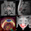
Fig. 2A–D
Fig. 2A–D
CT/MR/PET images are shown of fusion in the navigation display in Patient 19 with sacral chordoma involving S3 and S4. (A) Reformatted coronal view of CT images was fused with coronal view of T2-weighted MR images. (B) Reformatted sagittal view of CT images was fused with sagittal view of T1-weighted MR images. (C) Axial view of CT images was fused with axial view of PET images. The blue color in the PET image at the sacrum bone represented the tumor with hypermetabolic activities. (D) Anterior view of three-dimensional (3D) reconstruction of sacrum bone. The red structure represented the tumor extent that was determined and outlined from MR images. After multimodal image fusion, the segmented tumor volume was imported into the sacrum bone to form a 3D bone-tumor model. The two-dimensional reformatted images and the 3D model could be studied in detail and it greatly facilitated the surgical planning of the tumor resection. We resected the sacrum through a posterior approach under navigation guidance. We osteotomized the sacrum through right S2 and left S3 anterior foramina without sacrificing sacroiliac joint and could preserve both S2 and left S3 nerve roots. PET = positron emission tomography.
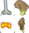
Fig. 3A–D
Fig. 3A–D
CAD custom prostheses are shown. Patient 20, a 6-year-old boy with distal femur osteosarcoma, (A) shows a joint-preserving extendable prosthesis and distal femur remaining epiphysis and (B) depicts the cross-section at the bone-implant junction. The gap between the edge of the prosthesis and the bone epiphysis represents the thickness of the femoral cartilage that could be outlined from the CT-MR fused image. For Patient 15, a 16-year-old girl with pelvic osteosarcoma, (CD) the surgical planning of PII and PIII resection and fitting of a custom pelvic prosthesis are shown.
Fig. 4A–D
Fig. 4A–D
(A) A coronal section of the CT images with incorporation of a CAD prosthesis for Patient 16, a 24-year-old man with right distal femur parosteal osteosarcoma, is shown. By using the CAD software, CAD data of custom prosthesis could be directly imported into the navigation system for planning of bone resection. The central cross represented the virtual marker (pedicle screw in the CT spine navigation software) that marked one of the locations of intended bone resection. (B) A sagittal section of the MR images showed the extent of the tumor. (C) A axial section of CT/MR image fusion at the intended resection of distal femur is shown. (D) A three-dimensional bone tumor model reconstructed from CT and MR image data sets is shown. The tumor volume was red. A joint-preserving resection with multiplanar osteotomies was planned at the distal femur and intended bone resections were marked with virtual screws. Reprinted with courtesy of Wong KC, Kumta SM, Tse LF, Ng EW, Lee KS. Image fusion for computer assisted tumor surgery (CATS). In: Ukimura O, ed. Image Fusion. 2011:373–390. Available at: http://www.intechopen.com/books/image-fusion/image-fusion-for-computer-assisted-tumor-surgery-cats-. Accessed January 12, 2011.
Fig. 5A–D
Fig. 5A–D
For Patient 14, a 21-year-old woman with left proximal femur low-grade chondrosarcoma, navigation images show intraoperative reformatted (A) coronal view (CT-MR fused image), (B) sagittal view (CT image), (C) axial view (CT-MR image), and (D) three-dimensional bone-tumor model. After registration with paired point and surface matching of the proximal femur bone, we verified the accuracy of the registration by running the tip of the navigation probe on the bone surface. The tip of the navigation probe was exactly at the bone surface on the virtual images. We could then proceed to execute the planned bone resection under navigation because the operative anatomy was real-time matching well with the virtual images.
Fig. 6A–B
Fig. 6A–B
For Patient 11, a 50-year-old man with recurrent chordoma and left pelvic metastases, PII and PIII resection and custom pelvic prosthesis were performed. The dimensions (length ab and cd) of the achieved bone resection at ilium were measured (A). They were then compared with the corresponding cross-section at the navigation planning (B). The difference of the dimension was ≤ 2 mm and the achieved bone resection was considered to be accurate as that planned.
Fig. 7A–E
Fig. 7A–E
Postoperative CT scans were performed in some patients with CAD custom prostheses. The postoperative CT images were fused with the preoperative CT images to validate the accuracy of CATS. The achieved positions of prostheses (yellow) were comparable to the planned ones (gray) in Patients 1 (A), 2 (B), 11 (C), 14 (E), and 15 (D).

References

    1. Anderson KC, Buehler KC, Markel DC. Computer assisted navigation in total knee arthroplasty: comparison with conventional methods. J Arthroplasty. 2005;20(Suppl 3):132–138. doi: 10.1016/j.arth.2005.05.009. - DOI - PubMed
    1. Bacci G, Ferrari S, Bertoni F, Ruggieri P, Picci P, Longhi A, Casadei R, Fabbri N, Forni C, Versari M, Campanacci M. Long-term outcome for patients with nonmetastatic osteosarcoma of the extremity treated at the Istituto Ortopedico Rizzoli according to the Istituto Ortopedico Rizzoli/Osteosarcoma-2 protocol: an updated report. J Clin Oncol. 2000;18:4016–4027. - PubMed
    1. Bergh P, Kindblom LG, Gunterberg B, Remotti F, Ryd W, Meis-Kindblom JM. Prognostic factors in chordoma of the sacrum and mobile spine: a study of 39 patients. Cancer. 2000;88:2122–2134. doi: 10.1002/(SICI)1097-0142(20000501)88:9<2122::AID-CNCR19>3.0.CO;2-1. - DOI - PubMed
    1. Cartiaux O, Docquier PL, Paul L, Francq BG, Cornu OH, Delloye C, Raucent B, Dehez B, Banse X. Surgical inaccuracy of tumor resection and reconstruction within the pelvis: an experimental study. Acta Orthop. 2008;79:695–702. doi: 10.1080/17453670810016731. - DOI - PubMed
    1. Cho HS, Kang HG, Kim HS, Han I. Computer-assisted sacral tumor resection. A case report. J Bone Joint Surg Am. 2008;90:1561–1566. doi: 10.2106/JBJS.G.00928. - DOI - PubMed

Publication types

MeSH terms