Randomized Controlled Trial
. 2012 Oct;222(4):355-64.
doi: 10.1007/s00221-012-3219-0. Epub 2012 Sep 5.

Magnified visual feedback exacerbates positional variability in older adults due to altered modulation of the primary agonist muscle

Affiliations
Randomized Controlled Trial

Magnified visual feedback exacerbates positional variability in older adults due to altered modulation of the primary agonist muscle

Harsimran S Baweja et al. Exp Brain Res. 2012 Oct.

Abstract

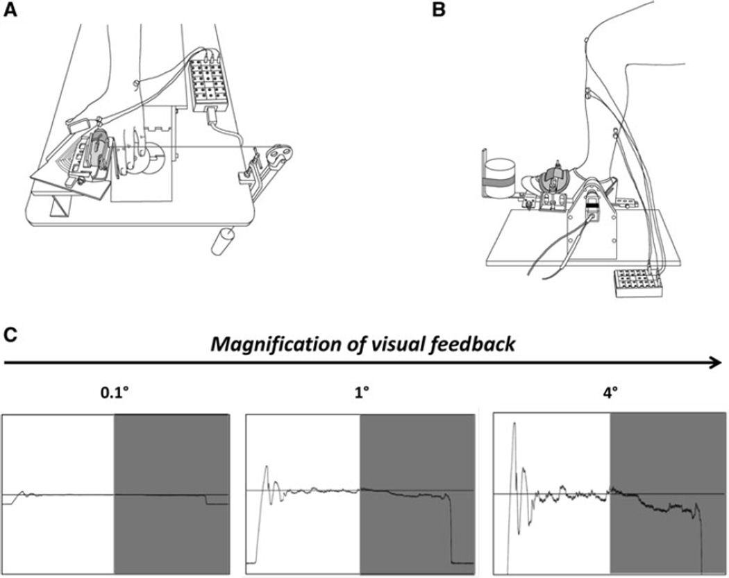
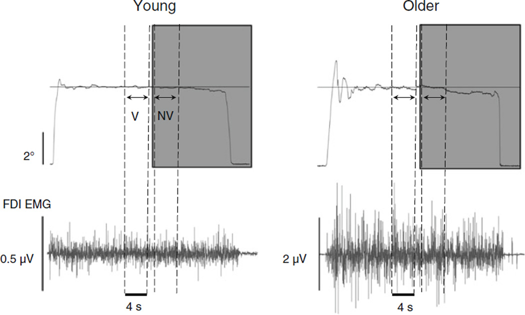
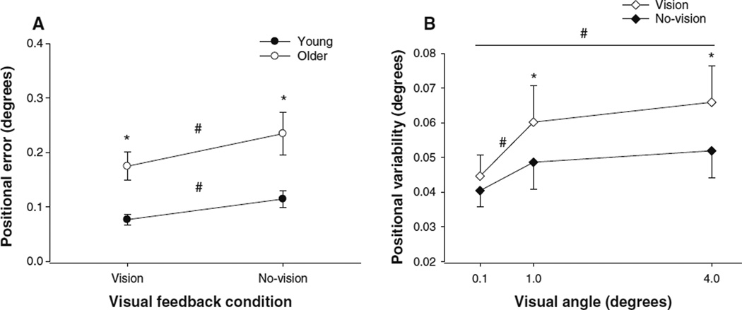
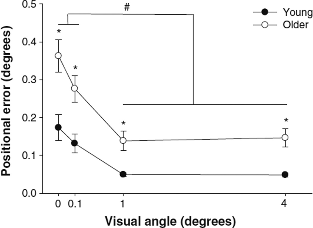
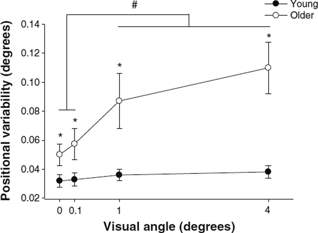
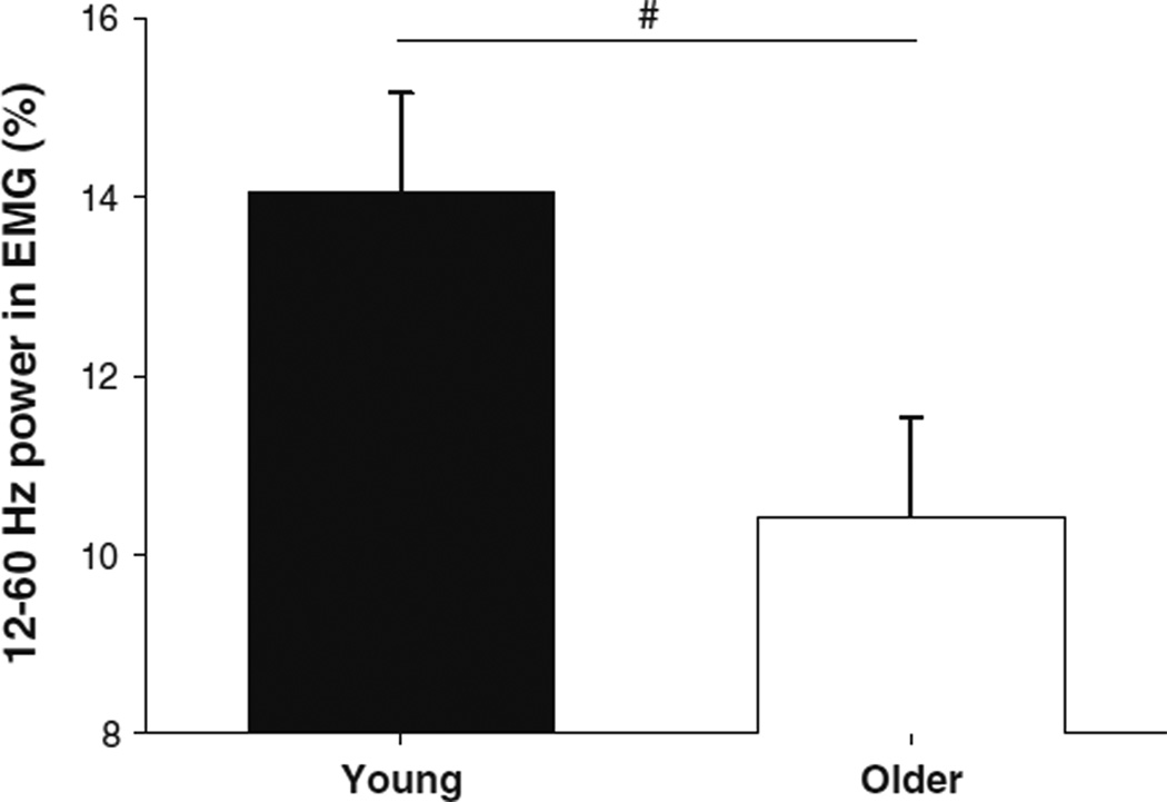
The purpose of this study was to determine whether magnified visual feedback during position-holding contractions exacerbates the age-associated differences in motor output variability due to changes in the neural activation of the agonist muscle in the upper and lower limb. Twelve young (18-35 years) and ten older adults (65-85 years) were instructed to accurately match a target position at 5° of index finger abduction and ankle dorsiflexion while lifting 10 % of their 1 repetition maximum (1RM) load. Position was maintained at three different visual angles (0.1°, 1°, and 4°) that varied across trials. Each trial lasted 25 s and visual feedback of position was removed from 15 to 25 s. Positional error was quantified as the root mean square error (RMSE) of the subject's performance from the target. Positional variability was quantified as the standard deviation of the position data. The neural activation of the first dorsal interosseus and tibialis anterior was measured with surface electromyography (EMG). Older adults were less accurate compared with young adults and the RMSE decreased significantly with an increase in visual gain. As expected, and independent of limb, older adults exhibited significantly greater positional variability compared with young adults that was exacerbated with magnification of visual feedback (1° and 4°). This increase in variability at the highest magnification of visual feedback was predicted by a decrease in power from 12 to 30 Hz of the agonist EMG signal. These findings demonstrate that motor control in older adults is impaired by magnified visual feedback during positional tasks.


Figures

Fig. 1
Fig. 1
a Experimental setup for testing the abduction–adduction of the index finger. The subject’s hand was flat on the platform and the forearm arm was placed in a form-fitting vacuum pillow. The forearm and hand were prone and position of hand and fingers was held stationary with metal plates. The index finger was placed in an orthosis to prevent flexion at the interphalangeal joints. The loads were attached medially at the level of the proximal interphalangeal joint on the finger orthosis and suspended using a pulley away from the subject. b Experimental setup for testing the ankle dorsiflexion/plantar flexion. The subject’s foot was placed in an adjustable foot plate and secured at the level of the metatarsals with a padded metal plate. The loads were attached directly in front of the foot. c Experimental conditions and the visual feedback angles for the experiment were 0.1° (left column), 1° (center column), and 4° (right column). Subjects were asked to match a position with the abduction–adduction of their index finger and the dorsiflexion/plantar flexion of their ankle at an amplitude of 5° range of motion. Each subject was instructed to match a constant position with abduction of the index finger or ankle dorsiflexion for 25 s. Visual feedback of the target and exerted position was given to the subjects from 0 to 15 s (visual feedback condition), whereas visual feedback of the target and exerted position was removed (gray bars) from 15 to 25 s (no-visual feedback condition) at all three gains
Fig. 2
Fig. 2
Representative example of the constant position task with the index finger. A representative trial from 1 young (left panel) and 1 older subject when matching a constant 5° position with a 10 % 1RM load. The position and EMG analysis was based on selected segments from each trial. The top row represents the force trace for the trials represented and the bottom row is the corresponding FDI EMG activity. The analysis was performed from 4.1 to 0.1 s prior to the removal of visual feedback (visual feedback condition; V) and 0.1 to 4.1 s after the removal of the visual feedback (no-visual feedback condition; NV)
Fig. 3
Fig. 3
a Positional error during different visual feedback conditions and b variability during different visual gain conditions. a The positional error by young (filled circles) and older (open circles) adults with vision and no-vision. Positional error at the 0.1° was significantly (#) higher during the no-vision condition. Older adults exhibited significantly greater (*) positional error when compared with young adults across all visual angles regardless of the effector used. b Subjects exhibited significantly greater (*) positional variability during the visual feedback condition (open diamonds) when compared with the no-vision condition (filled diamonds) with 1 and 4° regardless of the effector used. Furthermore, subjects exhibited significantly (#) lower positional variability at 0.1° when compared with variability at 1° and 4° visual angles during the vision condition
Fig. 4
Fig. 4
The interaction of aging and visual gain on positional error. The positional error produced by young (filled circles) and older (open circles) adults at four different visual angles (0°, 0.1°, 1°, and 4°). The positional error at 0° and 0.1° was significantly (#) higher than 1° and 4°. Older adults exhibited significantly greater (*) positional error when compared with young adults across all visual angles regardless of the effector used
Fig. 5
Fig. 5
The interaction of aging and visual gain on positional variability. The positional variability exhibited by young (filled circles) and older (open circles) adults at four different visual angles (0°, 0.1°, 1°, and 4°). The positional error at 0° and 0.1° was significantly (#) lower than 1° and 4°. Older adults exhibited significantly greater (*) positional error when compared with young adults across all visual angles regardless of the effector used
Fig. 6
Fig. 6
The effect of aging on EMG modulation from 12 to 60 Hz. Older adults (open bar) exhibited significantly lower (#) power from 12 to 60 Hz in the agonist EMG power spectrum when compared with young adults (filled bar) across all visual angles regardless of the effector used

Similar articles

Cited by

References

    1. Addison PS. The illustrated wavelet transform handbook. New York: Taylor & Francis Group; 2002.
    1. Baudry S, Maerz AH, Enoka RM. Presynaptic modulation of Ia afferents in young and old adults when performing force and position control. J Neurophysiol. 2010;103:623–631. - PMC - PubMed
    1. Baweja HS, Patel BK, Martinkewiz JD, Vu J, Christou EA. Removal of visual feedback alters muscle activity and reduces force variability during constant isometric contractions. Exp Brain Res. 2009;197:35–47. - PMC - PubMed
    1. Baweja HS, Kennedy DM, Vu JL, Vaillancourt DE, Christou EA. Greater amount of visual feedback decreases force variability by reducing force oscillations from 0–1 and 3–7 Hz. Eur J Appl Physiol. 2010a;108:935–943. - PMC - PubMed
    1. Baweja HS, Kwon MH, Glover SQ, Christou EA. Greater amount of visual feedback decreases error but not variability during movements and positional tasks with the finger and foot. San Diego: Society for Neuroscience; 2010b.

Publication types