. 2012 Nov;30(11):2548-60.
doi: 10.1002/stem.1214.

Subventricular zone localized irradiation affects the generation of proliferating neural precursor cells and the migration of neuroblasts

Affiliations

Subventricular zone localized irradiation affects the generation of proliferating neural precursor cells and the migration of neuroblasts

Pragathi Achanta et al. Stem Cells. 2012 Nov.

Abstract

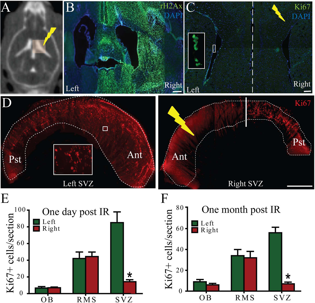
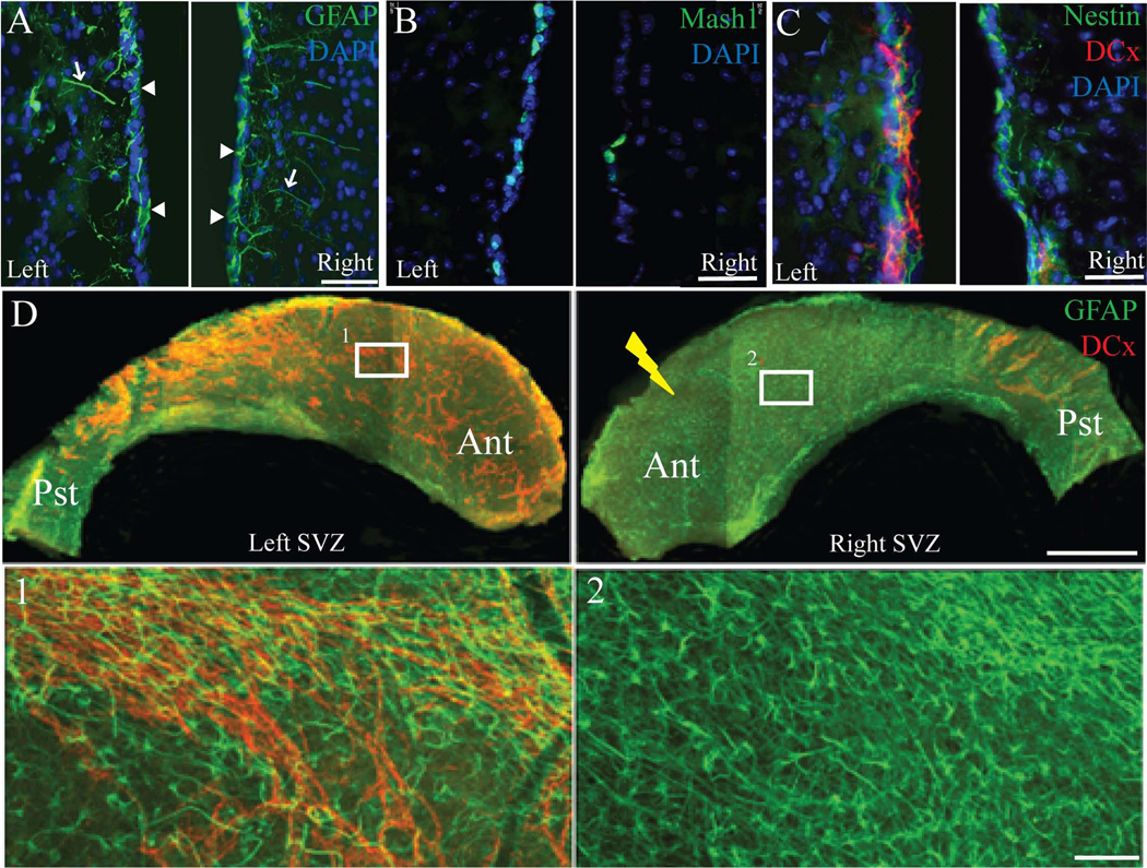
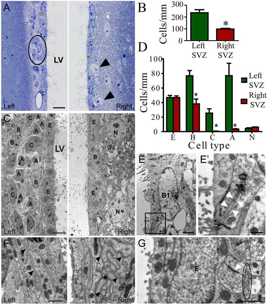
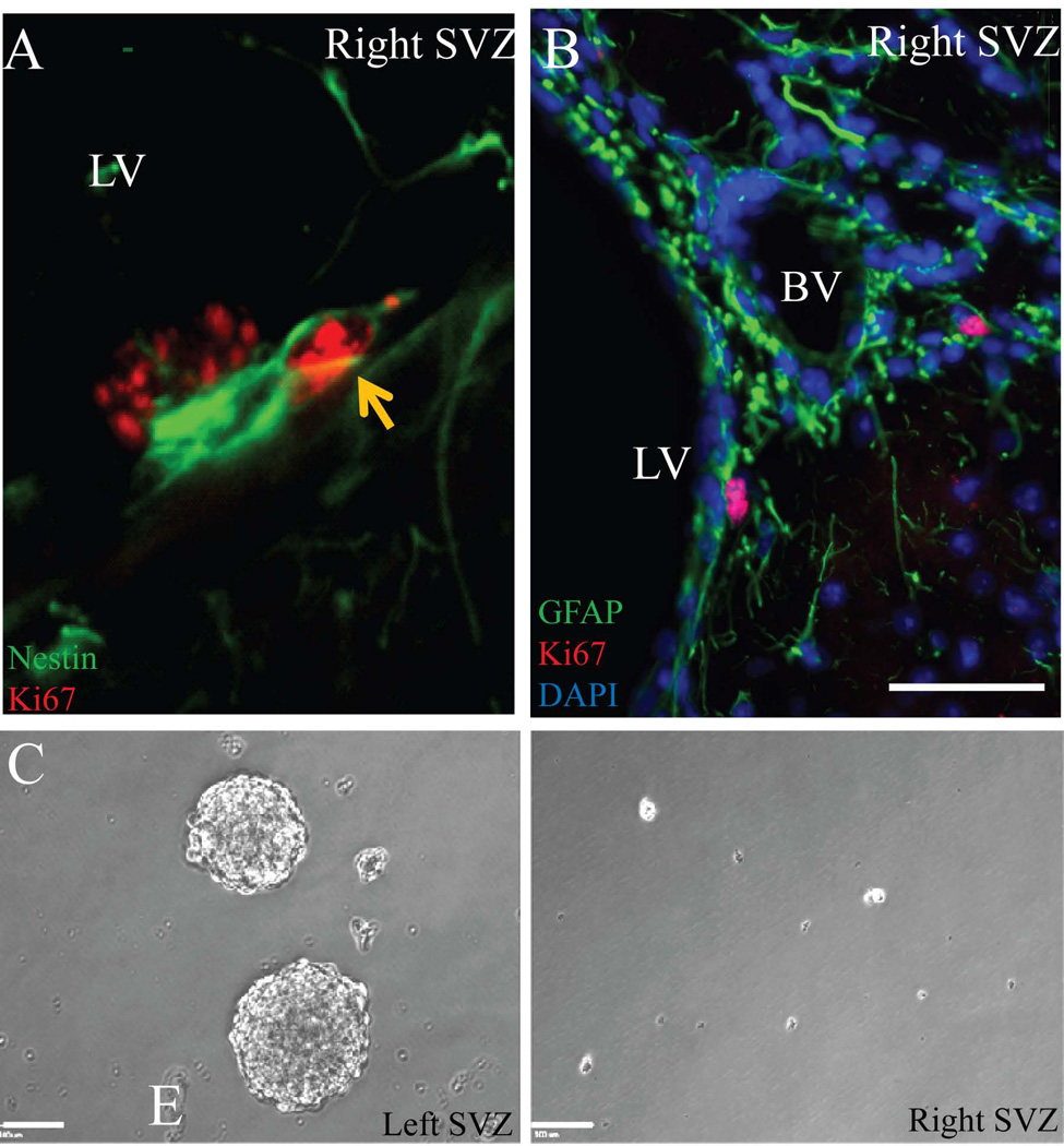
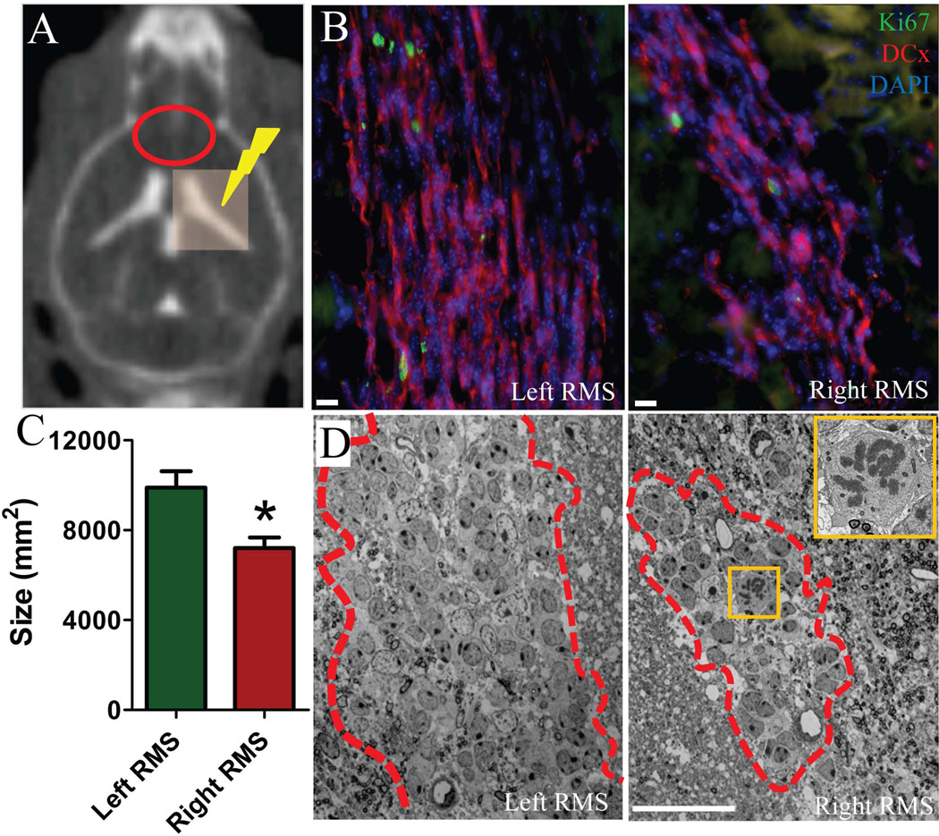
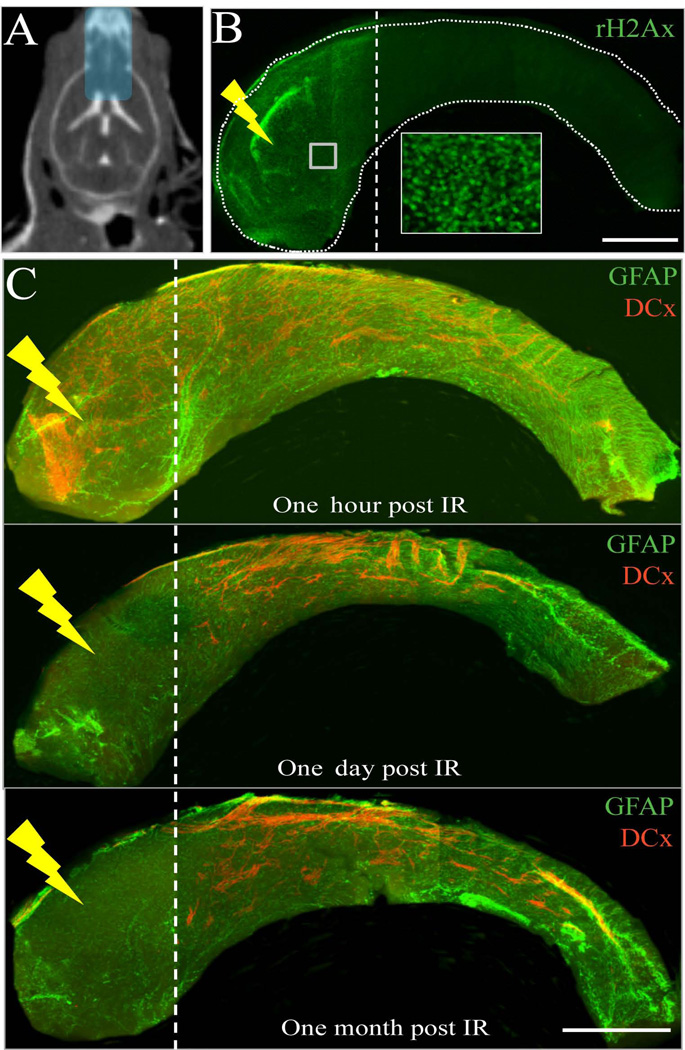
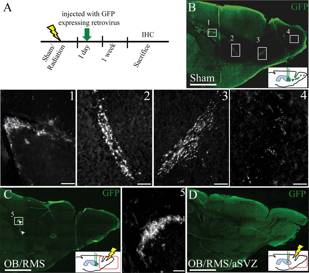
Radiation therapy is a part of the standard treatment for brain tumor patients, often resulting in irreversible neuropsychological deficits. These deficits may be due to permanent damage to the neural stem cell (NSC) niche, damage to local neural progenitors, or neurotoxicity. Using a computed tomography-guided localized radiation technique, we studied the effects of radiation on NSC proliferation and neuroblast migration in the mouse brain. Localized irradiation of the subventricular zone (SVZ) eliminated the proliferating neural precursor cells and migrating neuroblasts. After irradiation, type B cells in the SVZ lacked the ability to generate migrating neuroblasts. Neuroblasts from the unirradiated posterior SVZ did not follow their normal migratory path through the irradiated anterior SVZ. Our results indicate that the migrating neuroblasts were not replenished, despite the presence of type B cells in the SVZ post-irradiation. This study provides novel insights into the effects of localized SVZ radiation on neurogenesis and cell migration that may potentially lead to the development of new radiotherapy strategies to minimize damage to NSCs and neuroblast migration.


Conflict of interest statement

DISCLOSURES: The authors declare no conflict of interest and no financial disclosures.

Figures

Figure 1
Figure 1
Localized radiation of the right SVZ disrupts proliferation as compared to the left (control) side. A) Computed tomography (CT) image of a mouse after intrathecal iodine contrast showing ventricles and radiation plan with 3×3 mm beam covering the right ventricle. B) Immunohistochemistry (IHC) against γH2Ax (green) one hour after radiation confirmed that the 3×3 mm beam irradiated only the right lateral ventricle. C) IHC against Ki67 of a coronal section of the mouse brain one day post irradiation. Inset shows the Ki67+ cells (green) at higher magnification. D) Whole mount staining of SVZ against Ki67 one month post irradiation. Inset shows the Ki67+ cells (red) at higher magnification. The number of Ki67+ cells in the irradiated SVZ decreased significantly compared to the control SVZ after one day (E) and one month (F). Ant-Anterior, Pst-Posterior. Scale bars: 100µm (B–C), 1mm(D). *p<0.001.
Figure 2
Figure 2
Localized irradiation of the right SVZ eliminates the NPCs and migrating neuroblasts but not type B cells after one month. A) GFAP+ cells (green) in a coronal section of the mouse brain appeared to show no difference between the left and right SVZ. Type B cells were identified by their long GFAP processes (arrows) and their contact with the lateral ventricle (arrowheads). B) Fewer Mash-1+ NPCs (green) in the right SVZ with localized radiation compared to the control left SVZ. C) Nestin (green) and DCx (red) double labeling showed the presence of type- B cells but not migrating chains of neuroblasts upon localized radiation. D) GFAP (green) and DCx (red) immunohistochemistry of whole mount SVZ also confirmed the ablation of neuroblasts after localized radiation (right SVZ) after one month. Higher magnification images of left and right whole mount SVZ (insets) are shown in 1 and 2. Ant-Anterior, Pst-Posterior. Scale bars: 50µm (A–C), 1mm (D), 100µm (insets 1–2).
Figure 3
Figure 3
Cell organization of the non-irradiated left and irradiated right SVZ one month post-irradiation. A) Semithin sections of the left SVZ reveal typical cell organization with dark cells forming the migratory chains (circle) surrounded by expansions of astrocytes (light cells), located beneath the ependymal layer, while the irradiated SVZ lacks the proliferative populations, showing a monolayer of ependymal cells with neurons located close to the ventricle (arrow). B) Bar graph representing the number of cells/mm for the total cells in the SVZ, resulting in a cell reduction in the irradiated hemisphere. C) Electron micrographs depict details of each cell type remained in the left and right SVZ. Irradiated SVZ was reduced to a monolayer of ependymal cells with dispersed astrocytes and neurons without NPCs or neuroblasts. D) Bar graph representing the number of cells/mm for each SVZ cell type. E) Electron micrograph of a B1 cell contacting the lumen of the lateral ventricle of irradiated SVZ. Details of the primary cilia are shown at higher magnifications (E' arrowheads). F) Basal lamina in the right SVZ was more extensive after irradiation compared to the left SVZ (arrowheads). G) Amyelenic axons (circle) and synaptic contacts (arrowheads) next to ependymal cells in contact with ependymal cells in the irradiated SVZ. A-neuroblast, B-astrocyte, C-type C cell, N-neuron, LV-lateral ventricle, BV-blood vessel. Scale bars: 10µm (A), 5µm (C), 2µm (E), 500nm (E’), 1µm (F, G). * p<0.001
Figure 4
Figure 4
Residual type B cells in the irradiated right SVZ one month after radiation. A) Nestin (green) and Ki67 (red) double labeling show proliferating type B cells in the right SVZ (arrow). B) GFAP (green) and Ki67 (red) show proliferating type B cells (arrow) in close proximity to blood vessels. C) Neurospheres were formed from unirradiated control SVZ (left), while no neurospheres were formed from the 10-Gy irradiated SVZ (right). LV-lateral ventricle, BV-blood vessel. Scale bars: 50µm (B), 100µm (C)
Figure 5
Figure 5
Fewer migrating neuroblasts were observed in the RMS of the irradiated hemisphere after one month. A) Computed tomography (CT) image of a mouse after intrathecal iodine contrast showing ventricles and radiation plan with 3×3 mm beam covering the right ventricle. Red oval shows the region of RMS on CT. B) Ki67 (green) and DCx (red) immunolabeling in the RMS. Note a smaller number of migrating neuroblasts (DCx+/red cells) in the right RMS compared to the left RMS. C) Bar graph showing a significant reduction in the size of the RMS in the irradiated right hemisphere. D) Panoramic images of migratory chains and astrocytes forming the RMS (outlined with dashed line) under electron microscope. Note the difference of the RMS size. Inset shows a neuroblast in mitotic phase at higher magnification in the right RMS. Scale bars: 10µm (B), 20nm (D). * p<0.001.
Figure 6
Figure 6
Neuroblasts from the posterior SVZ do not migrate into the irradiated anterior SVZ. A) CT image of a mouse after intrathecal iodine contrast showing ventricles and treatment plan of 5×9 mm beam to irradiate anterior SVZ, RMS and OB. B) γH2Ax staining (green) of whole mount SVZ one hour post-irradiation validates the localized radiation treatment plan. Inset shows the γH2Ax+ cells at higher magnification. C) GFAP (green) and DCx (red) double labeling of SVZ whole mounts one hour, one day and one month after irradiation. Scale bars: 1mm (B–C).
Figure 7
Figure 7
Neuroblasts failed to migrate through the irradiated RMS to the OB. A) Experimental paradigm showing the time line for radiation, GFP expressing retroviral injections (green arrow), and sacrifice. B) Sagittal section of a non-irradiated mouse brain injected with GFP expressing retrovirus into the SVZ a week after injections, insets show the GFP labeled cells (green) in SVZ (1), descending limb of RMS (2), ascending limb of RMS (3), and olfactory bulb (4) at higher magnification. C) Sagittal section of a RMS/OB irradiated mouse brain with GFP labeled cells (green, shown in arrowheads) in SVZ (inset 5 at higher magnification) and no GFP labeled cells in the RMS+OB. The GFP+ cells in the SVZ appeared to be similar to the sham irradiated mice (inset 1 and 5). D) Sagittal section of a aSVZ/RMS/OB irradiated mouse brain with no GFP labeled cells in aSVZ/RMS/OB. Hypothetical presence of GFP+ cells in the SVZ, RMS and OB, a week after retroviral injections, in three different groups of mice that received sham or RMS/OB or aSVZ/RMS/OB radiation are shown in the insets of B, C, and D. Scale bars: 1mm (B–D), 100µm (1–5).

References

    1. Stupp R, Hegi ME, Mason WP, et al. Effects of radiotherapy with concomitant and adjuvant temozolomide versus radiotherapy alone on survival in glioblastoma in a randomised phase III study: 5-year analysis of the EORTC-NCIC trial. Lancet Oncol. 2009;10:459–466. - PubMed
    1. Stupp R, Mason WP, van den Bent MJ, et al. Radiotherapy plus concomitant and adjuvant temozolomide for glioblastoma. N Engl J Med. 2005;352:987–996. - PubMed
    1. Calabrese P, Schlegel U. Neurotoxicity of treatment. Recent Results Cancer Res. 2009;171:165–174. - PubMed
    1. Marazziti D, Baroni S, Catena-Dell'osso M, et al. Cognitive, Psychological and Psychiatric Effects of Ionizing Radiation Exposure. Curr Med Chem. 2012 - PubMed
    1. Silber JH, Radcliffe J, Peckham V, et al. Whole-brain irradiation and decline in intelligence: the influence of dose and age on IQ score. J Clin Oncol. 1992;10:1390–1396. - PubMed

Publication types