. 2012:3:1041.
doi: 10.1038/ncomms2036.

The amniote paratympanic organ develops from a previously undiscovered sensory placode

Affiliations

The amniote paratympanic organ develops from a previously undiscovered sensory placode

Paul O'Neill et al. Nat Commun. 2012.

Abstract

The paratympanic organ, a mechanosensory hair cell-containing pouch in the amniote middle ear, was first described 100 years ago, yet its origins remain unresolved. Homology with the anamniote spiracular organ is supported by association with homologous skeletal elements and similar central targets of afferent neurons, suggesting it might be a remnant of the water-dependent lateral line system, otherwise lost during the amniote transition to terrestrial life. However, this is incompatible with studies suggesting that it arises from the first epibranchial (geniculate) placode. Here we show that a previously undiscovered Sox2-positive placode, immediately dorsal to the geniculate placode, forms the paratympanic organ and its afferent neurons, which are molecularly and morphologically distinct from geniculate neurons. These data remove the only obstacle to accepting the homology of the paratympanic organ and spiracular organ. We hypothesize that the paratympanic organ/spiracular organ represents an ancient head ectoderm module, developmentally and evolutionarily independent of both lateral line and epibranchial placodes.


Figures
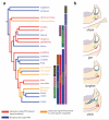
Figure 1
Figure 1. A spiracular organ or paratympanic organ is found in many vertebrate groups
(a) Vertebrate phylogeny highlighting the presence of a spiracular organ or paratympanic organ (PTO), constructed using data from refs. ,,. (NB Within the sarcopterygians, it is unknown whether or not coelacanths have a spiracular organ, so they are not represented in the tree.) (b) Cartoons demonstrating similarities in the development of the spiracular organ and PTO (in association with the first pharyngeal pouch) in different species, drawn from our data plus refs. ,. Dark blue: inner ear epithelium; green: geniculate ganglion; red: spiracular organ/PTO; pink: pharyngeal endoderm; purple, neural tube.
Figure 2
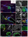
Figure 2. Sense organs of the first pharyngeal pouch
(a) Transverse haematoxylin and eosin (H&E) stained section of stage 40 (E14) chicken head highlighting position of paratympanic organ (PTO) and tympanic membrane. (b) Magnified view of PTO at stage 40 (E14). Mechanosensory hair cells (arrow) line the medial wall. (c) Transverse H&E stained section of shark spiracular organ at stage 33 (16-22 weeks). (d) Parasagittal section of shark head at stage 31 (60-80 days), highlighting the position of the spiracular organ in the first pharyngeal cleft. (e) Transverse section of stage 27 (E5) chicken embryo; the developing PTO is indicated at the tip of the first pharyngeal pouch. (f) Transverse section of stage 31 shark embryo (60-80 days). The forming spiracular organ is associated with the first pharyngeal cleft. (g) Schematic representation of isotopic quail/chick grafting procedure; excised quail ectoderm is transplanted to an equivalent position in chick hosts. (h) Transverse section of chimeric embryo following isotopic grafting. Quail cells are labelled with QCPN antibody (magenta) and contribute extensively to the PTO and geniculate ganglion (gg). (i) Magnified view of chimeric PTO at stage 34 (E8) demonstrating quail-derived hair cells and neurons of the “paratympanic extension of the geniculate ganglion” (pe). Scale bars: 100 μm (a), 50 μm (b,c,e,f,h,i), 500 μm (d). Myo7a, myosin VIIa (hair cell-specific marker); QCPN, quail-specific antibody (recognises peri-nuclear antigen); QN, quail neurite-specific antibody.
Figure 3
Figure 3. A distinct PTO placode
(a) Schematic indicating the approximate locations of wholemount images in panels b-e (box), and approximate plane of section for images in panels f-q (dotted line). (b) Whole mount Sox2 in situ hybridisation at stage 18 (65-69 hours) reveals a patch of Sox2-positive ectoderm (putative PTO placode, arrow) at the dorsocaudal edge of the first pharyngeal cleft. (c-e) Whole mount in situ hybridisation for the epibranchial placode markers Pax2, Sox3 and Delta1 at stage 18 (65-69 hours). (f-j) Transverse sections at the level shown in panel a, immunostained for Sox2 (green) and Sox3 (magenta), at stages 14 (50-53 hours), 18 (65-69 hours), 20 (70-72 hours), 22 (E3.5) and 24 (E4). (k) Transverse section at the level shown in panel a, immunostained for Sox2 and Pax2, at stage 24 (E4). (l-q) In situ hybridisation or immunostaining on sections of the PTO at stage 27 (E5) reveals expression of typical hair cell/sensory patch-associated markers (Atoh1, SOHo1, BMP4 and Prox1) and the hair cell-specific structural markers myosin VIIA (Myo7a) and Ptprq/Hair Cell Antigen (HCA). Scale bars: 50 μm.
Figure 4
Figure 4. The PTO placode gives rise to Brn3a-positive neurons
(a) Coronal section of PTO placode at stage 21 (E3.5), immunostained for the PTO placode marker Sox2 (blue), the neuronal marker Islet1 (green) and Brn3a (red). Dotted line on schematic insert indicates plane of section; axes indicated with arrows. (b-e) Magnified view of the boxed area in panel a, showing single channel and merged images. No Brn3a-positive cells are present at this stage. (f) Coronal section of PTO placode at stage 24 (E4), immunostained for Sox2 (blue), laminin (blue), Islet1 (green) and Brn3a (red). Brn3a-positive neurons (arrow) are closely associated with the Sox2-positive PTO placode. (g-j) Single channel and merged images of magnified view of boxed region in panel f, highlighting Brn3a-positive neurons (arrow) adjacent to a large breach in the basement membrane of the Sox2-positive PTO placode. (k) Coronal section of PTO at stage 27 (E5), immunostained for laminin (blue), Islet1 (green) and Brn3a (red). A discrete cluster of Brn3a-positive neurons (arrow) seem to be merging with the edge of the geniculate ganglion closest to the PTO. (l) Magnified view of PTO from panel k, showing both Brn3a and laminin in greyscale for clarity. Brn3a-positive cells can be seen delaminating from the PTO through gaps in the basement membrane. (m,n) Magnified view of boxed region from panel l, highlighting the delamination of Brn3a-positive cells. (o) Coronal section of PTO at stage 27 (E5), immunostained for Sox2 (green) and Brn3a (magenta), confirming the persistence of Sox2 protein in the PTO epithelium. Brn3a-positive cells are scattered throughout the epithelium. Scale bars: 50 μm (a,f,k,l,o), 25 μm (b-e,g-j,m,n).
Figure 5
Figure 5. PTO neurons populate the geniculate ganglion and also form a separate PTO ganglion
(a) Transverse section of geniculate ganglion at stage 24 (E4), following Phox2b in situ hybridisation to label epibranchial neurons. (b) Same section immunostained for Brn3a (magenta) and Islet1 (green), showing a cluster of Brn3a-positive neurons towards the edge of the geniculate ganglion in a region that (by comparison with panel a) seems Phox2b-negative. (c) Pseudo-coloured overlay of panels a and b highlighting the apparent segregation of Brn3a-positive and Phox2b-positive neurons. (d) Transverse section of geniculate ganglion at stage 31 (E7) immunostained for Brn3a (magenta) and Islet1 (green). Brn3a-positive neurons are still present within the geniculate ganglion. (e) A different transverse section at stage 31 (E7), immunostained for Brn3a (magenta) and Islet1 (green), showing that Brn3a-positive neurons are also detected in a small cluster between the PTO and geniculate ganglion (arrow). This PTO ganglion was previously described as the “paratympanic extension of the geniculate ganglion”. (f) At stage 35 (E9), H&E staining shows the axonal tracts connecting the geniculate ganglion and PTO. (g) The PTO ganglion resides within this axonal tract. (h,i) Magnified views of neuronal cell bodies within the geniculate ganglion and PTO ganglion, respectively, highlighting the smaller diameter of the PTO neurons. Scale bars: 100 μm (a-g), 50 μm (h,i). gg, geniculate ganglion.
Figure 6
Figure 6. The PTO nerve central projections overlap with inner ear afferents
Approximate dye-insertion sites indicated by corresponding arrows in panel k; magenta/green overlap shows as white. (a) Retrograde tracing at stage 28 (E5.5) from the cerebellum (rhombomere 1/2 boundary) (green) plus a caudal dye-insertion in the descending tract (magenta) shows projection of presumed PTO afferents as a distinct bundle within the facial nerve (arrow). gg, geniculate ganglion; tg, trigeminal ganglion; vg, vestibuloacoustic ganglion. (b,c) Stage 28 (E5.5) ganglia backfilled from a rostral (green) and caudal (magenta) dye-insertion into the hindbrain. In the geniculate ganglion, the rostral dye-insertion (green) labelled fibres in the greater petrosal nerve (gpn) and chorda tympani (ct), and a unique nerve lying between them, the PTO nerve (pton). [The caudal dye-insertion (magenta) labelled many afferents from the inner ear (VIII) and glossopharyngeal nerve (IX) but filled only a few geniculate ganglion neurons.] (c) Higher-power view of geniculate ganglion region from b. (d) At stage 30 (E6.5), labelling from the greater petrosal nerve alone (magenta) plus the inner ear (green) shows no overlap with inner ear afferents (VIII). st, solitary tract. (e-h) In contrast, labelling from the greater petrosal and PTO nerves (magenta), plus the inner ear (green), shows extensive overlap with inner ear afferents (VIII) at stage 30 (E6.5; e) and stage 28 (E5.5; f-h). fbm, facial branchiomotor neurons. (i) At stage 29 (E6), labelling from the PTO nerve (magenta) and inner ear (green) likewise shows projection of PTO afferents with inner ear afferents (VIII). (j) At stage 30 (E6.5), sequential labelling from the greater petrosal nerve (magenta) plus the geniculate ganglion (green) shows overlapping afferents in the solitary tract (st): the descending trigeminal tract (dV) and PTO afferents are only labelled with the geniculate ganglion dye-insertion (green). (k) Schematic showing the peripheral and central distribution of geniculate ganglion afferents: gustatory/viscerosensory afferents (green) to the greater petrosal (gpn) and chorda tympani (ctn) nerves and solitary tract; somatosensory afferents (blue) to the posterior auricular nerve (pan) and the descending tract of the trigeminal; PTO afferents (red) to the inner ear projection (VIII). (l) Coronal section at the approximate position indicated by the dashed line in panel k. Scale bars: 100 μm.
Figure 7
Figure 7. Competence to form the PTO and PTO neurons is dissociable from competence to form geniculate neurons
(a) Schematic summarising grafting of different regions of quail ectoderm into the peri-geniculate placode region of chicken hosts. Grafts from blue regions - prospective peri-geniculate (g) and otic (o) placodes - were competent to form both geniculate neurons and the PTO. Grafts from red regions - presumptive trigeminal placode (t), nodose placode (n) and trunk ectoderm (t) - were competent to form geniculate neurons but not the PTO. (b-d) Transverse sections through the PTO region of chimeric embryos after control isotopic grafts of quail peri-geniculate region ectoderm, immunostained for the quail perinuclear antigen (QCPN), the hair cell marker myosin VIIA (Myo7) and Brn3a, showing that peri-geniculate ectoderm contributes as expected to geniculate neurons, the PTO and Brn3a-positive PTO neurons. (e) In contrast, trunk ectoderm is competent to form geniculate neurons, but not the PTO. Similar results were obtained after grafting prospective trigeminal or nodose placode ectoderm. (f,g) Trunk ectoderm grafts also fail to form Brn3a-positive PTO neurons in either the geniculate or PTO ganglia. (h) In vitro culture of tissue from the otocyst generates hair cells after 5-7 days in floating culture (blue box in embryo schematic; lower panel showing cryosection immunostained for hair cell markers; blue bar in bar-chart). Tissue from the peri-geniculate placode region (first pharyngeal cleft) generates hair cells in floating culture only in the presence of BMPs (red box in embryo schematic; upper panel showing cryosection immunostained for hair cell markers; red bars in bar-chart). Tissue from the peri-petrosal placode region (second pharyngeal cleft) does not generate hair cells even in the presence of BMPs (green box in embryo schematic; see bar-chart for quantification), confirming the restriction of PTO-forming competence to the peri-geniculate region. Scale bars: 100 μm (b,c,e,f), 50 μm (d,g), 20 μm (h). HCA, hair cell antigen; Myo7a, myosin VIIa.
Figure 8
Figure 8. Overview of vertebrate placodes derived from the ‘posterior placodal area’
(a) Schematic summarising data from a cartilaginous fish, the shark Squalus acanthias, redrawn from ref. , suggesting the presence of a spiracular organ primordium (red) that is separate from the established series of six lateral line placodes (blue). (b) Schematic summarising data from a primitive ray-finned fish, the longnose gar, Lepisosteus osseus, redrawn from ref. , also showing a separate spiracular organ primordium. Similar results were also reported for the shortnose gar, Lepisosteus platystomus. (c) Schematic (modified from ref. 32) of a hypothetical generalised anamniote embryo, showing the proposed relationship between the otic vesicle (OV), lateral line placodes (blue), spiracular organ primordium (red), and epibranchial placodes (yellow). (d) Schematic of a pharyngula stage chicken embryo highlighting the position of the epibranchial (yellow) and PTO (red) placodes. AD, anterodorsal lateral line placode; AV, anteroventral lateral line placode; M, middle lateral line placode; O, otic lateral line placode; OV, otic vesicle; P, posterior lateral line placode; S, spiracular organ placode; ST, supratemporal lateral line placode.

Similar articles

Cited by

References

    1. Baker CVH, Bronner-Fraser M. Vertebrate cranial placodes I. Embryonic induction. Dev. Biol. 2001;232:1–61. - PubMed
    1. Schlosser G. Induction and specification of cranial placodes. Dev. Biol. 2006;294:303–51. - PubMed
    1. Schlosser G. Making senses: development of vertebrate cranial placodes. Int. Rev. Cell Mol. Biol. 2010;283:129–234. - PubMed
    1. von Bartheld CS, Giannessi F. The paratympanic organ: a barometer and altimeter in the middle ear of birds? J. Exp. Zool. B Mol. Dev. Evol. 2011;306:402–408. - PMC - PubMed
    1. von Bartheld CS. Functional morphology of the paratympanic organ in the middle ear of birds. Brain Behav. Evol. 1994;44:61–73. - PubMed

Publication types

MeSH terms

Substances