. 2013 Jan;31(1):104-16.
doi: 10.1002/stem.1215.

Surface tethered epidermal growth factor protects proliferating and differentiating multipotential stromal cells from FasL-induced apoptosis

Affiliations

Surface tethered epidermal growth factor protects proliferating and differentiating multipotential stromal cells from FasL-induced apoptosis

Melanie Rodrigues et al. Stem Cells. 2013 Jan.

Abstract

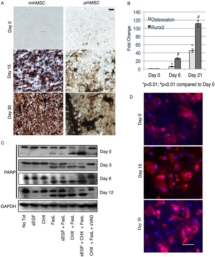
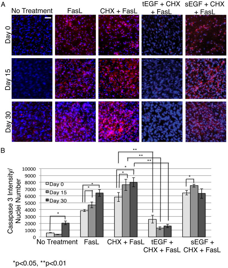
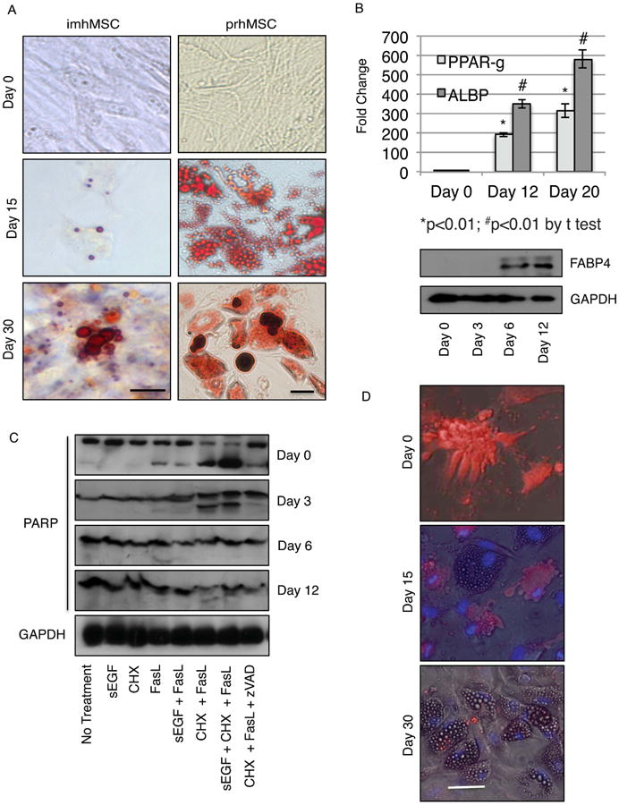
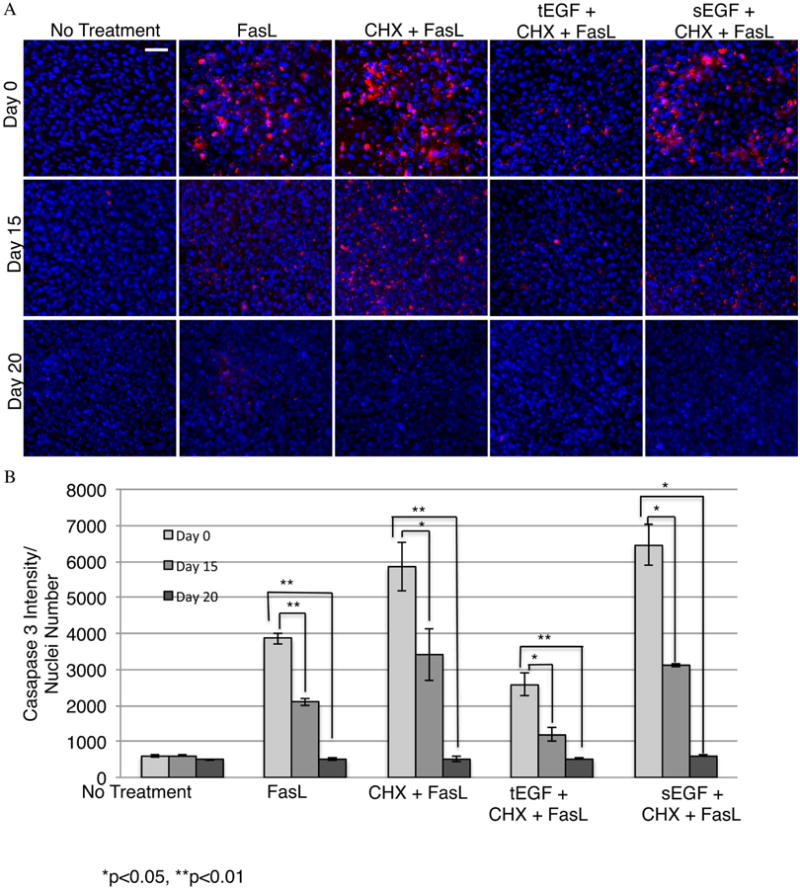
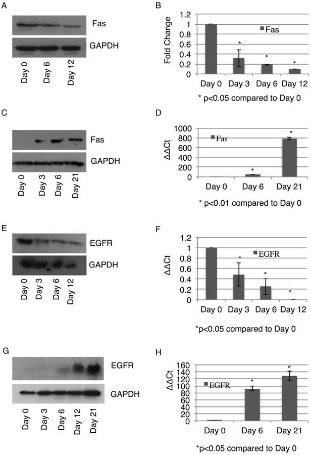
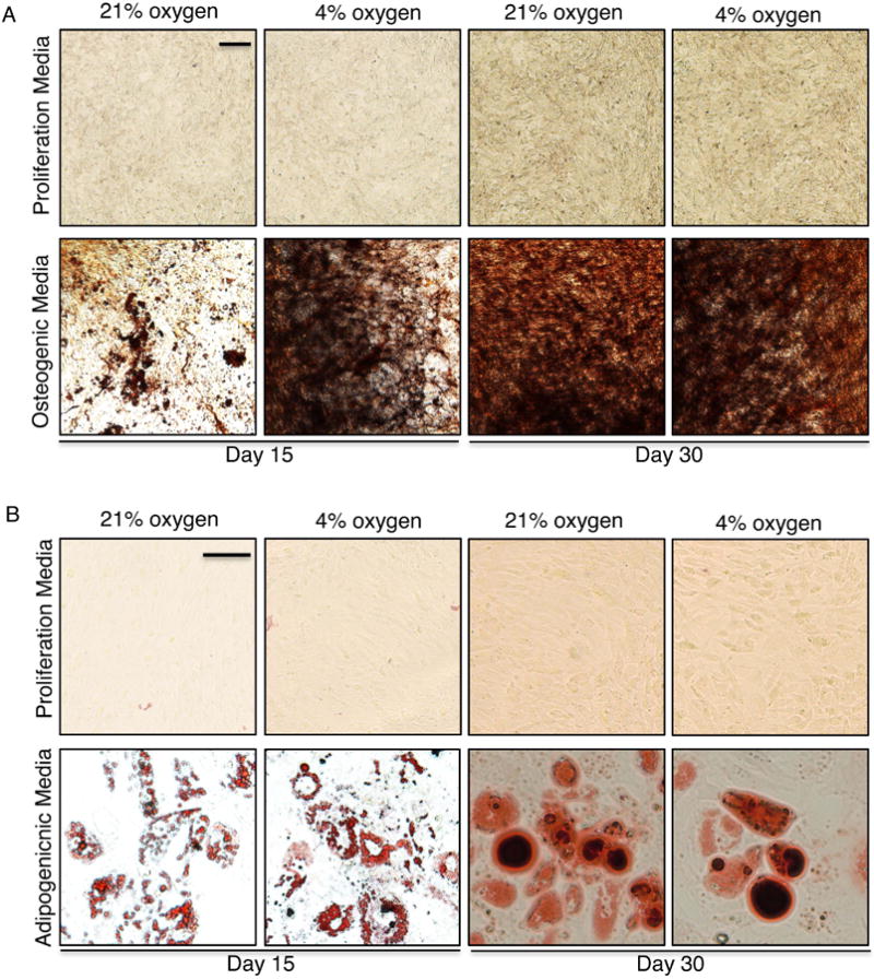
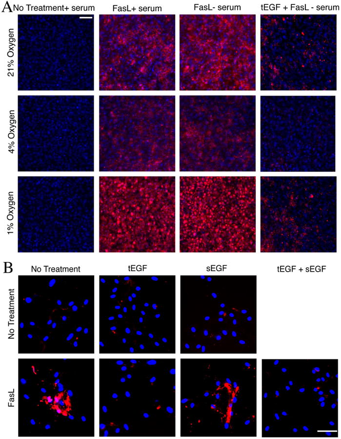
Multipotential stromal cells or mesenchymal stem cells (MSCs) have been proposed as aids in regenerating bone and adipose tissues, as these cells form osteoblasts and adipocytes. A major obstacle to this use of MSC is the initial loss of cells postimplantation. This cell death in part is due to ubiquitous nonspecific inflammatory cytokines such as FasL generated in the implant site. Our group previously found that soluble epidermal growth factor (sEGF) promotes MSC expansion. Furthermore, tethering EGF (tEGF) onto a two-dimensional surface altered MSC responses, by restricting epidermal growth factor receptor (EGFR) to the cell surface, causing sustained activation of EGFR, and promoting survival from FasL-induced death. sEGF by causing internalization of EGFR does not support MSC survival. However, for tEGF to be useful in bone regeneration, it needs to allow for MSC differentiation into osteoblasts while also protecting emerging osteoblasts from apoptosis. tEGF did not block induced differentiation of MSCs into osteoblasts, or adipocytes, a common default MSC-differentiation pathway. MSC-derived preosteoblasts showed increased Fas levels and became more susceptible to FasL-induced death, which tEGF prevented. Differentiating adipocytes underwent a reduction in Fas expression and became resistant to FasL-induced death, with tEGF having no further survival effect. tEGF protected undifferentiated MSC from combined insults of FasL, serum deprivation, and physiologic hypoxia. Additionally, tEGF was dominant in the face of sEGF to protect MSC from FasL-induced death. Our results suggest that MSCs and differentiating osteoblasts need protective signals to survive in the inflammatory wound milieu and that tEGF can serve this function.


Figures

Figure 1
Figure 1. MSC forming osteoblasts continue to be susceptible to inflammatory cytokines like FasL
Immortalized hMSC on mock surface (Magnification 20×, Scale bar 10μm) and prhMSC on tissue culture plastic (Magnification 20×, Scale bar 10μm) depositing hydroxyapatite by von Kossa (A). Change in expression of osteogenic markers by qPCR during osteogenic differentiation of imhMSC on mock surfaces (B). Immunoblot for full length and cleaved PARP in differentiating imhMSC grown on tissue culture plastic and treated with cytokines for 8 hours (C). Caspase 3 positive cells (red) and Hoescht 33342 (blue) stained prhMSC during progress of osteogenic differentiation (Magnification 10×, Scale bar 50m). Graphs of mean ± s.e.m and representative photomicrographs of cells, of two independent experiments.
Figure 2
Figure 2. tEGF protects differentiating osteogenic cells from death signals
Fluorochrome inhibitor of caspase assay (FLICA) stained imhMSC (Magnification 10×, Scale bar 50μm) grown on mock/tEGF surfaces under osteogenic conditions, after 8 hours of treatment with cytokines on Day 0/ Day 15/ Day 30 (A). Quantification of FLICA intensity normalized to cell numbers and optical background (B). Shown are representative photomicrographs of cells and graphs of mean ± s.e.m of two independent experiments.
Figure 3
Figure 3. MSC forming adipose cells become more resistant to inflammatory cytokines like FasL
Immortalized human MSC (imhMSC) on mock surface (Magnification 40×, Scale bar 10μm) and isolated primary human MSC (prhMSC) on tissue culture plastic (Magnification 20×, Scale bar 10μm) differentiating into adipose cells by Oil Red O (A). Change in expression of adipogenic markers by qPCR and immunoblot during adipogenic differentiation of imhMSC on mock surfaces (B). Immunoblot for full length and cleaved PARP in differentiating imhMSC grown on tissue culture plastic and treated with cytokines for 8 hours (C). Caspase 3 positive cells (red) and Hoescht 33342 (blue) stained overlaid with phase contrast images of prhMSC during progress of adipogenic differentiation (Magnification 10×, 50μm). Graphs of mean ± s.e.m and representative photomicrographs of cells, of two independent experiments.
Figure 4
Figure 4. tEGF does not alter resistance of adipose cells to death signals
Fluorochrome inhibitor of caspase assay (FLICA) stained imhMSC (Magnification 10×, Scale bar 50μm) grown on mock/tEGF surfaces under adipogenic conditions, after 8 hours of treatment with cytokines on Day 0/ Day 15/ Day 20 (A). Quantification of FLICA intensity normalized to cell numbers and optical background (B). Shown are representative photomicrographs of cells and graphs are of mean ± s.e.m of two independent experiments.
Figure 5
Figure 5. Changes in protein receptor levels during differentiation contribute to altered reactions to death signals
Change in levels of Fas in differentiating adipocytes grown on tissue culture plastic by immunoblot (A) and real time qPCR (B). Change in levels of Fas in differentiating osteoblasts grown on tissue culture plastic by immunoblot (C) and real time qPCR (D). Change in levels of EGFR in differentiating adipocytes grown on tissue culture plastic by immunoblot (E) and real time qPCR (F). Change in levels of EGFR in differentiating osteoblasts grown on tissue culture plastic by immunoblot (E) and real time qPCR (F). Representative immunoblots and graphs are of mean ± s.e.m from two independent experiments.
Figure 6
Figure 6. tEGF does not inhibit osteogenesis or adipogenesis in the presence of differentiation media; Faster osteogenesis occurs at 4% oxygen
Primary hMSC (Magnification 20×, Scale bar 50μm) on tEGF surface in the presence of proliferation or osteogenic media at 21% or 4% oxygen, stained for von Kossa (A). Immortalized hMSC (Magnification 40×, Scale bar 10μm) on tEGF surface in the presence of proliferation or adipogenic media at 21% or 4% oxygen, stained for Oil Red O (B)
Figure 7
Figure 7. Tethered EGF is protective of MSC under FasL treatment, serum deprivation, low oxygen, and in the presence of sEGF
(A) Undifferentiated MSCs are more susceptible to FasL and serum deprivation mediated cell death at 1% oxygen, which tEGF is able to ablate. Fluorescent caspase inhibitor (FLICA) stained undifferentiated imhMSC (Magnification 10×, Scale bar 10μm) grown on tissue culture plastic/tEGF surfaces after 24 hours with or without serum and/ or 8 hours of treatment with FasL. (B) Presence of sEGF does not inhibit the protection rendered by tEGF to FasL treated prhMSCs (Magnification 10×, Scale bar 50μm)

Similar articles

Cited by

References

    1. Pittenger MF, Mackay AM, Beck SC, et al. Multilineage potential of adult human mesenchymal stem cells. Science. 1999;284(5411):143–147. - PubMed
    1. Jones E, Yang X. Mesenchymal stem cells and bone regeneration: current status. Injury. 2011;42(6):562–568. - PubMed
    1. Caplan AI, Dennis JE. Mesenchymal stem cells as trophic mediators. J Cell Biochem. 2006;98(5):1076–1084. - PubMed
    1. Caplan AI. Why are MSCs therapeutic? New data: new insight. J Pathol. 2009;217(2):318–324. - PMC - PubMed
    1. Uccelli A, Prockop DJ. Why should mesenchymal stem cells (MSCs) cure autoimmune diseases? Curr Opin Immunol. 2010;22(6):768–774. - PubMed

Publication types