. 2012 Sep;68(Pt 9):1232-41.
doi: 10.1107/S0907444912026236. Epub 2012 Aug 18.

The structure of Enterococcus faecalis thymidylate synthase provides clues about folate bacterial metabolism

Affiliations

The structure of Enterococcus faecalis thymidylate synthase provides clues about folate bacterial metabolism

Cecilia Pozzi et al. Acta Crystallogr D Biol Crystallogr. 2012 Sep.

Abstract

Drug resistance to therapeutic antibiotics poses a challenge to the identification of novel targets and drugs for the treatment of infectious diseases. Infections caused by Enterococcus faecalis are a major health problem. Thymidylate synthase (TS) from E. faecalis is a potential target for antibacterial therapy. The X-ray crystallographic structure of E. faecalis thymidylate synthase (EfTS), which was obtained as a native binary complex composed of EfTS and 5-formyltetrahydrofolate (5-FTHF), has been determined. The structure provides evidence that EfTS is a half-of-the-sites reactive enzyme, as 5-FTHF is bound to two of the four independent subunits present in the crystal asymmetric unit. 5-FTHF is a metabolite of the one-carbon transfer reaction catalysed by 5-formyltetrahydrofolate cyclo-ligase. Kinetic studies show that 5-FTHF is a weak inhibitor of EfTS, suggesting that the EfTS-5-FTHF complex may function as a source of folates and/or may regulate one-carbon metabolism. The structure represents the first example of endogenous 5-FTHF bound to a protein involved in folate metabolism.


Figures

Figure 1
Figure 1
Major folate-dependent pathways in bacterial one-carbon metabolism. The scheme gives the names of the enzymes involved (in italics). The gene names of the enzymes for E. coli are given in parentheses. The chemical formulae and the names of the substrates and the products of the reactions are also indicated.
Figure 2
Figure 2
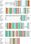
Sequence alignment among nine different species. Identical residues are highlighted in cyan, residues with conservative changes are highlighted in orange and residues with similar changes are highlighted in yellow. Residues located at the monomer–monomer interface of the EfTS enzyme are marked by an italic letter i, while active-site residues are marked by an italic letter a.
Figure 3
Figure 3
(a) Quaternary structure of EfTS. The two subunits of the heterodimer differ in the conformation of the small domain owing to the presence of a ligand bound to one of the active-site cavities. The ligand-bound subunit (magenta) is in a closed conformation, while the unbound subunit (light blue) is in an open conformation. The ligand is represented as sticks in CPK colours. (b) Cartoon representation of the tertiary fold of the EfTS B subunit with secondary structure labelled. The small domain is yellow, while the large domain has β-strands coloured light blue and α-helices coloured red. Unstructured loops are in grey, type 3 turns are in pink, type 4 turns are in tan, type 5 turns are in coral and β-bulges are in green, as is the location of the S–S bridge.
Figure 4
Figure 4
(a) Least-squares superimposition of the large domain of the EfTS–5-­FTHF complex (light-blue β-strands and red α-helices) with the analogous domain of the binary complex LcTS–dUMP (magenta; unpublished data from our laboratory). The similarity of the structures of the two enzymes can be appreciated as well as the occlusion of the dUMP-binding site by the chemical modification of EfTS Cys197. (b) Least-squares superimposition of the small domain of the EfTS–5-FTHF complex (yellow) with the analogous domain of the binary complex LcTS–dUMP (magenta; unpublished data from our laboratory) and the small domain of EcTS (blue; unpublished data from our laboratory). The similarity of the EfTS and LcTS small domains contrasts with the completely different structure of the small domain of EcTS.
Figure 5
Figure 5
Stick representation of the 5-FTHF ligand bound to the B and D subunits of EfTS with superimposed electron-density map (blue wire at 1.5σ) computed with 2F oF c coefficients and refined phases. The β-­mercaptoethanol-modified Cys197 is also shown as sticks, together with other relevant residues in the active-site cavity.

Similar articles

Cited by

References

    1. Anderson, A. C., O’Neil, R. H., DeLano, W. L. & Stroud, R. M. (1999). Biochemistry, 38, 13829–13836. - PubMed
    1. Anguera, M. C., Suh, J. R., Ghandour, H., Nasrallah, I. M., Selhub, J. & Stover, P. J. (2003). J. Biol. Chem. 278, 29856–29862. - PubMed
    1. Benvenuti, M. & Mangani, S. (2007). Nature Protoc. 2, 1633–1651. - PubMed
    1. Berger, S. H., Berger, F. G. & Lebioda, L. (2004). Biochim. Biophys. Acta, 1696, 15–22. - PubMed
    1. Birdsall, B., Burgen, A. S., Hyde, E. I., Roberts, G. C. & Feeney, J. (1981). Biochemistry, 20, 7186–7195. - PubMed

Publication types

Associated data