Review
. 2013 Jan;38(1):138-66.
doi: 10.1038/npp.2012.125. Epub 2012 Sep 5.

The dynamics of DNA methylation in schizophrenia and related psychiatric disorders

Affiliations
Review

The dynamics of DNA methylation in schizophrenia and related psychiatric disorders

Dennis R Grayson et al. Neuropsychopharmacology. 2013 Jan.

Abstract

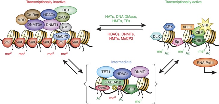
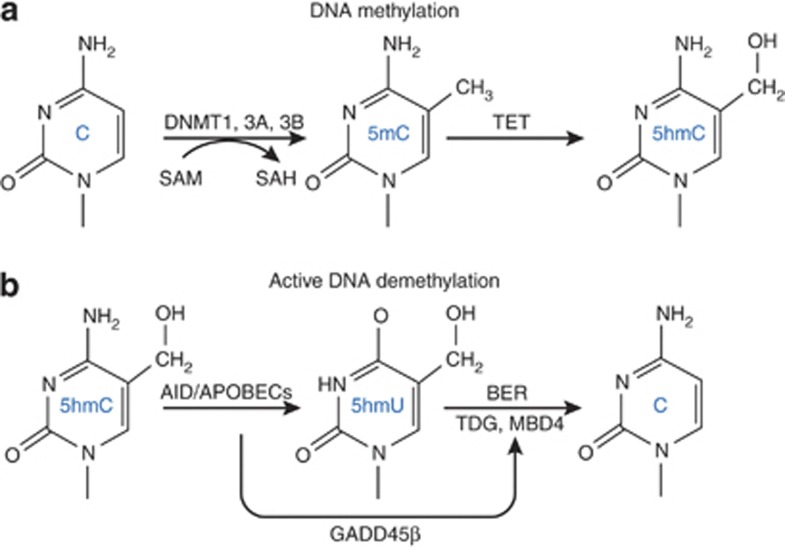
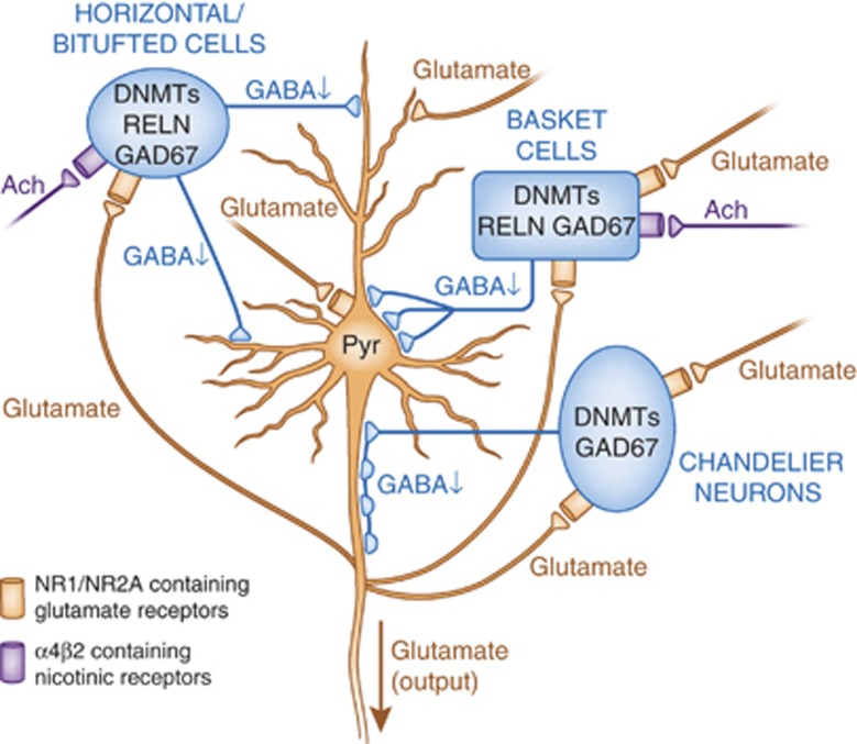
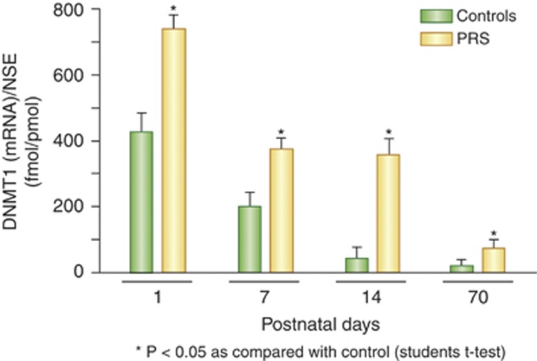
Major psychiatric disorders such as schizophrenia (SZ) and bipolar disorder (BP) with psychosis (BP+) express a complex symptomatology characterized by positive symptoms, negative symptoms, and cognitive impairment. Postmortem studies of human SZ and BP+ brains show considerable alterations in the transcriptome of a variety of cortical structures, including multiple mRNAs that are downregulated in both inhibitory GABAergic and excitatory pyramidal neurons compared with non-psychiatric subjects (NPS). Several reports show increased expression of DNA methyltransferases in telencephalic GABAergic neurons. Accumulating evidence suggests a critical role for altered DNA methylation processes in the pathogenesis of SZ and related psychiatric disorders. The establishment and maintenance of CpG site methylation is essential during central nervous system differentiation and this methylation has been implicated in synaptic plasticity, learning, and memory. Atypical hypermethylation of candidate gene promoters expressed in GABAergic neurons is associated with transcriptional downregulation of the corresponding mRNAs, including glutamic acid decarboxylase 67 (GAD67) and reelin (RELN). Recent reports indicate that the methylation status of promoter proximal CpG dinucleotides is in a dynamic balance between DNA methylation and DNA hydroxymethylation. Hydroxymethylation and subsequent DNA demethylation is more complex and involves additional proteins downstream of 5-hydroxymethylcytosine, including members of the base excision repair (BER) pathway. Recent advances in our understanding of altered CpG methylation, hydroxymethylation, and active DNA demethylation provide a framework for the identification of new targets, which may be exploited for the pharmacological intervention of the psychosis associated with SZ and possibly BP+.


Figures

Figure 1
Figure 1
Proteins bound to DNA and histones cooperate in facilitating transitions between active and inactive chromatin states. Schematic representation of the transitions between a transcriptionally inactive promoter (left) and a transcriptionally active state (right). The transcriptionally inactive state is characterized by DNA methylation and the binding of various repressor proteins, including DNA methyltransferase 1 (DNMT1) and 3A, methyl-binding domain proteins (MBDs, MeCP2), co-repressors, and modified histones associated with repressive chromatin marks (H3K9me2, H3K9me3, H3K27me2, H3K27me3, etc.). The intermediate state (shown in brackets) is stable and ‘poised' for either repression or activation. In the intermediate state, the DNA/protein complex is characterized by the binding of DNMT1 to unmethylated CpGs and ten-eleven translocase-1 (TET-1) bound to 5-methylcytosines (5mCs) and 5-hyroxymethylcytosine (5hmCs). In the transitional phase, DNMT1 is associated with histone deacetylases (HDACs) and excess DNMT3A shifts this towards the inactive state (left). The binding of TET1 to hydroxymethylated CpGs in this same intermediate state reinforces stable repression until the entry of GADD45β, which recruits proteins required for DNA demethylation (deaminases and glycosylases). DNA demethylation is accompanied by additional histone modifications (mediated by HATs and HMTs). Hydroxymethylated CpGs are further modified and removed. In this model, HDAC inhibitors facilitate a disruption of the inactive state and depending on the availability of GADD45β, DNA demethylation ensues (Kundakovic et al, 2009; Guidotti et al, 2011). In the active (open) state, various transcription factors (TFs) bind and occupy their specific DNA recognition sites enabling transcription. The specific TFs involved depend on the gene being activated and the neuronal phenotype (West and Greenberg, 2011). Some of the transcription factors are shown bound to the intermediate state (such as CREB, which upon phosphorylation (P) recruits the histone acetyltransferase CBP). Transcriptionally active promoters are represented as an open chromatin structure characterized by the presence of acetylated (H3K9ac, H3K14ac) and methylated (eg, H3K4me1, H3K4me3, H3K9me1, H3K27me1, H3K79me1, etc) histones. The model highlights repressive roles for DNMT1 and TET1, which depends upon the availability of accessory proteins (DNMT3A and GADD45β, respectively) to modify their function in postmitotic neurons. Based on localization studies of DNMT1 in GABAergic neurons (Kadriu et al, 2011) and GADD45β in pyramidal neurons (Gavin et al, 2012), these mechanisms are likely unique to specific types of neurons depending on neurotransmitter phenotype. ARX, aristaless-related homeobox; bHLH, basic helix-loop-helix transcription factors; CBP, CREB-binding protein; Co-Rep, co-repressor proteins; CREB, cyclic AMP response element-binding protein; D, deaminase; DLX, distal-less homeobox; DMAP1, DNA methyltransferase 1-associated protein; DNA DMase, DNA demethylase; G, gycosylase; HATs, Histone acetyl transferases; HMTs, histone methyl transferases; HP1, hetrochromatin protein 1; me1, monomethyl; me2, dimethyl; me3, trimethyl; MeCP2, methyl CpG-binding protein 2; P, phosphoryl group; RB1, retinoblastoma 1; SP1, promoter-specific transcription factor; SRF, serum response factor; TFs, transcription factors.
Figure 2
Figure 2
DNA methylation and demethylation are in a dynamic balance in neurons. The top panel (a) shows key steps associated with DNA methylation. DNA methyltransferases (DNMTs) catalyze the methylation of the fifth position of the pyrimidine ring of cytosine in CpG dinucleotides. S-adenosylmethionine (SAM) serves as the methyl donor that is converted to S-adenosylhomocysteine (SAH) following methyl group transfer. 5-methylcytosine (5mC) can be hydroxylated in a subsequent reaction catalyzed by members of the ten-eleven translocase (TET) family of methylcytosine dioxygenases. TET1–3 are 2-oxoglutarate-Fe(II) oxygenases, which hydroxylate 5mC to 5-hydroxymethylcytosine (5hmC). TETs 1 and 3 contain a –CXXC- domain, which binds with high affinity to clustered, unmethylated CpG dinucleotides. Structural analyses of DNMT1 show that it also contains a similar –CXXC- domain (see Text Box 2). The bottom panel (b) shows steps involved with the removal of the methyl group from 5hmC. The first step is an oxidative deamination of 5hmC to produce 5-hydroxymethyluridine (5hmU) by the AID/APOBEC family of deaminases. Activation-induced cytidine deaminase (AID) is also a member of the apolipoprotein B mRNA-editing catalytic polypeptides that deaminate 5mC and 5hmC to form thymine and 5hmU, respectively. These intermediates are subsequently processed by the uracil-DNA glycosylase (UDG) family that includes thymine-DNA glycosylase (TDG, MBD4) and single-strand-selective monofunctional uracil-DNA glycosylase 1 (SMUG1). These latter steps are collectively part of the base excision repair glycosylases (BER) that may also generate additional reactive intermediates such as 5-formylcytosine and 5-carboxylcytosine (Wu and Zhang, 2011). GADD45β is an activity-induced neuronal immediate early gene that facilitates active DNA demethylation (Ma et al, 2009a).
Figure 3
Figure 3
DNA methyltransferase (DNMT) overexpression leads to the downregulation of mRNAs in GABAergic neurons, increased methylation, and reduced gamma-aminobutyric acid (GABA) output (hypofunction). Schematic representation of the principal neuronal circuits in the cortex showing the reciprocal interaction between GABAergic innervation of pyramidal neurons and glutamatergic innervation of horizontal/bitufted, basket, and chandelier GABAergic interneurons. The GABAergic promoter downregulation in schizophrenia (SZ) and bipolar disorder with psychosis (BP+) patients is characterized by increased DNMT1 and 3A, and reduced GAD67, RELN and a variety of interneuron markers (Fung et al, 2010). These neurons also exhibit compromised expression of additional genes associated with inhibitory neuron function including NR1/NR2-containing-NMDA selective glutamate receptors and α4β2-containing nicotinic receptors. Glutamatergic inputs (shown in red) are meant to exhibit the excitatory input that arises from proximal pyramidal neurons or additional brain regions such as the thalamus. Ach, acetylcholine; Pyr, pyramidal neuron.
Figure 4
Figure 4
Co-localization of DNA methyltransferase 1 (DNMT1) and RELN immunoreactivities with GAD67 expression in mouse cortical neurons. The enhanced green fluorescent protein (GFP) was knocked into the GAD67 start codon to create GAD67+/−mice (Tamamaki et al, 2003). In these mice, GFP is expressed from the GAD67 promoter so that GFP immunoreactivity could be used to mark GAD67-positive (GABAergic) neurons. Fixed sections were incubated with their respective primary and secondary antibodies (Kadriu et al, 2011). Top panels show the co-localization of GFP immunoreactivity (GAD67) (a) and RELN (b) immunoreactivity. The merged signal shows co-localization (c). Similarly, GFP (GAD67) immunoreactivity (d) co-localizes with DNMT1 (e) as shown in f. Scale bar=40 μm. For details regarding immunohistochemistry, see Kadriu et al (2011).
Figure 5
Figure 5
DNA methyltransferase 1 (DNMT1) is elevated after birth and progressively declines with age in frontal cortex of control mice. Compared with controls, DNMT1 elevation is greater in offspring of prenatally stressed mice (PRS) even at 2 months of age. For the prenatal stress paradigm, pregnant mice were restrained in a transparent tube for 30 min twice daily from day 7 of pregnancy to delivery (Matrisciano et al, 2011). At the indicated times, RNA was harvested and analyzed for DNMT1 mRNA levels. *p<0.05, Student's t-test vs control values. Figure 5 is modified from Matrisciano et al (2012a).

References

    1. Aapola U, Kawasaki K, Scott HS, Ollila J, Vihinen M, Heino M, et al. Isolation and initial characterization of a novel zinc finger gene, DNMT3L, on 21q22.3, related to the cytosine-5-methyltransferase 3 gene family. Genomics. 2000;65:293–298. - PubMed
    1. Aapola U, Lyle R, Krohn K, Antonarakis SE, Peterson P. Isolationand initial characterization of the mouse Dnmt3l gene. Cytogenet Cell Genet. 2001;92:122–126. - PubMed
    1. Abdolmaleky HM, Cheng KH, Faraone SV, Wilcox M, Glatt SJ, Gao F, et al. Hypomethylation of MB-COMT promoter is a major risk factor for schizophrenia and bipolar disorder. Hum Mol Genet. 2006;15:3132–3145. - PMC - PubMed
    1. Abdolmaleky HM, Cheng KH, Russo A, Smith CL, Faraone SV, Wilcox M, et al. 2005Hypermethylation of the reelin (RELN). promoter in the brain of schizophrenic patients: a preliminary report Am J Med Genet B Neuropsychiatr Genet 13460–66.The first paper to provide biochemical evidence that the reelin promoter is hypermethylated in the brain of schizophrenia patients. - PubMed
    1. Abdolmaleky HM, Thiagalingam S. Can the schizophrenia epigenome provide clues for the molecular basis of pathogenesis. Epigenomics. 2011;3:679–683. - PubMed