Review
. 2012:747:55-73.
doi: 10.1007/978-1-4614-3229-6_4.

Death by caspase dimerization

Affiliations
Review

Death by caspase dimerization

Sarah H MacKenzie et al. Adv Exp Med Biol. 2012.

Abstract

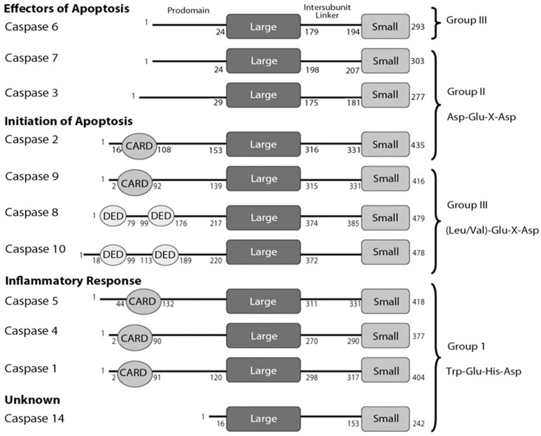
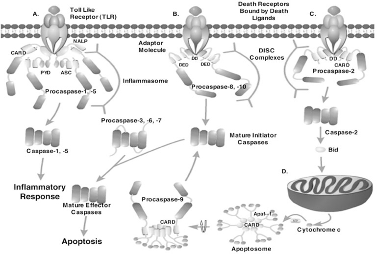
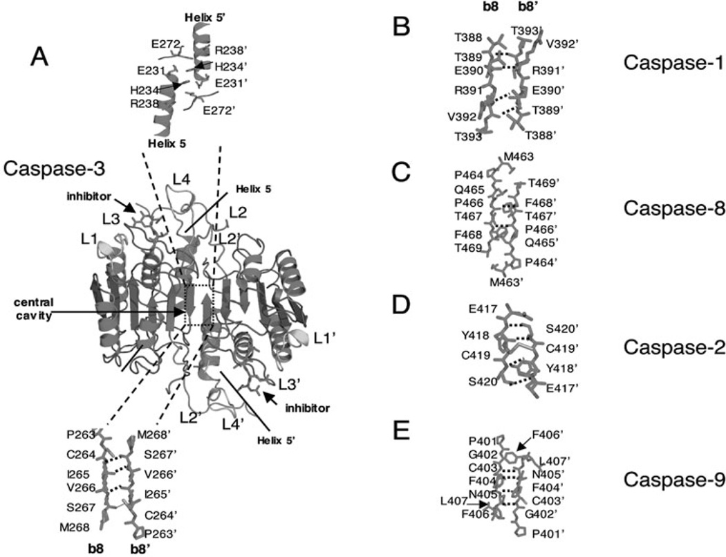
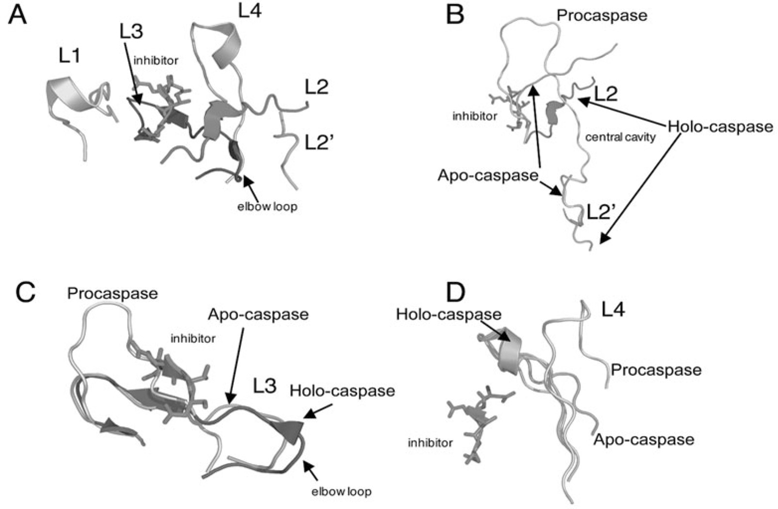
Controlled cell death, or apoptosis, occurs in response to many different environmental stimuli. The apoptotic cascade that occurs within the cell in response to these cues leads to morphological and biochemical changes that trigger the dismantling and packaging of the cell. Caspases are a family of cysteine-dependent aspartate-directed proteases that play an integral role in the cascade that leads to apoptosis. Caspases are grouped as either initiators or effectors of apoptosis, depending on where they enter the cell death process. Prior to activation, initiator caspases are present as monomers that must dimerize for full activation whereas effector caspases are present as dimeric zymogens that must be processed for full activation. The stability of the dimer may be due predominately to the interactions in the dimer interface as each caspase has unique properties in this region that lend to its specific mode of activation. Moreover, dimerization is responsible for active site formation because both monomers contribute residues that enable the formation of a fully functional active site. Overall, dimerization plays a key role in the ability of caspases to form fully functional proteases.


Figures

Figure 1
Figure 1
Human caspase organization. Caspases are grouped on the left according to function and on the right according to the recognition sequence of the substrate. Each caspase has an N-terminal prodomain, where some contain either a CARD (caspase recruitment domain) or DED (death effector domain) motif, followed by the large subunit (LARGE), an intersubunit linker and the small subunit (SMALL). The numbers on each caspase molecule refers to the length of each specific domain, which was determined using the NCBI domain organization database (http://www.ncbi.nlm.nih.gov/).
Figure 2
Figure 2
The caspase cascade. A) Inflammatory caspase activation: a ligand binds to a toll-like receptor (TLR), which signals a NALP protein to bind to the TLR. ASC interacts with the pyrin domain (PYD) of NALP via PYD:PYD interactions. The CARD domain of ASC interacts with the CARD domain of procaspase-1 and the CARD domain of NALP interacts with the CARD domain of procaspase-5, forming the inflammasome. The inflammasome complex promotes dimerization of caspases-1 and -5, leading to their activation and the inflammatory response. B) The extrinsic apoptotic pathway: a death ligand binds to a death receptor, which signals an adaptor molecule to bind to the receptor via death domain (DD) interactions. The DED motif of the adaptor molecule interacts with the DED of procaspases-8 and -10, forming a DISC complex. Dimerization (mechanism unknown) results in maturation and full activity. Caspases-8 and -10 then process executioner caspases. C) Procaspase-2, a unique caspase, is activated when a ligand binds to a death receptor, which signals an adaptor molecule to bind via interactions with the death domain. The CARD of the adaptor molecule interacts with the CARD of procaspase-2 to promote dimerization in a DISC-like complex. Upon removal of the prodomain, caspase-2 cleaves Bid, a protein responsible for the increased permeability of the mitochondria. D) The intrinsic apoptotic pathway: an increase in the cytosolic concentration of cytochrome c leads to the formation of the apoptosome. The apoptosome is composed of Apaf-1 monomers that form a heptameric structure when cytochrome c binds to the WD40 motifs of Apaf-1, in an ATP-dependent manner, leading to interactions of the CARDs. The CARD of procaspase-9 then interacts with the CARD of Apaf-1, increasing the local concentration of procaspase-9 monomers and thereby promoting dimerization and activation. Caspase-9 then processes effector caspases, which leads to apoptosis. Effector caspases are activated by cleavage of their prodomain and intersubunit linker.
Figure 3
Figure 3
Comparison of caspase dimer interfaces. A) Structure of caspase-3 (PDB entry 2J30). α-helices 5 and 5’ and active site loops L1, L2, L2’, L3 and L4 are labeled. The prime (‘) indicates residues from the second heterodimer. Residues in β-strands 8 and 8’ are shown below the structure and ionic interactions between residues in helices 5 and 5’ are shown above the structure rotated by 180° to view sidechains. Amino acids in the dimer interface (β-strands 8 and 8’) of caspase-1 (B) (PDB entry 2HBQ), caspase-8 (C) (PDB entry 1QTN), caspase-2 (D) (PDB entry 1PYO) and of caspase-9 (E) (PDB entry 1JXQ) are shown. For A-E, the dashed lines indicate main chain hydrogen bonds between β-strands 8 and 8’. Structures were generated using Pymol (Delano Scientific LLC, Palo Alto, CA).
Figure 4
Figure 4
Active site loop movements upon maturation of caspase-7. A) Active site loops 1–4 and 2’. B) Movements in L2 and L2’ upon maturation and substrate binding. Holo-caspase-7 (PDB entry 1F1J), apo-caspase-7 (PDB entry 1K86), procaspase-7 (PDB entry 1GQF). Note that in procaspase-7 the intersubunit linker encompasses both L2 and L2’. C) Movements in L3 upon maturation. D) Movements in L4 upon maturation. For A-D, the inhibitor bound to holo-caspase-7 is indicated. Structures were generated using Pymol (Delano Scientific LLC, Palo Alto, CA).
Figure 5
Figure 5
Active site rearrangements of caspases-1 and -3. A) Movements in L2 and L3 of caspase-1 upon substrate binding. The following colors are used. Green: apo-caspase-1 (PDB entry 1SC1), blue: holo-caspase-1 with malonate bound (PDB entry 1SC3). B) Different rotamer configurations for Gln283 in the ligand-free (blue) or ligand-bound (green) conformations of caspase-1 and loop movements in L2. Ala285 refers to the catalytically inactive mutant of caspase-1. C) Movements of Arg286 upon substrate binding result in intercalation of the side chain between Cys331 and Pro335, from active site loop 3, forming a new salt bridge with Glu390 from the dimer interface. The red sphere indicates a water molecule between Glu390 and Glu390’. D) In caspase-3 (PDB entry 2J30), the charge of Arg164 (equivalent to Arg286 in caspase-1) is neutralized by Glu124, which is situated above the dimer interface. For B and C, the secondary structure and active site loops are colored the same as those in Figures 3A and 4A and side chains are colored using the cpk color mode. Structures were generated using Pymol (Delano Scientific LLC, Palo Alto, CA). A color version of this image is available at www.landesbioscience.com/curie.
Figure 6
Figure 6
Active site rearrangements of caspase-9 upon inhibitor binding. A) Structure of caspase-9 (PDB entry 1JXQ) with inhibitor bound to one active site. The structure is colored as in Figure 3 except the loop insertion between β-strands 3 and 4 is shown in green. B) Upon inhibitor binding to one active site, loop rearrangements result in movement of the Tyr345 side chain, on β-strand 7, away from the active site and toward the protein interior, causing the side chain of Phe404 to move toward the dimer interface. Due to steric constraints in the interface, these movements can occur only in one heterodimer, so the second active site remains disorganized. C) Interactions among amino acids in the elbow loop (Phe348-Phe351) and the second heterodimer (Phe246’, Pro338’ and Phe406’). For A-C, the secondary structure and active site loops are colored the same as those in Figures 3A and 4A and side chains are colored using the cpk color mode. Structures were generated using Pymol (Delano Scientific LLC, Palo Alto, CA). A color version of this figure is available at www.landesbioscience.com/curie.
Figure 7
Figure 7
Structure of caspase-7 with FICA bound in the dimer interface (PDB entry 1SHL). Binding of inhibitor in the dimer interface displaces the side chain of Tyr223 and results in disorganized active site loops L2, L3 and L4. L2’ occupies the central cavity, as observed for apo-caspase-7 (see Fig. 4B). The secondary structure and active site loops are colored the same as those in Figures 3A and 4A and side chains are colored using the cpk color mode. Structures were generated using Pymol (Delano Scientific LLC, Palo Alto, CA).

References

    1. Jacobson MD, Weill M, Raff MC. Programmed cell death in animal development. Cell. 1997;88:347–354. - PubMed
    1. Fadeel B, Orrenius S. Apoptosis: a basic biological phenomenon with wide-ranging implications in human disease. J Internal Med. 2005;258:479–517. - PubMed
    1. Kabore AF, Johnston JB, Gibson SB. Changes in the apoptotic and survival signaling in cancer cells and their potential therapeutic implications. Current Cancer Drug Targets. 2004;4:147–163. - PubMed
    1. Fulda S, Debatin K-M. Targeting apoptosis pathways in cancer therapy. Current Cancer Drug Targets. 2004;4:569–576. - PubMed
    1. Meng XW, Lee S-H, Kaufmann SH. Apoptosis in the treatment of cancer: a promise kept? Curr Op Cell Biol. 2006;18:668–676. - PubMed

Publication types

MeSH terms

LinkOut - more resources