. 2012 Nov;32(21):4482-92.
doi: 10.1128/MCB.00872-12. Epub 2012 Sep 4.

HtrA1 is a novel antagonist controlling fibroblast growth factor (FGF) signaling via cleavage of FGF8

Affiliations

HtrA1 is a novel antagonist controlling fibroblast growth factor (FGF) signaling via cleavage of FGF8

Goo-Young Kim et al. Mol Cell Biol. 2012 Nov.

Abstract

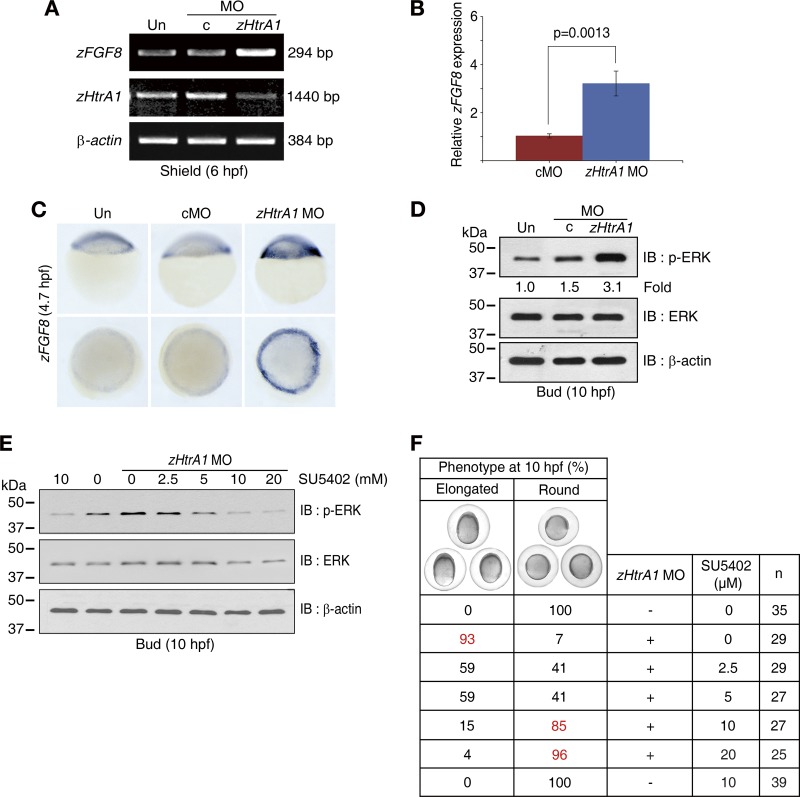
Accumulating evidence suggests that HtrA1 (high-temperature requirement A1) is involved in modulating crucial cellular processes and implicated in life-threatening diseases, such as cancer and neuropathological disorders; however, the exact functions of this protease in vivo remain unknown. Here, we show that loss of HtrA1 function increases fibroblast growth factor 8 (FGF8) mRNA levels and triggers activation of FGF signaling, resulting in dorsalization in zebrafish embryos. Notably, HtrA1 directly cleaves FGF8 in the extracellular region, and this cleavage results in decreased activation of FGF signaling, which is essential for many physiological processes. Therefore, HtrA1 is indispensable for dorsoventral patterning in early zebrafish embryogenesis and serves as a key upstream regulator of FGF signaling through the control of FGF levels. Furthermore, this study offers insight into new strategies to control human diseases associated with HtrA1 and FGF signaling.


Figures

Fig 1
Fig 1
zHtrA1 is the true zebrafish ortholog of human HtrA1. (A) Schematic view of human and zebrafish HtrA1 showing the signal peptide (SP), insulin-like growth factor binding protein-binding (IGFBP), Kazal inhibitor (KI), serine protease, and PDZ domains. The table indicates amino acid identity between the serine protease domains. (B) Rapid affinity purification of the HtrA1 proteins. (C) Endoproteolytic activity of the HtrA1 serine proteases. GST-HtrA1 proteins (0.75 μM) were incubated for the indicated times at 37°C with β-casein (10 μM) as an exogenous substrate. (D) Induction of cell death in an HtrA1 serine protease-dependent manner. The extent of cell death was calculated by dividing the number of round cells with nuclear condensation and fragmentation (dead cells [arrowheads]) by the total number of GFP-positive cells and is expressed as a percentage. “Mock” refers to cells transfected with a control plasmid expressing only GFP. DIC, differential interference contrast; M-HtrA2, mature HtrA2 used as a positive control for cell death. Bar, 10 μm. (E) Stage-specific expression of zHtrA1. Total RNA was extracted from zebrafish embryos at the indicated developmental time points, and RT-PCR was carried out with the zHtrA1 primer set and normalized to β-actin used as an internal control. (F to H) Spatial expression of zHtrA1 at the indicated developmental time points. WISH was performed on zebrafish embryos at different developmental stages using a DIG-labeled zHtrA1 antisense RNA probe, corresponding to nucleotide positions 1 to 784 of zHtrA1. hpf, hour postfertilization; s, somite; ea, embryonic axis; pf, pectoral fin bud.
Fig 2
Fig 2
Knockdown of zHtrA1 causes dorsalization during zebrafish embryogenesis. (A and B) Knockdown of zHtrA1. Zebrafish embryos were injected with the indicated MO at the one-cell stage. Aberrant splicing caused by the zHtrA1 MO in the zHtrA1 pre-mRNA was assessed by RT-PCR (see Fig. S3A and Table S2 in the supplemental material). Un, uninjected; cMO, control MO. (B) Morphological changes visible in the zHtrA1 KD embryos. The dorsal (D) and ventral (V) regions of all embryos were oriented in the same direction. Phenotypic frequencies (percent) were calculated by dividing the number of dorsalized embryos (P) by the total number of embryos. The zHtrA1-ATG MO was designed to prevent translation of the zHtrA1 protein (see Fig. S4D in the supplemental material). Injection with zHtrA1-ATG MO led to dorsalized phenotypes, consistent with phenotypes obtained with zHtrA1 MO (see Fig. S4E and F). (C) Expression levels of the dorsoventral patterning markers. WISH was performed on the shield-stage embryos with probes for chd and gsc. Arrowheads indicate the dorsal territories. (D and E) Expansion of dorsally derived somites in the zHtrA1 KD embryos. WISH was performed on 7-somite-stage embryos with both the myoD (somatic mesoderm) and pax2.1 probes (7 embryos per group) (D). Dorsal view, anterior to the top. (E) The sizes of the fourth somite (double-headed arrow in panel D) are presented as means ± SEM (*, P < 0.0001 compared with control group). One-way ANOVA was used for statistical analyses.
Fig 3
Fig 3
Loss of zHtrA1 function activates the FGF signaling pathway. (A to C) The upregulation of zFGF8 in zHtrA1 KD embryos. The levels of zFGF8 mRNA were assessed by RT-PCR with the zFGF8 O primer set (A) and quantitative real-time PCR (qRT-PCR) (7 experiments were performed; more than 8 embryos per group were used for each experiment) with the 5-U1 primer set (B) (see Table S2 in the supplemental material). Data are means ± SEM. (C) WISH was performed with the zFGF8 probe (11 embryos per group). Top and bottom panels show lateral and animal pole views, respectively. (D) FGF8-induced ERK activation in the zHtrA1 KD embryos. At 10 hpf, embryonic lysates were resolved by 10% SDS-PAGE, and activation of ERK was assessed by IB with anti-p-ERK PAb. The levels of p-ERK were normalized to ERK, and the values are presented as fold change relative to the untreated control. (E and F) Specificity of FGF8-induced ERK activation in the zHtrA1 KD embryos. The zHtrA1 MO was injected at the one-cell stage, and then embryos were treated with SU5402 at 2.5 hpf (initial stage of zFGF8 expression). At 10 hpf, embryonic lysates were resolved by 10% SDS-PAGE, followed by IB for p-ERK (E). Phenotypic rescue of elongated embryos was morphologically analyzed under a stereo dissection microscope (F). Phenotypic frequencies (percent) were calculated by dividing the number of elongated (or round) forms of embryos by the total number of embryos (n).
Fig 4
Fig 4
FGF8 positively regulates FGF8 expression through an FGF signaling feedback loop. (A) A schematic diagram of the zFGF8 mRNA. All of the indicated PCR products were amplified by the corresponding primer sets (see Table S2 in the supplemental material). (B) In vitro-transcribed zFGF8 (nucleotides 418 to 1710, 0.1 fmol/embryo) was injected into one-cell stage embryos, and transcriptional activation of endogenous zFGF8 was analyzed by qRT-PCR (4 experiments were performed; more than 13 embryos per group were used for each experiment) with the indicated primer sets. Data are means ± SEM (*, P < 0.02, and **, P < 0.01 compared with control group). (C) Recombinant human FGF8 protein (1.6 fmol/embryo) was injected into one-cell-stage embryos, and endogenous zFGF8 RNA was detected by WISH (8 embryos per group) with a zFGF8 antisense probe at 28 hpf (arrowheads, zFGF8-expressed somite borders). (D) Abrogation of the increase in FGF8 transcript levels by blockade of FGF signaling with SU5402. Embryos were treated with SU5402 at 2.5 hpf, and levels of indicated target transcripts were analyzed by qRT-PCR (3 experiments were performed; more than 15 embryos per group were used for each experiment) with the indicated primer sets. Data are means ± SEM (***, P < 0.001 compared with control group). (E) Working model of the FGF8-mediated positive feedback loop of the FGF signaling cascade. FGF8 binds to FGFR in the extracellular region and thereby activates the mitogen-activated protein kinase (MAPK) cascade (3, 42, 43). Notably, the FGF8 gene is transcriptionally activated via FGF signaling. The newly synthesized FGF8 protein is secreted into the extracellular region.
Fig 5
Fig 5
HtrA1 negatively regulates the FGF signaling pathway by cleaving FGF8. (A and B) Inhibition of zFGF8 expression and FGF signaling in the embryos overexpressing the active serine protease zHtrA1. zHtrA1 mRNA (0.3 fmol/embryo) was injected into one-cell-stage embryos. (A) The levels of zFGF8 transcript were assessed by WISH (3 embryos per group). (B) ERK activation was determined by the extent of p-ERK. (C and D) Direct cleavage of FGF8 by HtrA1. The indicated GST fusion proteins were incubated with zebrafish (C) or human (D) [35S]Met-labeled FGF8 for 3 h at 37°C. (E) Schematic diagram of the CM assay designed to assess the effect of extracellular HtrA1 on the regulation of the FGF signaling pathway. All CM were collected from HEK293 cells transfected with plasmids encoding HtrA1 at 24 h posttransfection or with siRNAs at 48 h posttransfection. HEK293 cells, which were cultured in serum-free medium for 6 h, were treated with the CM plus FGF8 for 30 min. The effect of the enzyme-substrate pair HtrA1 and FGF8 on the FGF signaling pathway in the extracellular region was assessed by the extent of p-ERK. (F and G) Inhibition of FGF signaling by HtrA1 in the extracellular region. Serum-starved HEK293 cells were treated with the indicated CM supplemented with FGF8 for 30 min. Cell lysates were resolved by 15% SDS-PAGE, followed by IB with the indicated antibodies.
Fig 6
Fig 6
Restoration of zFGF8-induced FGF signaling activation to a normal state in the presence of active zHtrA1. zFGF8 (0.1 fmol) and zHtrA1 (0.3 fmol) RNAs were coinjected into one-cell-stage embryos. (A) p-ERK was assessed by IB at 10 hpf. (B) Rescued phenotype frequencies (percent) were calculated by dividing the number of round embryos by the total number of embryos (n) at 10 hpf. (C) Embryo size from the anterior to the posterior pole, presented as means ± SEM (*, P < 0.001 compared with control group). (D) Working model of HtrA1 as a novel antagonist in the FGF8-mediated positive feedback loop of FGF signaling. HtrA1 cleaves FGF8 in the extracellular region, which may be required to maintain FGF8 and FGF signaling at a constant level.

Similar articles

Cited by

References

    1. Baldi A, et al. 2002. The HtrA1 serine protease is down-regulated during human melanoma progression and represses growth of metastatic melanoma cells. Oncogene 21:6684–6688 - PubMed
    1. Beenken A, Mohammadi M. 2009. The FGF family: biology, pathophysiology and therapy. Nat. Rev. Drug Discov. 8:235–253 - PMC - PubMed
    1. Bottcher RT, Niehrs C. 2005. Fibroblast growth factor signaling during early vertebrate development. Endocr Rev. 26:63–77 - PubMed
    1. Bottcher RT, Pollet N, Delius H, Niehrs C. 2004. The transmembrane protein XFLRT3 forms a complex with FGF receptors and promotes FGF signalling. Nat. Cell Biol. 6:38–44 - PubMed
    1. Chamberland A, et al. 2009. Identification of a novel HtrA1-susceptible cleavage site in human aggrecan: evidence for the involvement of HtrA1 in aggrecan proteolysis in vivo. J. Biol. Chem. 284:27352–27359 - PMC - PubMed

Publication types

MeSH terms