. 2013 Jan 1;22(1):1-17.
doi: 10.1093/hmg/dds371. Epub 2012 Sep 4.

Mutations in FKBP10, which result in Bruck syndrome and recessive forms of osteogenesis imperfecta, inhibit the hydroxylation of telopeptide lysines in bone collagen

Affiliations

Mutations in FKBP10, which result in Bruck syndrome and recessive forms of osteogenesis imperfecta, inhibit the hydroxylation of telopeptide lysines in bone collagen

Ulrike Schwarze et al. Hum Mol Genet. .

Abstract

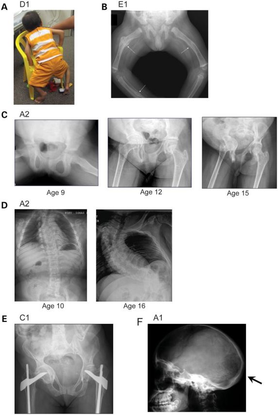
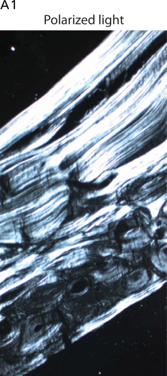
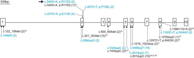
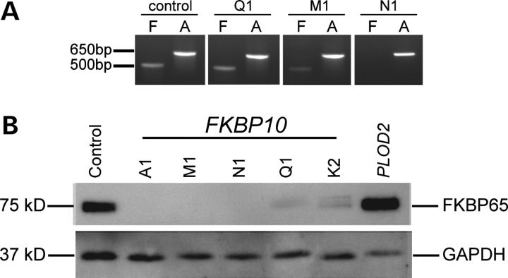
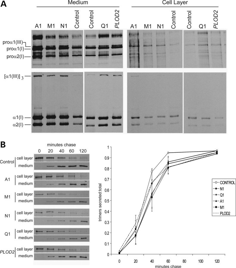
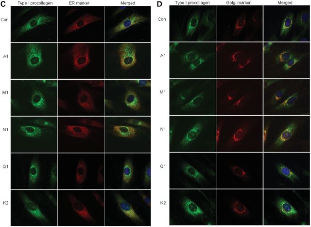
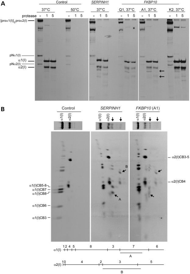
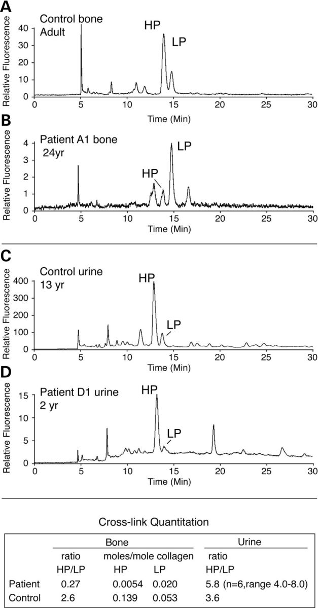
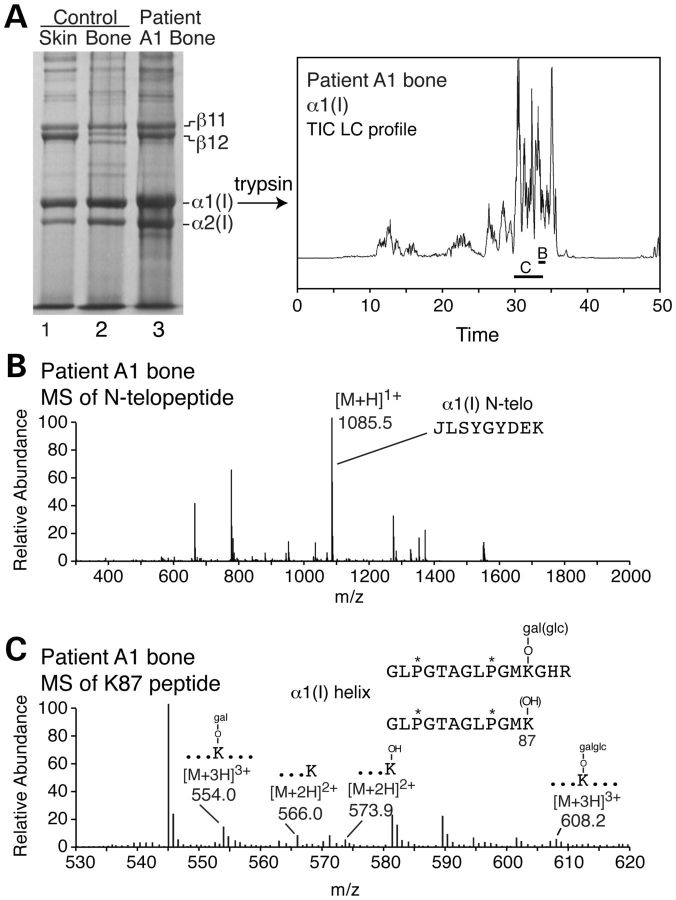
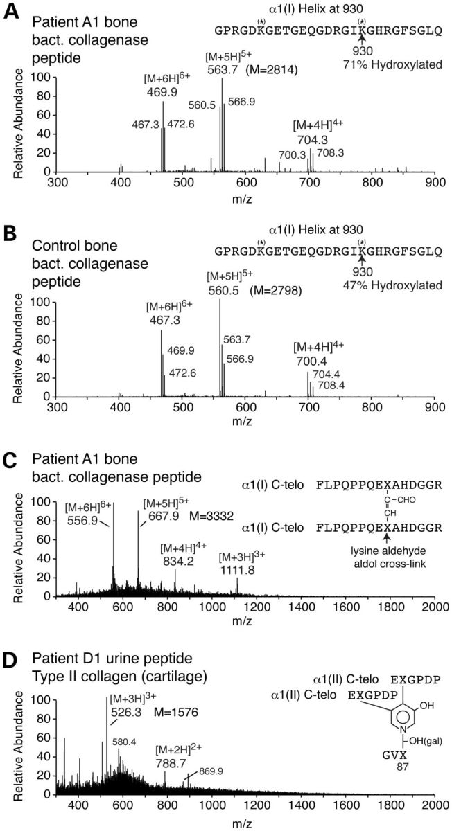
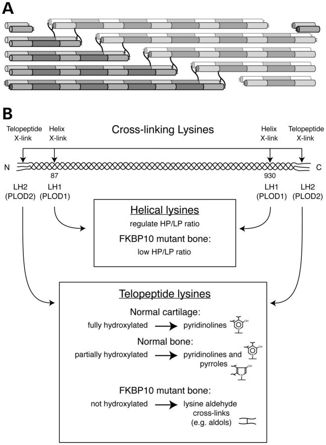
Although biallelic mutations in non-collagen genes account for <10% of individuals with osteogenesis imperfecta, the characterization of these genes has identified new pathways and potential interventions that could benefit even those with mutations in type I collagen genes. We identified mutations in FKBP10, which encodes the 65 kDa prolyl cis-trans isomerase, FKBP65, in 38 members of 21 families with OI. These include 10 families from the Samoan Islands who share a founder mutation. Of the mutations, three are missense; the remainder either introduce premature termination codons or create frameshifts both of which result in mRNA instability. In four families missense mutations result in loss of most of the protein. The clinical effects of these mutations are short stature, a high incidence of joint contractures at birth and progressive scoliosis and fractures, but there is remarkable variability in phenotype even within families. The loss of the activity of FKBP65 has several effects: type I procollagen secretion is slightly delayed, the stabilization of the intact trimer is incomplete and there is diminished hydroxylation of the telopeptide lysyl residues involved in intermolecular cross-link formation in bone. The phenotype overlaps with that seen with mutations in PLOD2 (Bruck syndrome II), which encodes LH2, the enzyme that hydroxylates the telopeptide lysyl residues. These findings define a set of genes, FKBP10, PLOD2 and SERPINH1, that act during procollagen maturation to contribute to molecular stability and post-translational modification of type I procollagen, without which bone mass and quality are abnormal and fractures and contractures result.


Figures

Figure 1.
Figure 1.
Clinical and radiographic variability in patients with FKBP10 mutations. (A) Subject D1 (age 2½ years). He is of short stature (<3rd centile) and is unable to plantar flex either foot or extend the knees because of contractures. By the age of 3 he had sustained no fractures. (B) Radiograph from subject E1 age 3 months. Fractures were noted in utero and limb contractures were present at birth. At age 3 months he had a skull fracture and multiple rib and long bone fractures (arrows). (C) Development of severe acetabular protrusion during adolescence in A2. (D) The development of scoliosis in A2. Her first fracture occurred at the age of 16 years. (E) Subject C1 age 35. Note marked acetabular protrusion on the right. She sustained femoral fractures at age 11 and 13, but has had no fractures since. (F) Skull findings of playtbasia, relative macrocephaly and Wormian bones (arrow) in A1.
Figure 2.
Figure 2.
Bone histology. Undecalcified transiliac bone biopsy from A1 at age 17. Under polarized light a normal lamellar pattern is evident, which distinguishes the pattern from that seen in bone from individuals with OI type VI that results from mutations in SERPINF1.
Figure 3.
Figure 3.
Location of mutations in FKBP10. The gene diagram is drawn to scale. Mutations identified in this study are shown in cyan and those identified by others in black. Missense mutations are indicated above the diagram. Frame shift and nonsense mutations and a single splice site mutation are depicted below. The numbers in parentheses indicate the number of alleles. The superscripts link the published mutations to the respective references. The arrow pointing at ‘c.344G > A, p. R115Q (2)’ indicates that the cross-linking data were published by Bank et al. (24).
Figure 4.
Figure 4.
Analysis of the effect of mutations in FKBP10 and PLOD2 on the stability of mRNA and FKBP65, the protein product of FKBP10. (A) Mutations in FKBP10 have different effects on mRNA stability that depend on the nature of the mutation. RNA was isolated from the cells from Q1 (c.337G > A, p.Glu113Lys), M1 (c.743dupC, p.Gln249Thrfs*12), and N1 (c.831dupC, p.Gly278Argfs*95), cDNA was synthesized and then amplified for 30 cycles with primers in exons 3 and 6. The first lane, F, for each sample represents the FKBP10 amplification, and the second lane, A, represents actin. In the presence of the missense mutation (Q1), the mRNA is stable and similar in abundance to that in the control. In the M1 sample there is residual mRNA, and there is no measurable stable mRNA from the cells from N1. (B) Mutations in FKBP10 result in unstable or diminished FKBP65 protein. Western blot analysis showed loss of the FKBP65 protein in individuals with frameshifts due to single base duplications in FKBP10 (A1 [c.948dupT, p.Ile317Tyrfs*56], M1, and N1), and marked reduction of protein in individuals with missense mutations (Q1 and K2). Compound heterozygosity for a missense and nonsense mutation in PLOD2 has no effect on the amount of FKBP65 present.
Figure 5.
Figure 5.
Effects of different mutations in FKBP10 on synthesis and intracellular transport of type I procollagen through the cell. (A) Type I procollagen is not overmodified in cells from individuals with frameshift (A1, M1, N1), missense (Q1) mutations in FKBP10 or biallelic mutations in PLOD2. Cells were labeled with [3H]-proline for 16 h and macromolecules in the culture medium and in the cell layer were harvested and separated after reduction of disulfide bonds (top panels) or treated with pepsin to remove the amino- and carboxyl-terminal propeptides and separated under non-reducing conditions. (B) There is a modest delay in secretion of type I procollagen from cells with mutations in FKBP10 and PLOD2. Cells were labeled for 1 h with [3H]-proline then chased for up to 2 h with unlabeled proline. At 60 min, the rate of secretion of the trimers of type I procollagen showed a delay in the patient cells compared with control (standard t-test, P-value of 0.046), quantitated in the graph to the right. (C and D) Distribution of type I procollagen between the RER (C) and the Golgi (D). Complete loss of FKBP65 as a result of frameshift mutations in FKBP10 (A1, M1, N1) and partial loss of protein that results from missense mutations (Q1, K2) all lead to slight retention of type I procollagen in the RER. Aggregates of type I procollagen reported by Alanay et al. (10) are not evident in these images.
Figure 5.
Figure 5.
Effects of different mutations in FKBP10 on synthesis and intracellular transport of type I procollagen through the cell. (A) Type I procollagen is not overmodified in cells from individuals with frameshift (A1, M1, N1), missense (Q1) mutations in FKBP10 or biallelic mutations in PLOD2. Cells were labeled with [3H]-proline for 16 h and macromolecules in the culture medium and in the cell layer were harvested and separated after reduction of disulfide bonds (top panels) or treated with pepsin to remove the amino- and carboxyl-terminal propeptides and separated under non-reducing conditions. (B) There is a modest delay in secretion of type I procollagen from cells with mutations in FKBP10 and PLOD2. Cells were labeled for 1 h with [3H]-proline then chased for up to 2 h with unlabeled proline. At 60 min, the rate of secretion of the trimers of type I procollagen showed a delay in the patient cells compared with control (standard t-test, P-value of 0.046), quantitated in the graph to the right. (C and D) Distribution of type I procollagen between the RER (C) and the Golgi (D). Complete loss of FKBP65 as a result of frameshift mutations in FKBP10 (A1, M1, N1) and partial loss of protein that results from missense mutations (Q1, K2) all lead to slight retention of type I procollagen in the RER. Aggregates of type I procollagen reported by Alanay et al. (10) are not evident in these images.
Figure 6.
Figure 6.
Mutations in FKBP10 affect protease sensitivity of the type I collagen triple helix. (A) Secreted type I procollagen molecules from cells with FKBP10 mutations are more sensitive to proteolytic digestion than those from control cells, with the apparent cleavage sites similar to those with mutations in SERPINH1. Proteins in medium from cultured dermal fibroblasts labeled overnight with [3H]-proline were digested for 1 or 5 min with a combination of trypsin and chymotrypsin at 37°C without prior cooling of the sample. Procollagens secreted into the culture medium by control cells and then pretreated at 50°C for 15 min were completely degraded following treatment with trypsin/chymotrypsin while procollagens from medium left at 37°C had protease-resistant triple-helical domains. A subset of type I procollagen molecules secreted from patient cells were cleaved asymmetrically at 37°C (the black arrows indicate fragments seen in affected cells and either not seen or in low abundance in the control cells). (B) Characterization by cyanogen bromide cleavage of products of type I procollagen following trypsin/chymotrypsin treatment. The gel lanes from runs equivalent to those in (A) were excised and the bands cleaved with cyanogen bromide. The larger of the new bands, which migrated just below α2(I) was derived from α1(I). The α1(I)CB7 fragment (residues 551–821 of the triple-helical domain) is missing and replaced by fragment A. The size of the fragment indicates that the parent peptide had been cleaved at or near the mammalian collagenase cleavage site (775–776 in the triple-helical domain). The smaller fragment in the parent gel was derived from α2(I). The α2(I)CB3-5 fragment (residues 357–1014 in the triple-helical domain) was shortened, fragment B and the estimated size indicated that it had been cleaved in the region of the collagenase cleavage site (residues 776–777 of the triple-helical domain).
Figure 7.
Figure 7.
HP and LP pyridinoline cross-links in hydrolysates of bone from an individual with a frameshift mutation in FKBP10, control human bone and urine samples from affected and control individuals. In patient A1 bone (B and table) the ratio of HP/LP is reversed from normal (A) and the pyridinoline cross-link content (moles/mole collagen) is ∼10% of that in control adult bone. Compared with control (C) the HP/LP ratio is higher in urine from D1 (D). The table shows the mean and range for the HP/LP ratios in urine samples from N1, N2, B1, B4, B5 and D1. Note that the total amount of pyridinoline cross-links in urine from individuals with mutations in FKBP10 is markedly lower than that seen in control urine (see the fluorescence scale difference between C and D).
Figure 8.
Figure 8.
Electrospray mass spectrometry of tryptic peptides from cross-linking sites in bone type I collagen from an individual (A1) with a frameshift mutation in FKBP10. (A) SDS–PAGE analysis of collagen extracted from control skin, control bone and demineralized patient bone (A1). The pattern of the dimeric cross-linked forms (β11 and β12) in A1 bone is more like that in control skin than in control bone. Individual chains were digested with trypsin and the peptides separated by LCMS. Peptides in the regions marked B and C were further analyzed as shown in sections B and C. (B) The amino-terminal telopeptide from α1(I) contained a non-hydroxylated lysyl residue which was not seen in control bone (latter data not shown). (C) The hydroxylation and glycosylation status of Lys-87 cross-linking site in α1(I) reflected marked heterogeneity among molecules.
Figure 9.
Figure 9.
Electrospray mass spectrometry of a bacterial collagenase-derived peptide from the α1(I) Lys-930 cross-linking site of bone type I collagen and a urinary peptide from individuals with a frameshift mutation in FKBP10. (A and B) Peptide pools containing the Lys-930 cross-linking lysine were isolated by C8 reverse-phase HPLC from bacterial collagenase digests of A1and control adult bone collagen and profiled for post-translational variants. In the A1 bone Lys-930 (A) was 71% hydroxylated versus 47% in control bone (B). The lysine at 918 was less hydroxylated than the one at position 930 in control and A1 bone. (C) The digest of A1 bone collagen contained a peptide (M = 3332 Da), not found in normal bone, that proved to be the lysine aldol dimer of two α1(I)-C-telopeptide fragments (C). The parent ion charge envelope of the latter is shown (556.96+, 667.95+, etc) from which the MS/MS fragmentation patterns (not shown) and molecular mass established the structure shown. (D) The structure of a cross-linked peptide pool from the urine of D1 is derived from the C-telopeptide cross-linking domain of type II collagen and contains exclusively an HP cross-link. This peptide was identified in control children's urine (29) and the 526.33+ and 788.72+ ions are of the unglycosylated HP peptide and 580.44+ and 869.92+ are the galactosyl (mono-glycosylated) HP version.
Figure 10.
Figure 10.
Pathway of cross-link formation in bone type I collagen and cartilage type II collagen. (A) The symmetrically placed four sites of lysine-based cross-linking in types I and II collagen molecules, two telopeptide and two triple-helical, interact to form cross-links in fibrils. (B) Two lysyl hydroxylases regulate collagen cross-linking in bone. LH1 is primarily responsible for hydroxylation of the K87 and K930 sites in the triple-helical domain, and LH2 hydroxylates the telopeptide lysines. With mutations in FKBP10 there is selective underhydroxylation of telopeptide lysines in bone type I collagen but not cartilage type II collagen, similar to the situation in Bruck syndrome caused by PLOD2 mutations (18,24).

Similar articles

Cited by

References

    1. Lapunzina P., Aglan M., Temtamy S., Caparrós-Martín J., Valencia M., Letón R., Martínez-Glez V., Elhossini R., Amr K., Vilaboa N., et al. Identification of a frameshift mutation in Osterix in a patient with recessive osteogenesis imperfecta. Am. J. Hum. Genet. 2010;87:110–114. - PMC - PubMed
    1. Baldridge D., Schwarze U., Morello R., Lennington J., Bertin T., Pace J., Pepin M., Weis M., Eyre D., Walsh J., et al. CRTAP and LEPRE1 mutations in recessive osteogenesis imperfecta. Hum. Mutat. 2008;29:1435–1442. - PMC - PubMed
    1. Morello R., Bertin T., Chen Y., Hicks J., Tonachini L., Monticone M., Castagnola P., Rauch F., Glorieux F., Vranka J., et al. CRTAP is required for prolyl 3- hydroxylation and mutations cause recessive osteogenesis imperfecta. Cell. 2006;127:291–304. - PubMed
    1. Marini J., Cabral W., Barnes A. Null mutations in LEPRE1 and CRTAP cause severe recessive osteogenesis imperfecta. Cell Tissue Res. 2010;339:59–70. - PMC - PubMed
    1. Cabral W., Chang W., Barnes A., Weis M., Scott M., Leikin S., Makareeva E., Kuznetsova N., Rosenbaum K., Tifft C., et al. Prolyl 3-hydroxylase 1 deficiency causes a recessive metabolic bone disorder resembling lethal/severe osteogenesis imperfecta. Nat. Genet. 2007;39:359–365. - PMC - PubMed

Publication types

Supplementary concepts