. 2012 Sep 18;109(38):15307-11.
doi: 10.1073/pnas.1201632109. Epub 2012 Sep 4.

Role of genes linked to sporadic Alzheimer's disease risk in the production of β-amyloid peptides

Affiliations

Role of genes linked to sporadic Alzheimer's disease risk in the production of β-amyloid peptides

Jitin Bali et al. Proc Natl Acad Sci U S A. .

Abstract

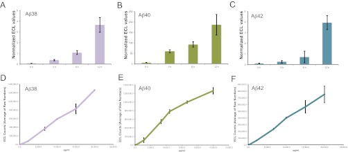
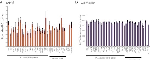
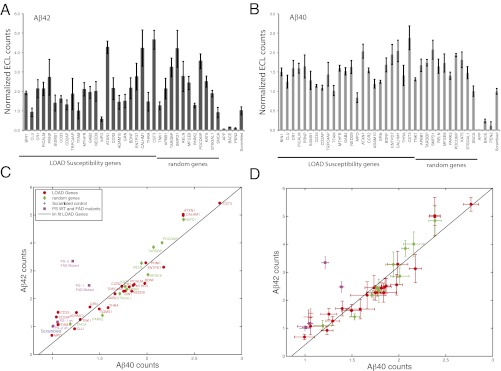
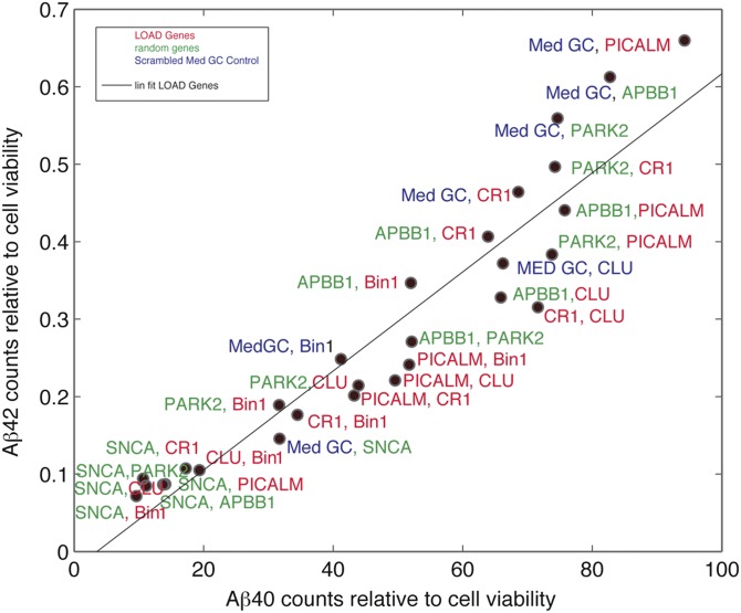
Alzheimer's disease (AD) is characterized by the presence of toxic protein aggregates or plaques composed of the amyloid β (Aβ) peptide. Various lengths of Aβ peptide are generated by proteolytic cleavages of the amyloid precursor protein (APP). Mutations in many familial AD-associated genes affect the production of the longer Aβ42 variant that preferentially accumulates in plaques. In the case of sporadic or late-onset AD, which accounts for greater than 95% of cases, several genes are implicated in increasing the risk, but whether they also cause the disease by altering amyloid levels is currently unknown. Through loss of function studies in a model cell line, here RNAi-mediated silencing of several late onset AD genes affected Aβ levels is shown. However, unlike the genes underlying familial AD, late onset AD-susceptibility genes do not specifically alter the Aβ42/40 ratios and suggest that these genes probably contribute to AD through distinct mechanisms.


Conflict of interest statement

The authors declare no conflict of interest.

Figures

Fig. 1.
Fig. 1.
(A–C) Quantification of cell culture derived and synthetic Aβ peptides. Aβ38 (A), 40 (B), and 42 (C) levels in supernatant of HeLa swAPP cells after 0 h, 3 h, 6 h, and 12 h were analyzed using the triplex ECL assay (MSD). The values are given as ECL counts normalized to cell viability. (A–C): Linearity of the detection for each of the analytes in the multiplex ECL assay platform was determined for different concentrations of synthetic Aβ38 (D), Aβ40 (E), and Aβ42 (F) peptides.
Fig. 2.
Fig. 2.
β-Cleavage of APP is not specifically affected by genes involved in LOAD susceptibility. (A) HeLa swAPP cells were transfected with siRNAs to the corresponding genes, and the supernatant was analyzed for sAPPβ 72 h after transfection (with 3 h medium exchange) using duplex ECL array (MSD). The values are given as ECL counts normalized to cell viability and relative to that of a scrambled (MedGC) control. The genes (BIN1, CLU, CR1, PICALM, CD33, and CD2AP) are among the top 10 AlzGene meta-analysis results (ranking is based on P value, and top 10 have P values <0.00001) listed in the AlzGene database. APP, BACE1, Pen-2, and scrambled (MedGC) serve as both transfection and assay controls. (B) Cell viability: HeLa swAPP cells were incubated for 72 h after siRNA transfection and subjected to a cell viability assay using AlamarBlue. Results are shown as fluorescence counts using a microplate absorbance/fluorometer plate reader (Molecular Devices Spectramax Gemini XS). Effects of positive (KIF11) and negative (scrambled) controls are shown among the other corresponding silenced genes. Note that only Kif11 silencing produces dramatic effects in cell viability.
Fig. 3.
Fig. 3.
Genes involved in LOAD susceptibility do not affect Aβ42/40 levels. HeLa- swAPP cells were transfected with the corresponding siRNAs, and the medium was analyzed for (A) Aβ42 and (B) Aβ40. The values are given as ECL counts normalized to cell viability and relative to that of medium GC scrambled control. Aβ42 vs. Aβ40 plot without (C) and with (D) error bars. Aβ42 ECL counts, normalized to Aβ42 counts of the scrambled control (Scrambled), plotted vs. normalized Aβ40 counts for AD risk genes [LOAD Genes (red ●)], random control genes (green ◆). Aβ42 and Aβ40 counts of FAD genes, PS1, and PS2 mutations along with the wild type controls (PS1 WT, PS2 WT) are indicated by a pink ■, and normalized to those of mock transfected control. A linear fit was added as a guide. Error bars in A and B are SD and in (D) are SEM. Note that only the FAD mutants of PS1 and PS2 alter the Aβ42 to Aβ40 ratio (C and D).
Fig. 4.
Fig. 4.
Epistatic interactions among the genes involved in the risk of AD do not affect Aβ42/40 levels. HeLa-swAPP cells were transfected with siRNAs targeting the corresponding genes and the medium was analyzed for Aβ42 and Aβ40. The values are given as ECL counts normalized to cell viability. MedGC represents scrambled siRNA control. Aβ42 vs. Aβ40 plot in which normalized Aβ42 counts were plotted vs. normalized Aβ40 counts for AD risk genes (gene symbols are represented in red), random control genes (gene symbols are represented in green), and MedGC control (represented in blue). A linear fit was added as a guide.

References

    1. Brookmeyer R, Johnson E, Ziegler-Graham K, Arrighi HM. Forecasting the global burden of Alzheimer's disease. Alzheimer's Dementia. 2007;3:186–191. - PubMed
    1. Frisoni GB, Hampel H, O’Brien JT, Ritchie K, Winblad B. Revised criteria for Alzheimer’s disease: What are the lessons for clinicians? Lancet Neurol. 2011;10:598–601. - PubMed
    1. Ankarcrona M, Mangialasche F, Winblad B. Rethinking Alzheimer’s disease therapy: Are mitochondria the key? J Alzheimers Dis. 2010;20(Suppl 2):S579–S590. - PubMed
    1. Small SA, Gandy S. Sorting through the cell biology of Alzheimer’s disease: Intracellular pathways to pathogenesis. Neuron. 2006;52:15–31. - PMC - PubMed
    1. Mangialasche F, Solomon A, Winblad B, Mecocci P, Kivipelto M. Alzheimer’s disease: Clinical trials and drug development. Lancet Neurol. 2010;9:702–716. - PubMed

Publication types