. 2012 Sep 18;109(38):15229-34.
doi: 10.1073/pnas.1213726109. Epub 2012 Sep 4.

Molecular basis for recognition of methylated and specific DNA sequences by the zinc finger protein Kaiso

Affiliations

Molecular basis for recognition of methylated and specific DNA sequences by the zinc finger protein Kaiso

Bethany A Buck-Koehntop et al. Proc Natl Acad Sci U S A. .

Abstract

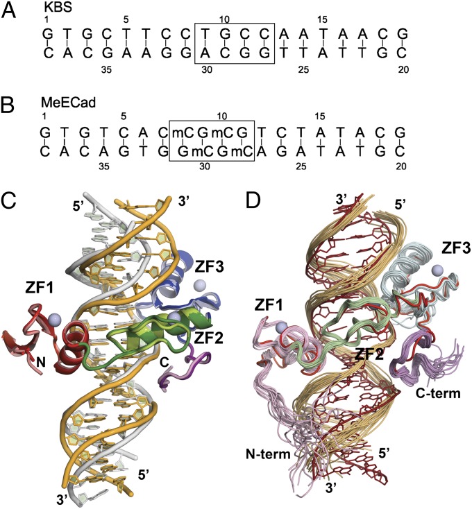
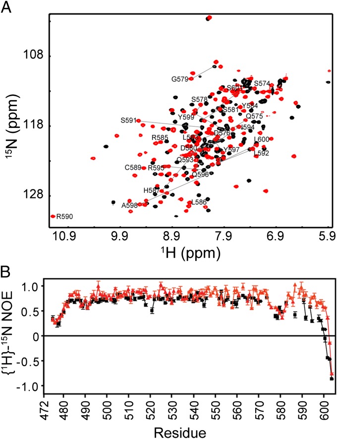
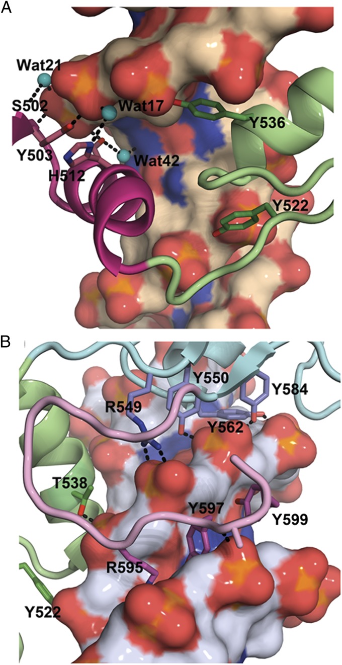
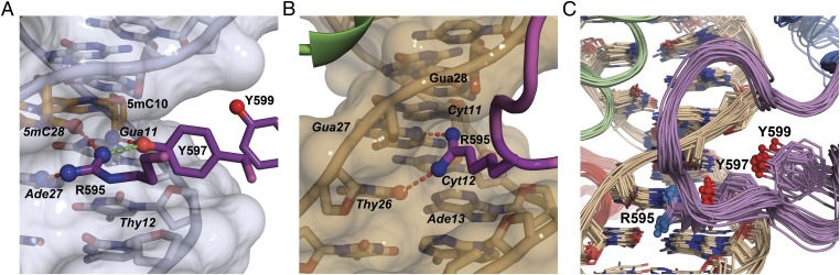
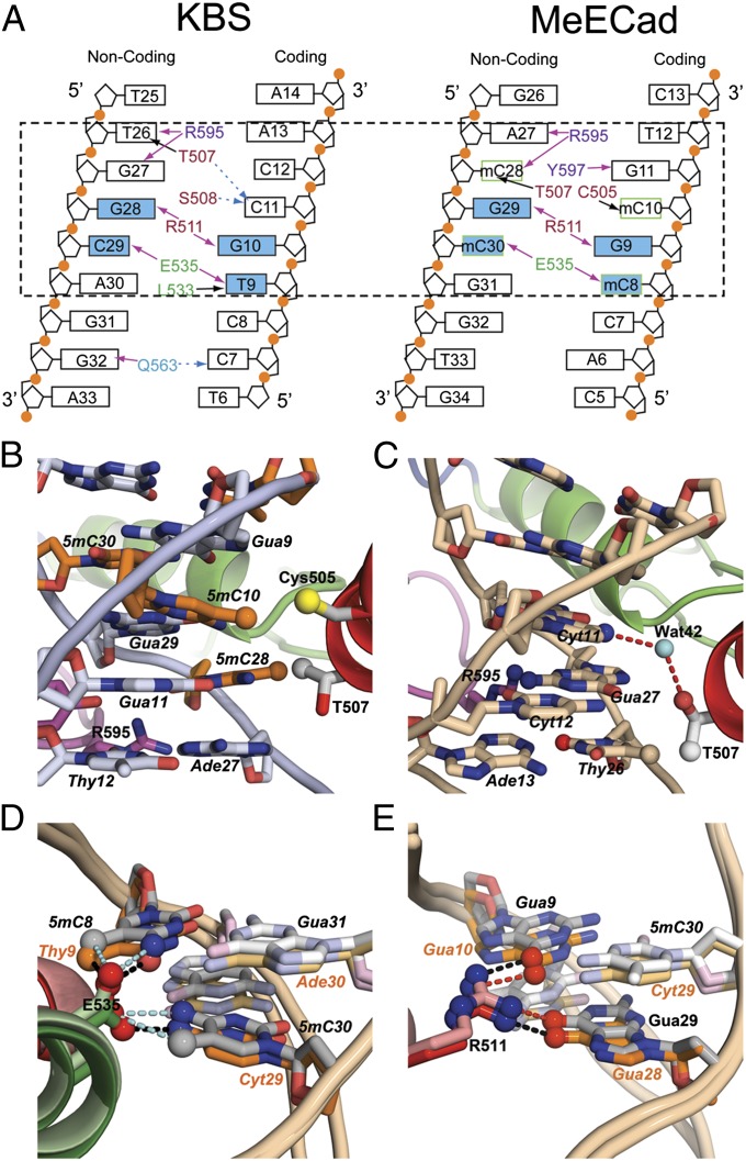
Methylation of CpG dinucleotides in DNA is a common epigenetic modification in eukaryotes that plays a central role in maintenance of genome stability, gene silencing, genomic imprinting, development, and disease. Kaiso, a bifunctional Cys(2)His(2) zinc finger protein implicated in tumor-cell proliferation, binds to both methylated CpG (mCpG) sites and a specific nonmethylated DNA motif (TCCTGCNA) and represses transcription by recruiting chromatin remodeling corepression machinery to target genes. Here we report structures of the Kaiso zinc finger DNA-binding domain in complex with its nonmethylated, sequence-specific DNA target (KBS) and with a symmetrically methylated DNA sequence derived from the promoter region of E-cadherin. Recognition of specific bases in the major groove of the core KBS and mCpG sites is accomplished through both classical and methyl CH···O hydrogen-bonding interactions with residues in the first two zinc fingers, whereas residues in the C-terminal extension following the third zinc finger bind in the opposing minor groove and are required for high-affinity binding. The C-terminal region is disordered in the free protein and adopts an ordered structure upon binding to DNA. The structures of these Kaiso complexes provide insights into the mechanism by which a zinc finger protein can recognize mCpG sites as well as a specific, nonmethylated regulatory DNA sequence.


Conflict of interest statement

The authors declare no conflict of interest.

Figures

Fig. 1.
Fig. 1.
(A) Sequence of oligonucleotide containing the KBS motif (TCCTGCNA). The box shows the core sequence equivalent to the methylated DNA site shown in B. (B) Sequence of the MeECad oligonucleotide containing two symmetrically methylated CpG sites (boxed). (C) Overlay of the Kaiso:KBS (dark colors, yellow DNA) and Kaiso:MeECad (light colors, gray DNA) X-ray crystal structures, superimposed on the protein backbone. The three Cys2His2 zinc finger DNA-binding domains are colored red (ZF1), green (ZF2), and blue (ZF3), and the zinc atoms are shown as gray spheres. The C-terminal extension is colored purple. In the crystal structure of the KBS complex, the protein backbone is ordered between residues 482–597, whereas the MeECad complex is ordered between residues 481–600. (D) Superposition of the crystal structure (red protein and DNA) and the 20 lowest-energy structures in the NMR ensemble of the Kaiso:KBS complex. The structures are superimposed on their protein backbones. The zinc finger domains in the NMR structures are colored pink (N-terminal extension and ZF1), pale green (ZF2), pale blue (ZF3), and magenta (C-terminal extension). This figure was prepared using PYMol.
Fig. 2.
Fig. 2.
(A) 1H-15N HSQC spectra of free (black) and KBS-bound (red) Kaiso. Selected cross peaks are labeled to indicate the large changes in chemical shift that occur as the C-terminal extension becomes structured upon binding to DNA. (B) {1H}-15N heteronuclear NOEs for free Kaiso (black squares) and Kaiso in complex with KBS (red triangles). The small values of the heteronuclear NOE and the nearly random coil chemical shifts for residues 575–604 in the free protein indicate that this region is disordered in the absence of DNA.
Fig. 3.
Fig. 3.
(A) DNA backbone contacts between ZF1 (magenta) and ZF2 (green) and the coding strand of the KBS complex. Cyan spheres indicate water molecules that mediate hydrogen bonding to the phosphate backbone. (B) Interactions of ZF2 (green) and ZF3 (blue) with the backbone of the noncoding strand in the MeECad complex. ZF3 is anchored to the DNA backbone by a network of hydrogen bonds from a cluster of tyrosines (Tyr-550, Tyr-562, and Tyr-584) and from the Arg-549 guanidinium group. The side chains of Arg-595, Tyr-597, and Tyr-599 in the C-terminal extension (pink) project deeply into the narrowed minor groove. This figure was prepared using PyMol.
Fig. 4.
Fig. 4.
Minor groove interactions. (A) View of minor groove protein:DNA contacts for the MeECad complex, showing hydrogen bonding (dashed red lines) between the Arg-595 and Tyr-597 side chains and the bases of A27, 5mC28 (orange), and G11. A buttressing hydrogen bond between Tyr-597 and Arg-595 is shown in green. The protein backbone is cut away for clarity. (B) View of minor groove contacts in the KBS complex showing hydrogen bonding of the Arg-595 side chain to T26 and G27. (C) DNA contacts made by C-terminal extension [purple backbone with nitrogen (red) and oxygen (blue) molecules] in NMR structures of the Kaiso:KBS complex, showing the insertion of Arg-595, Tyr-597, and Tyr-599 side chains into the minor groove (DNA in beige with nitrogen in blue and oxygen in red). This figure was prepared using PyMol.
Fig. 5.
Fig. 5.
Kaiso:KBS and Kaiso:MeECad base-specific interactions. (A) Summary of major groove interactions. Recognition of MeECad is mediated by major groove interactions with only the four 5mC:G base pairs; additional minor groove hydrogen bonds extend the binding site to A27. The contact surface for KBS is larger because of the bending of the DNA around the core recognition sequence. The dashed box highlights the core contact site in the two DNA sequences. Residues are colored red, green, blue, and purple to denote their location in ZF1, ZF2, ZF3, and the C-terminal extension, respectively. Residues in the C-terminal extension (purple) make minor groove contacts. Black arrows denote hydrophobic interactions, pink arrows indicated classical hydrogen bonds, and dashed blue arrows indicate water-mediated hydrogen bonds. A complete summary of all DNA base and backbone contacts is shown in Fig. S2 and Table S3. (B) Cys-505 and Thr-507 form a hydrophobic pocket that accommodates the methyl groups (orange spheres) of 5mC10 and 5mC28. The 5-methylcytosines are highlighted in orange. (C) Base-specific interactions of Thr-507 in the KBS complex. The red lines show water-mediated hydrogen bonds to the N4-amino of C11. The water is shown as a cyan sphere. (D) Hydrogen-bonding interactions involving Glu-535 in the core mCpG- (gray) and KBS- (orange) binding sites. (E) Hydrogen-bonding interactions involving Arg-511 in the core mCpG- (gray) and KBS- (orange) binding sites. The Kaiso color scheme is as in Fig. 1C. This figure was prepared using PyMol.

References

    1. Miranda TB, Jones PA. DNA methylation: The nuts and bolts of repression. J Cell Physiol. 2007;213:384–390. - PubMed
    1. Robertson KD. DNA methylation and human disease. Nat Rev Genet. 2005;6:597–610. - PubMed
    1. Clouaire T, Stancheva I. Methyl-CpG binding proteins: Specialized transcriptional repressors or structural components of chromatin? Cell Mol Life Sci. 2008;65:1509–1522. - PMC - PubMed
    1. Bostick M, et al. UHRF1 plays a role in maintaining DNA methylation in mammalian cells. Science. 2007;317:1760–1764. - PubMed
    1. Klose RJ, Bird AP. Genomic DNA methylation: The mark and its mediators. Trends Biochem Sci. 2006;31:89–97. - PubMed

Publication types

LinkOut - more resources