. 2012 Sep 25;109(39):E2625-34.
doi: 10.1073/pnas.1213813109. Epub 2012 Sep 4.

Fusion activation by a headless parainfluenza virus 5 hemagglutinin-neuraminidase stalk suggests a modular mechanism for triggering

Affiliations

Fusion activation by a headless parainfluenza virus 5 hemagglutinin-neuraminidase stalk suggests a modular mechanism for triggering

Sayantan Bose et al. Proc Natl Acad Sci U S A. .

Abstract

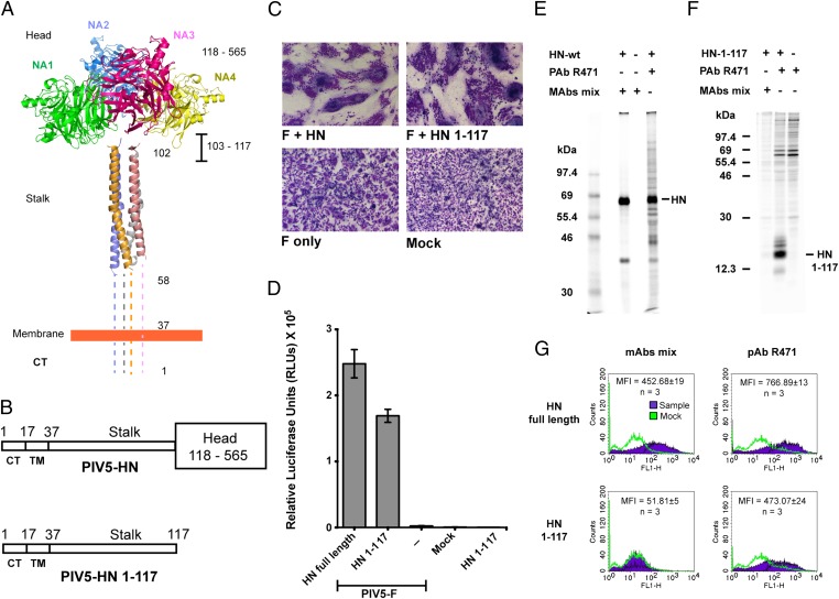
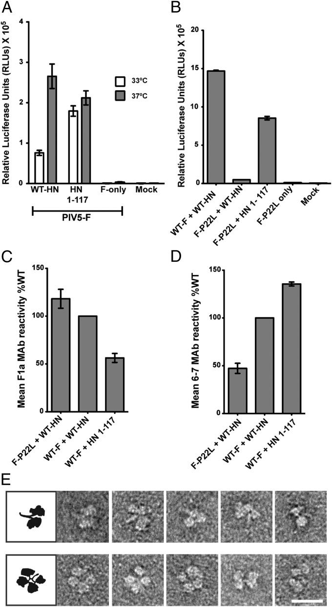
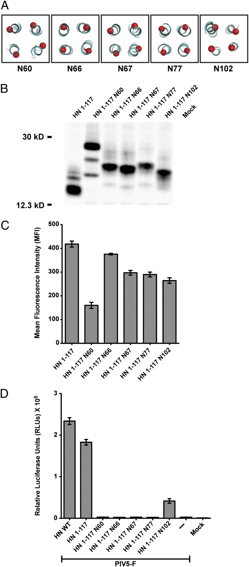
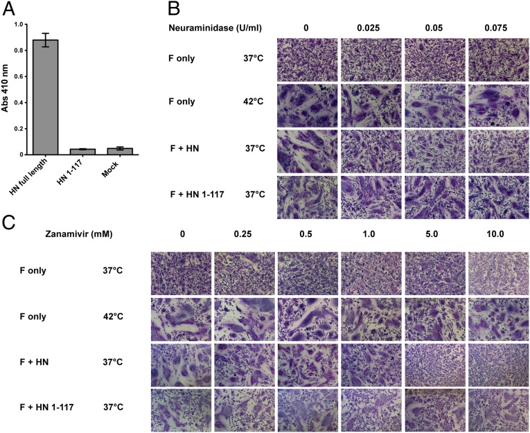
The Paramyxoviridae family of enveloped viruses enters cells through the concerted action of two viral glycoproteins. The receptor-binding protein, hemagglutinin-neuraminidase (HN), H, or G, binds its cellular receptor and activates the fusion protein, F, which, through an extensive refolding event, brings viral and cellular membranes together, mediating virus-cell fusion. However, the underlying mechanism of F activation on receptor engagement remains unclear. Current hypotheses propose conformational changes in HN, H, or G propagating from the receptor-binding site in the HN, H, or G globular head to the F-interacting stalk region. We provide evidence that the receptor-binding globular head domain of the paramyxovirus parainfluenza virus 5 HN protein is entirely dispensable for F activation. Considering together the crystal structures of HN from different paramyxoviruses, varying energy requirements for fusion activation, F activation involving the parainfluenza virus 5 HN stalk domain, and properties of a chimeric paramyxovirus HN protein, we propose a simple model for the activation of paramyxovirus fusion.


Conflict of interest statement

The authors declare no conflict of interest.

Figures

Fig. 1.
Fig. 1.
The PIV5 HN stalk domain is sufficient to activate F and mediate cell–cell fusion. (A) Structural view of the PIV5 HN full-length protein based on X-ray crystal structures of the tetrameric PIV5 HN globular head domains (16) and the 4HB of the PIV5 HN stalk domain (21). Regions for which there is no known structure (amino acids 1–58 and 103–117) are represented by dotted lines. CT, cytoplasmic tail; NA1–NA4, globular neuraminidase head domains. (B) Schematic representation of the PIV5 HN full-length WT protein (amino acids 1–565) (Upper) and the PIV5 HN stalk protein (amino acids 1–117) lacking the globular head domain (amino acids 118–565) (Lower). CT, cytoplasmic tail; TM, transmembrane domain. (C) Representative micrographs of syncytia showing cell–cell fusion in BHK-21 cells transfected with F and WT HN or HN 1–117 stalk 18 h posttransfection. (D) Luciferase reporter assay of cell–cell fusion. (E and F) Radioimmunoprecipitation of [35S]-TranS–labeled transfected 293T cell lysates, using PIV5 HN pAb R471 or a mixture of mAbs to the PIV5 HN globular head. (E) WT HN protein analyzed on a 10% (wt/vol) SDS/PAGE gel. (F) HN 1–117 stalk protein analyzed on a 15% (wt/vol) SDS/PAGE gel. Numbers to the left of each panel are molecular masses in kDa. (G) Representative graphs showing the detection of proteins at the cell surface of 293T cells transfected with WT HN or HN 1–117 stalk using flow cytometry. Surface proteins were detected using a mixture of HN mAbs or HN pAb R471. Average mean fluorescence intensities (MFI) across three independent experiments are shown.
Fig. 2.
Fig. 2.
Absence of the PIV5 HN globular head domain energetically favors fusion activation. (A) Fusion activation at 33 °C (white bars) and 37 °C (gray bars) by the PIV5 HN WT and HN 1–117 stalk protein. Fusion was measured by a luciferase assay. (B) Luciferase reporter assay showing relative activation of a hypofusogenic mutant of PIV5 F (F-P22L) by WT PIV5 HN protein in comparison with activation by the PIV5 HN 1–117 stalk protein. (C and D) Detection of pre- and postfusion PIV5 F and PIV5 F P22L proteins on the surface of cells cotransfected with WT PIV5 HN or the PIV5 HN 1–117 stalk, using flow cytometry. The amounts of surface antigen detected by an F-prefusion conformation–specific mAb, F1a, (C) and an F-postfusion conformation–specific mAb, 6-7, (D) are shown as a percentage MFI of the WT F + WT HN sample (n = 3). (E) Representative electron micrographs of purified PIV5 HN ectodomain showing different conformations of the dimer-of-dimer globular heads with respect to the stalk domain. (Upper) Conformations of HN in which the stalk is exposed (four-heads-up–like arrangements). (Lower) Examples of HN with the heads juxtaposed with the stalk domain (four-heads-down–like arrangements). (Scale bar, 20 nm.)
Fig. 3.
Fig. 3.
N-linked carbohydrate chains in the putative F-interacting region of PIV5 HN 1–117 block fusion activation. (A) Schematic representation of positions of residues on the PIV5 HN 1–117 stalk to which sites for the addition of N-linked carbohydrate chains were added by mutagenesis. The mutants are named according to the residue carrying the N-glycosylation. (B) Migration pattern of N-glycosylation mutants in the PIV5 HN 1–117 stalk on a 15% (wt/vol) SDS PAGE gel. Polypeptides were immunoprecipitated from radiolabeled lysates of transfected 293T cells using PIV5 HN pAb R471. Numbers on the left are molecular masses in kDa. (C) Cell-surface expression of PIV5 HN 1–117 N-glycosylation mutants determined using HN pAb R471 as measured by flow cytometry (n = 3). (D) Fusion activation of PIV5 F by the HN 1–117 stalk and its N-glycosylation mutants. Fusion was quantified using a luciferase reporter assay as described in Experimental Procedures.
Fig. 4.
Fig. 4.
F activation mediated by the PIV5 HN 1–117 stalk does not depend on engagement of the sialic acid receptor. (A) Receptor-binding activity of the PIV5 HN 1–117 stalk. Chicken RBCs bound onto surfaces of transfected 293T cells were quantified by lysing the bound RBCs after extensive PBS washes and measuring absorbance of chicken hemoglobin at 410 nm. (B and C) BHK-21 cells transfected with PIV5 F only or cotransfected with PIV5 F and PIV5 HN or PIV5 HN 1–117 stalk were incubated with C. perfringens neuraminidase or zanamivir at 37 °C or 42 °C. Cells were fixed, stained, and imaged 18 h posttransfection. (B and C) Fusion activation in the presence of 0 U/mL, 0.025 U/mL, 0.05 U/mL, and 0.075 U/mL C. perfringens neuraminidase to remove most sialic acid linkages (B) and with the addition of 0 mM, 0.25 mM, 0.5 mM, 1 mM, 5 mM, and 10 mM zanamivir to inhibit PIV5 HN catalytic sites specifically (C).
Fig. 5.
Fig. 5.
Chimeras and NDV HN stalk activation. (A) Schematic representation of the chimeric protein PIV5-NDV-HN, consisting of the PIV5 HN stalk (amino acids 1–117) followed by the NDV HN head (amino acids 124–571). (B) Surface expression of PIV5-NDV-HN as measured by flow cytometry using a mixture of NDV HN 4a and 2b mAbs and expressed as a percentage of WT NDV HN expression. (C) Receptor-binding activity of PIV5-NDV-HN compared with PIV5 HN and NDV HN and expressed as a percentage of WT NDV HN bound to chicken RBCs at 4 °C. (D) Neuraminidase activity of the PIV5-NDV-HN chimera compared with PIV5 HN and NDV HN proteins, as a measure of fluorescent product formation, expressed as a percentage of WT NDV HN. (E) Fusion promotion of the PIV5-NDV-HN chimera as measured by luciferase reporter assays at 33 °C and 37 °C.
Fig. 6.
Fig. 6.
Structurally conserved loops in the paramyxovirus HN globular head domains have important regulatory roles in fusion and receptor binding. (A) Bottom view of the PIV5 HN four-heads-up form showing the positions of N121 (green), N125 (green), and P233 (red) residues on all four monomers. The dimer-of-dimer interface is indicated by a black box. (B) Side view of the four-heads-down form as a structural alignment of PIV5 HN globular heads (aquamarine) with the NDV HN structure (orange) showing the head/stalk interface (black boxes). (C) Magnified top view of B. Corresponding PIV5 HN residues (blue) and NDV HN residues (red) are highlighted in the head–stalk interface. (D and E) Expression of PIV5 HN, N121A, and N125A (D) and P233L mutant polypeptides (E) immunoprecipitated from radiolabeled cell lysates with PIV5 HN pAb R471. (F) Surface expression of the PIV5 HN globular head mutants expressed as a percentage of WT PIV5 HN surface expression using PIV5 HN pAb R471 and flow cytometry. (G) Receptor-binding activity of mutant proteins expressed as a percentage of WT PIV5 HN bound to chicken RBCs at 4 °C. (H) Luciferase reporter assay for fusion showing fusion promotion by the N121A, N125A, and P233L mutants cotransfected with PIV5-F, expressed as a percentage of WT PIV5-F and WT PIV5 HN fusion.
Fig. 7.
Fig. 7.
Schematic model of paramyxovirus F activation based on putative structural rearrangements in the receptor-binding protein. (A) The receptor-binding protein in the four-heads-down position. The formation of the contacts in the head–stalk interface prevents physical interaction of F with the stalk region of the receptor-binding protein. (B) Creation of the contacts in the dimer-of-dimer interface of the receptor-binding protein in the four-heads-up position moves the heads upwards, exposing the stalk. This exposure allows F to interact with the HN stalk, causing F triggering. Black balls indicate the sialic acid binding site residues in the four neuraminidase head domains (colored red, green, blue, and yellow) of paramyxovirus HN. Prefusion F is represented by the PIV5 prefusion F structure (purple) (6). Dotted lines represent regions of paramyxovirus F and HN proteins for which no structural data are available. CT, cytoplasmic tail.
Fig. P1.
Fig. P1.
Glycoproteins F and HN mediate paramyxovirus entry into target cells. The model of paramyxovirus fusion proposed here suggests that the receptor-binding protein (represented by HN crystal structures) remains in the “four-heads-down” form (Left) when not bound to the sialic acid receptor. In this conformation, F cannot interact with HN. On binding receptor (Right) via the active sites (black balls), the arrangement of the globular heads (red, blue, green, and yellow) changes, exposing the stalk 4HB. The stalk contains the F-activating region, which now can interact with F, causing F to merge the viral and cellular membranes. Dotted lines represent regions of the F and HN proteins for which atomic structures have not been determined for any paramyxovirus.

References

    1. Heminway BR, Yu Y, Galinski MS. Paramyxovirus mediated cell fusion requires co-expression of both the fusion and hemagglutinin-neuraminidase glycoproteins. Virus Res. 1994;31:1–16. - PubMed
    1. Horvath CM, Paterson RG, Shaughnessy MA, Wood R, Lamb RA. Biological activity of paramyxovirus fusion proteins: Factors influencing formation of syncytia. J Virol. 1992;66:4564–4569. - PMC - PubMed
    1. Hu XL, Ray R, Compans RW. Functional interactions between the fusion protein and hemagglutinin-neuraminidase of human parainfluenza viruses. J Virol. 1992;66:1528–1534. - PMC - PubMed
    1. Morrison T, McQuain C, McGinnes L. Complementation between avirulent Newcastle disease virus and a fusion protein gene expressed from a retrovirus vector: Requirements for membrane fusion. J Virol. 1991;65:813–822. - PMC - PubMed
    1. Yao Q, Hu X, Compans RW. Association of the parainfluenza virus fusion and hemagglutinin-neuraminidase glycoproteins on cell surfaces. J Virol. 1997;71:650–656. - PMC - PubMed

Publication types

LinkOut - more resources