. 2012 Sep 18;109(38):15336-41.
doi: 10.1073/pnas.1201701109. Epub 2012 Sep 4.

In vivo reprogramming of Sox9+ cells in the liver to insulin-secreting ducts

Affiliations

In vivo reprogramming of Sox9+ cells in the liver to insulin-secreting ducts

Anannya Banga et al. Proc Natl Acad Sci U S A. .

Abstract

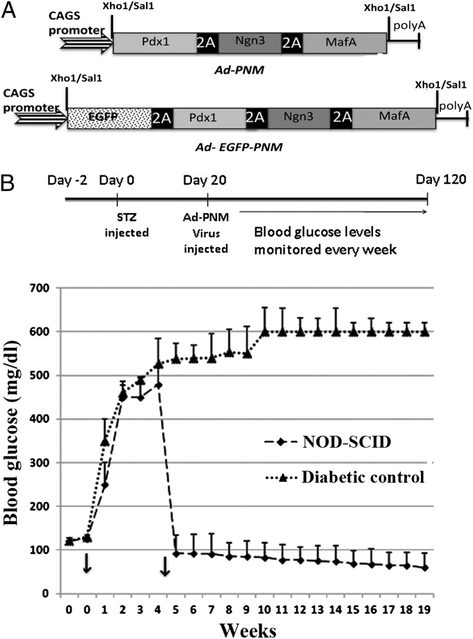
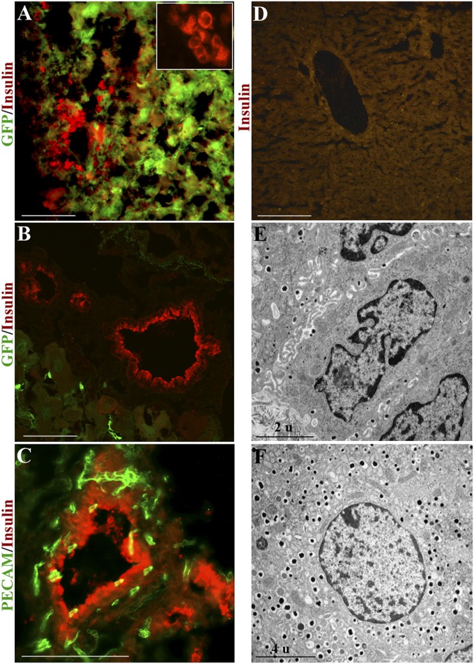
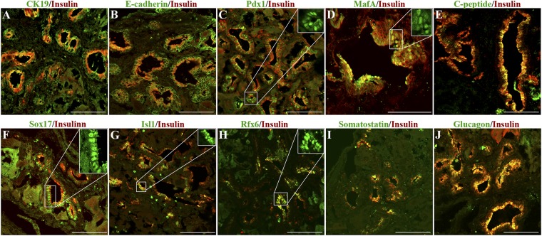
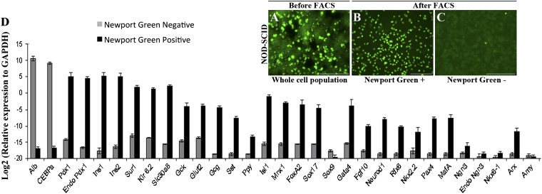
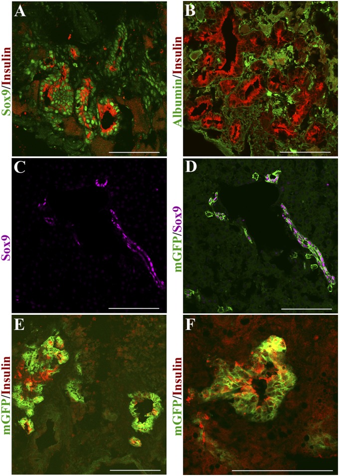
In embryonic development, the pancreas and liver share developmental history up to the stage of bud formation. Therefore, we postulated that direct reprogramming of liver to pancreatic cells can occur when suitable transcription factors are overexpressed. Using a polycistronic vector we misexpress Pdx1, Ngn3, and MafA in the livers of NOD-SCID mice rendered diabetic by treatment with streptozotocin (STZ). The diabetes is relieved long term. Many ectopic duct-like structures appear that express a variety of β-cell markers, including dense core granules visible by electron microscopy (EM). Use of a vector also expressing GFP shows that the ducts persist long after the viral gene expression has ceased, indicating that this is a true irreversible cell reprogramming event. We have recovered the insulin(+) cells by cell sorting and shown that they display glucose-sensitive insulin secretion. The early formed insulin(+) cells can be seen to coexpress SOX9 and are also labeled in mice lineage labeled for Sox9 expression. SOX9(+) cells are normally found associated with small bile ducts in the periportal region, indicating that the duct-like structures arise from this source. This work confirms that developmentally related cells can be reprogrammed by suitable transcription factors and also suggests a unique therapy for diabetes.


Conflict of interest statement

The authors declare no conflict of interest.

Figures

Fig. 1.
Fig. 1.
Persistent normoglycemic levels are maintained following Ad-PNM delivery. (A) Diagrams of the vectors. (B) Long-term maintenance of normal blood glucose levels compared with diabetic controls (n = 5 per curve). Arrows show the time of STZ injection (week 0) and the time of Ad-PNM delivery (between weeks 4 and 5). The attenuation in blood glucose level is observed a few days after Ad-PNM delivery.
Fig. 2.
Fig. 2.
Ectopic insulin+ ducts in the liver. (A) Insulin+ cells appear as small clusters and are mostly EGFP+ 1 wk after Ad-EGFP-PNM. Inset shows the presence of insulin in the cytoplasm (red color only). (B) Absence of EGFP from the ductal structures after 4 wk. (C) Platelet endothelial cell adhesion molecule (PECAM) staining of the insulin+ ducts shows the presence within the duct walls of small blood vessels. (D) Absence of insulin staining in control liver of diabetic mouse. (E) TEM shows the presence of dense core granules in the cytoplasm of duct cells. (F) Dense core granules in β cell of a normal mouse islet. In this and other figures, pictures represent at least three independent experiments. (Scale bars, 100 μm.)
Fig. 3.
Fig. 3.
Ectopic ducts express markers for β cells. (A) CK-19. (B) E-cadherin. (C) PDX1. (D) MAFA. (E) C-peptide. (F) SOX17. (G) ISL1. (H) RFX6. (I) Somatostatin. (J) Glucagon. (Scale bars, 100 μm.) Cases shown in F and I are from mice dosed with Ad-EGFP-PNM, so there are some patches of cytoplasmic GFP visible in addition to the antibody fluorescence. All cases are shown 12 wk after Ad-PNM. Insets show the nuclear localization of transcription factors.
Fig. 4.
Fig. 4.
Isolation of insulin+ cells by Newport Green staining and FACS. Newport Green-stained cells before sorting (A) and after sorting (B and C). (D) qRT-PCR analysis of sorted cells. Ct values were normalized to Gapdh gene expression within the same cDNA sample. Results are presented as fold increase based on Gapdh gene expression and the values on the y axis represent log2 multiples of the Gapdh value, which is represented by 0. The data are means of three independent experiments on livers collected 12 wk after Ad-PNM administration. (Scale bars, 100 μm.)
Fig. 5.
Fig. 5.
Insulin+ cells secrete insulin in response to glucose. (A) Plasma glucose. (B) Serum insulin levels following i.p. injection of glucose (n = 3). (C) Measurement of glucose-stimulated insulin release from mouse islets. (D) Measurement of glucose-stimulated insulin release from the Newport Green+ and Newport Green cells, sorted from liver of Ad-PNM–treated mice. Results are mean ± SE (n = 3).
Fig. 6.
Fig. 6.
Origin of ectopic ducts from SOX9+ cells. (A) Insulin+ ducts costain for SOX9. (B) They do not costain for albumin. Specimens 10 d after Ad-PNM administration. (C and D) Control Sox9-CreERT2; mTmG mouse labeled by tamoxifen treatment at E15.5 shows GFP in cells of the small bile ducts. SOX9 is shown in lilac, representing far red; GFP is green. (E and F) Insulin+ structures induced by Adeno-PNM arise from GFP-labeled cells. Insulin is shown in red, representing far red. Two mice were analyzed. (Scale bars, 100 μm.)

References

    1. Ieda M, et al. Direct reprogramming of fibroblasts into functional cardiomyocytes by defined factors. Cell. 2010;142:375–386. - PMC - PubMed
    1. Vierbuchen T, et al. Direct conversion of fibroblasts to functional neurons by defined factors. Nature. 2010;463:1035–1041. - PMC - PubMed
    1. Huang P, et al. Induction of functional hepatocyte-like cells from mouse fibroblasts by defined factors. Nature. 2011;475:386–389. - PubMed
    1. Sekiya S, Suzuki A. Direct conversion of mouse fibroblasts to hepatocyte-like cells by defined factors. Nature. 2011;475:390–393. - PubMed
    1. Takahashi K, Yamanaka S. Induction of pluripotent stem cells from mouse embryonic and adult fibroblast cultures by defined factors. Cell. 2006;126:663–676. - PubMed

Publication types