. 2012 Sep 18;109(38):15455-60.
doi: 10.1073/pnas.1206061109. Epub 2012 Sep 4.

Skeletal parasympathetic innervation communicates central IL-1 signals regulating bone mass accrual

Affiliations

Skeletal parasympathetic innervation communicates central IL-1 signals regulating bone mass accrual

Alon Bajayo et al. Proc Natl Acad Sci U S A. .

Abstract

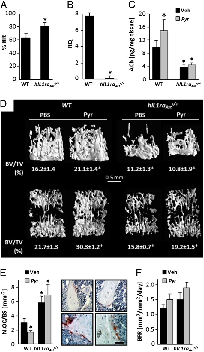
Bone mass accrual is a major determinant of skeletal mass, governed by bone remodeling, which consists of bone resorption by osteoclasts and bone formation by osteoblasts. Bone mass accrual is inhibited by sympathetic signaling centrally regulated through activation of receptors for serotonin, leptin, and ACh. However, skeletal activity of the parasympathetic nervous system (PSNS) has not been reported at the bone level. Here we report skeletal immune-positive fibers for the PSNS marker vesicular ACh transporter (VAChT). Pseudorabies virus inoculated into the distal femoral metaphysis is identifiable in the sacral intermediolateral cell column and central autonomic nucleus, demonstrating PSNS femoral innervation originating in the spinal cord. The PSNS neurotransmitter ACh targets nicotinic (nAChRs), but not muscarinic receptors in bone cells, affecting mainly osteoclasts. nAChR agonists up-regulate osteoclast apoptosis and restrain bone resorption. Mice deficient of the α(2)nAChR subunit have increased bone resorption and low bone mass. Silencing of the IL-1 receptor signaling in the central nervous system by brain-specific overexpression of the human IL-1 receptor antagonist (hIL1ra(Ast)(+/+) mice) leads to very low skeletal VAChT expression and ACh levels. These mice also exhibit increased bone resorption and low bone mass. In WT but not in hIL1ra(Ast)(+/+) mice, the cholinergic ACh esterase inhibitor pyridostigmine increases ACh levels and bone mass apparently by inhibiting bone resorption. Taken together, these results identify a previously unexplored key central IL-1-parasympathetic-bone axis that antagonizes the skeletal sympathetic tone, thus potently favoring bone mass accrual.


Conflict of interest statement

The authors declare no conflict of interest.

Figures

Fig. 1.
Fig. 1.
Parasympathetic innervation in bone. (A and B) VAChT-positive nerve fibers (arrows) in distal femoral metaphysis. BT, bone trabeculae. (A) Low- and (B) high-power view. (C) Propagation of immunoreactive pseudorabies virus from distal femoral metaphysis to sacral spinal cord segments of intermediolateral column (arrow) and central autonomic nucleus (arrowhead). (Insets) Higher magnification of boxed zones. (Scale bars, 50 μm.)
Fig. 2.
Fig. 2.
Expression of nicotinic receptors in bone cells. (A) RT-PCR analysis of nicotinic receptor subunits in primary cultures of newborn mouse calvarial osteoblasts (Ob) bone marrow-derived monocytes (mono) and osteoclasts (Oc). Ct, positive control of whole brain or skeletal muscle RNA extract. (B–E) Immunocytochemical staining for nicotinic receptor subunits in osteoclasts and osetoblasts. (B and D) Osteoclasts in culture; red, subunit (B) α2-subunit, (D) β2-subunit; green, actin; blue, nuclei. (C and E) in-vivo immunohistochemistry of femoral trabecular bone; arrows, osteoblasts; double arrows, osteoclasts: (C) α2-subunit, (E) β2-subunit. (Scale bars, 20 μm.)
Fig. 3.
Fig. 3.
Low bone mass phenotype in α2nAChR−/− mice. (A) Bone volume density (BV/TV) and 3D μCT images from representative animals with median BV/TV values; (B and C) Bone remodeling parameters in distal femoral metaphysic: (B) osteoclast number (scale bar, 20 μm); (C) serum C-telopeptide of type I collagen (CTX). Data are mean ± SE obtained in six to nine mice per condition. *t test, P < 0.05 vs. WT controls.
Fig. 4.
Fig. 4.
nAChR activation enhances osteoclast apoptosis and inhibits mineralized matrix resorption. (A–J) Effect of CCh in bone marrow derived osteoclastogenic cultures. (A and B) Intact osteoclasts derived from WT (black bars) and α2nAChR−/− (gray bars) mice. (C) WT apoptotic osteoclasts. (D) In situ images of intact and apoptotic osteoclasts in respective control and CCh-treated cultures. (Scale bar, 500 μm.) (E) Apoptotic osteoclasts treated with CCh and mecamylamine. (F–I) Real-time RT-PCR analysis of osteoclastic genes in intact osteoclasts. (F) Trap5b; (G) Bcl2L1; (H) Bid; (I) Cathepsin K; (J) caspase 3 activity. pNA, p-nitroaniline. (K–M) Pit formation in osteoclastogenic cultures of intact cells grown on dentin slices. (K) Pit number, (L) average pit size, (M) toluidine blue staining of vehicle and CCh treated dentin slices. (Scale bar, 100 μm.) Data are mean ± SE obtained in five to six (A–E, K, and L) or three (F–J) culture wells per condition. *P < 0.05, vs. WT vehicle-treated controls (Veh). (A, C, E, K, and L) one-way ANOVA; (B) two-way ANOVA; (F–J) t test.
Fig. 5.
Fig. 5.
Cardiac and skeletal parasympathetic signaling is regulated by central IL-1 receptor activity. (AF) Reduced parasympathetic tone in hIL1raAst+/+ mice. (A) Percentage of pretreatment heart rate following propranolol administration; (B) VAChT mRNA levels in distal femoral metaphysis. (C–F) Cholinergic and bone remodeling parameters in vehicle and pyridostigmine-treated mice: (C) ACh levels in distal femoral metaphysis; (D) μCT representative images of distal femoral metaphyses (Upper), and L3 vertebral bodies (Lower) from animals with median volume density (BV/TV) values and actual BV/TV determination. (E and F) Analysis of bone resorption and bone formation in distal femoral metaphysis. (E) Osteoclast number (Oc.N/BS) and representative images of TRAP-stained sections from WT (Upper) and hIL1raAst+/+ (Lower) mice treated with vehicle (Left) or pyridostigmine (Right). (Scale bar, 80 μm.) (F) Bone formation rate (BFR). Data are mean ± SE obtained in 5–10 mice per condition. *P < 0.05 vs. WT vehicle treated controls. (A and B) t test; (C and F) two-way ANOVA.
Fig. 6.
Fig. 6.
Model of autonomic regulation of bone mass accrual. Skeletal sympathetic tone, which inhibits bone formation and stimulates bone resorption, is centrally regulated by leptin, serotonin, and acetylcholine (19). It is antagonized by parasympathetic activity controlled by brain IL-1 signaling and results in enhanced bone formation and restrained bone resorption.

References

    1. Rodan GA, Martin TJ. Therapeutic approaches to bone diseases. Science. 2000;289:1508–1514. - PubMed
    1. Elefteriou F, et al. Leptin regulation of bone resorption by the sympathetic nervous system and CART. Nature. 2005;434:514–520. - PubMed
    1. Katayama Y, et al. Signals from the sympathetic nervous system regulate hematopoietic stem cell egress from bone marrow. Cell. 2006;124:407–421. - PubMed
    1. Yirmiya R, et al. Depression induces bone loss through stimulation of the sympathetic nervous system. Proc Natl Acad Sci USA. 2006;103:16876–16881. - PMC - PubMed
    1. Tam J, et al. The cannabinoid CB1 receptor regulates bone formation by modulating adrenergic signaling. FASEB J. 2008;22:285–294. - PubMed

Publication types

LinkOut - more resources