

Rituximab plus Ifosfamide, Carboplatin and Etoposide for T-Cell/Histiocyte-Rich B-Cell Lymphoma Arising in Nodular Lymphocyte-Predominant Hodgkin's Lymphoma
- PMID: 22949903
- PMCID: PMC3433022
- DOI: 10.1159/000341562
Rituximab plus Ifosfamide, Carboplatin and Etoposide for T-Cell/Histiocyte-Rich B-Cell Lymphoma Arising in Nodular Lymphocyte-Predominant Hodgkin's Lymphoma
Abstract
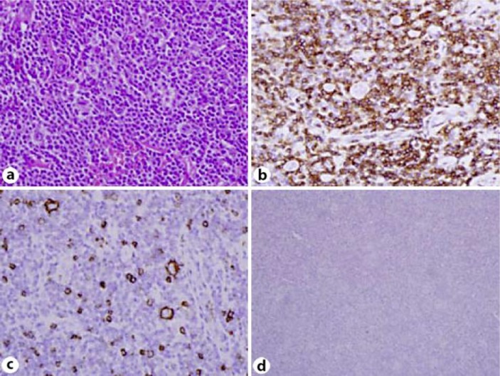
A small subset of patients with nodular lymphocyte-predominant Hodgkin's lymphoma (NLPHLs) develop a non-Hodgkin lymphoma either concurrently or subsequently, usually T-cell/histiocyte-rich B-cell lymphomas (T/HRBCL), which are subtypes of diffuse large B-cell lymphomas (DLBCL). The standard treatment of DLBCL patients is rituximab-based chemotherapy with cyclophosphamide, adriamycin, vincristine and prednisolone. However, the administration of this chemotherapy regimen to patients with DLBCL arising in NLPHL brings concern about the cardiac toxicity of anthracycline because the majority of these patients had already received anthracycline-based chemotherapy with doxorubicin, bleomycin, vinblastine and dacarbazine at the time of NLPHL. Herein, we report 2 patients with sequential transformation of NLPHL to T/HRBCL. They initially presented with limited-stage NLPHL and subsequently developed T/HRBCL after 16 and 8 months, respectively. At the time of T/HRBCL, they were treated with rituximab, ifosfamide, carboplatin and etoposide, and complete responses were obtained.
Keywords: Chemotherapy; Nodular lymphocyte-predominant Hodgkin's lymphoma; T-cell/histiocyte-rich B-cell lymphoma; Transformation.
Figures


References
-
- Poppema S. Lymphocyte-predominance Hodgkin's disease. Semin Diagn Pathol. 1992;9:257–264. - PubMed
-
- Chan WC. Cellular origin of nodular lymphocyte-predominant Hodgkin's lymphoma: immunophenotypic and molecular studies. Semin Hematol. 1999;36:242–252. - PubMed
-
- Lee AI, LaCasce AS. Nodular lymphocyte predominant Hodgkin lymphoma. Oncologist. 2009;14:739–751. - PubMed
-
- Rudiger T, Gascoyne RD, Jaffe ES, et al. Workshop on the relationship between nodular lymphocyte predominant Hodgkin's lymphoma and T cell/histiocyte-rich B cell lymphoma. Ann Oncol. 2002;13((suppl 1)):44–51. - PubMed
-
- Bennett MH, MacLennan KA, Vaughan Hudson G, Vaughan Hudson B. Non-Hodgkin's lymphoma arising in patients treated for Hodgkin's disease in the BNLI: a 20-year experience. British National Lymphoma Investigation. Ann Oncol. 1991;2((suppl 2)):83–92. - PubMed
Publication types
LinkOut - more resources
Full Text Sources
