. 2012;5(6):503-11.
Epub 2012 Jul 29.

Morphoproteomics provides support for TGF-β pathway signaling in the osteoclastogenesis and immune dysregulation of osteolytic Langerhans cell histiocytosis

Affiliations

Morphoproteomics provides support for TGF-β pathway signaling in the osteoclastogenesis and immune dysregulation of osteolytic Langerhans cell histiocytosis

Sanda Alexandrescu et al. Int J Clin Exp Pathol. 2012.

Abstract

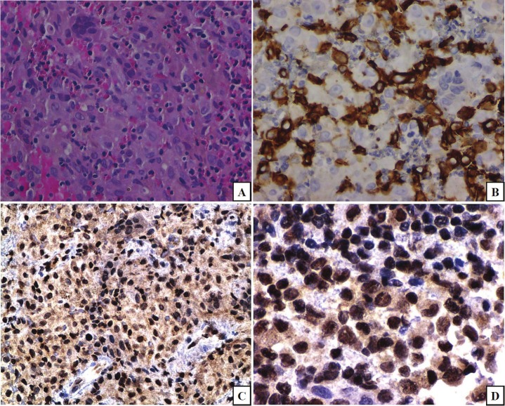
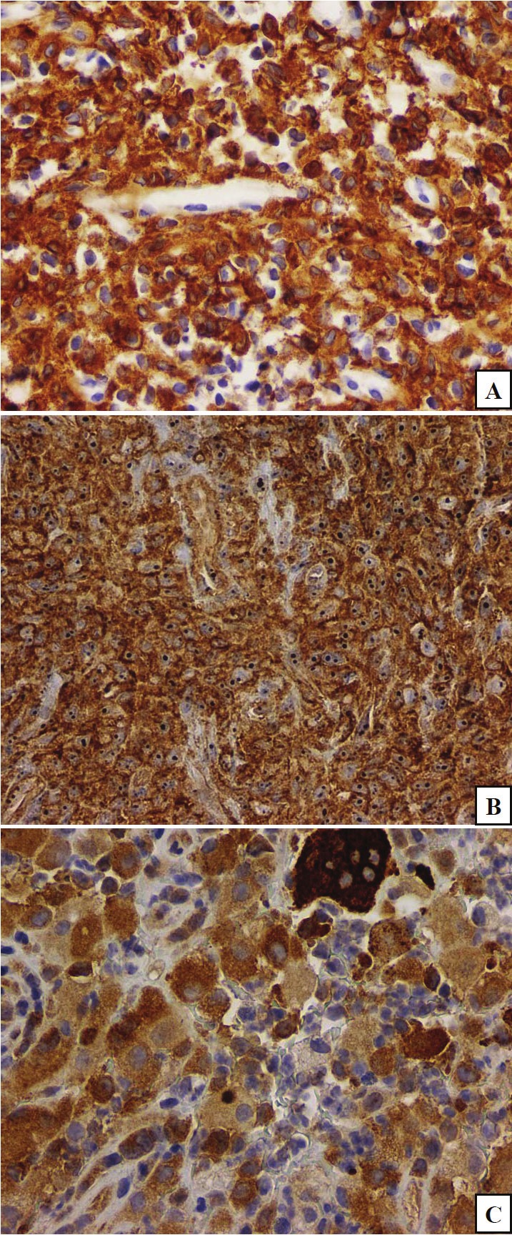
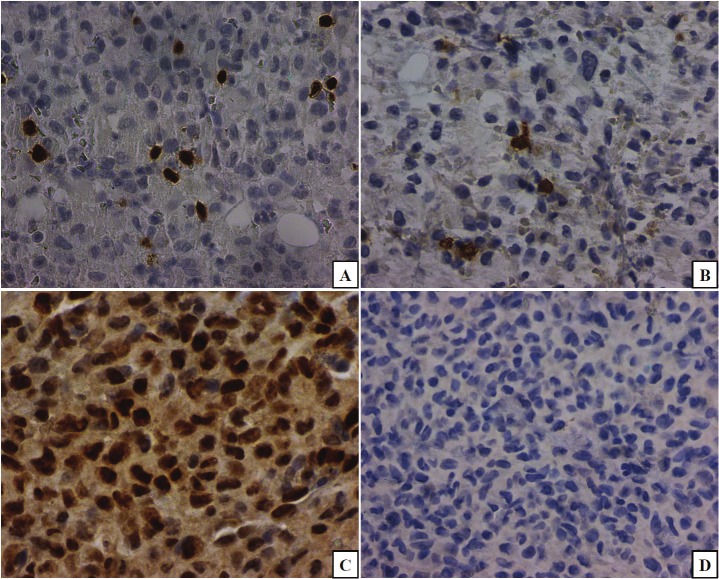
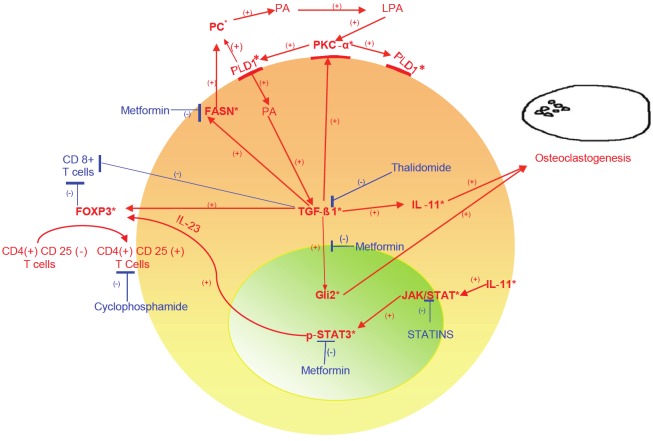
Langerhans cell histiocytosis (LCH) has a challenging and still unclear pathogenesis. A body of literature points to impaired maturation of the lesional dendritic cells, and to immune dysregulation in the form of increased FoxP3 cells. Various cytokine abnormalities such as expression of transforming growth factor (TGF)-β have been reported, as well as abnormalities in lipid content in LCH cells. Morphoproteomic techniques were applied to identify the signal transduction pathways that could influence histogenesis and immune regulation in osteolytic LCH. Five pediatric cases of osteolytic LCH were examined, using antibodies against CD1a, S100, CD68, CD8, FoxP3, phosphorylated (p)-STAT3 (Tyr705), protein kinase C (PKC)-α, phospholipase (PL)D1, fatty acid synthase (FASN), and zinc finger protein, Gli2. Positive and negative controls were performed. A FoxP3(+)/CD8(+) cell ratio was calculated by counting the FoxP3+ and CD8+ cells in 10 high power fields for each case. There is induction of sonic hedgehog (SHH) mediators consistent with TGF-β signaling pathway through Smad3-dependent activation of Gli2, findings supported by the plasmalemmal and cytoplasmic expression of PKC-α and PLD1, and nuclear expression of Gli2, in lesional cells. The FoxP3+/CD8+ cell ratio is increased, ranging from 1.7-7.94. There is moderate cytoplasmic expression of FASN in most of the Langerhans cells, a finding that supports previously published phospholipid abnormalities in LCH and is consistent with PKC-α/PLD1/TGF-β signaling. With our study, we strongly suggest that the TGF-β cell signaling pathway is a major player in the pathogenesis of LCH, leading to non-canonical induction of nuclear Gli2 expression, thereby contributing to osteoclastogenesis in LCH histiocytes. It could also cause a state of immune frustration in LCH, by inducing the transformation of CD4(+)CD25(-) cells into CD4(+)/FoxP3(+) cells. This coincides with the clinical evidence of a response to thalidomide in patients with osteolytic LCH, given its reported ability to reduce TGF-beta 1 and FoxP3 cells. Such TGF-β signaling in osteoclastogenesis and immune dysregulation, and the presence of FASN in the majority of cells, have additional therapeutic implications for osteolytic LCH.

Keywords: Langerhans cell histiocytosis; Morphoproteomics; TGF-β; histogenesis; osteoclastogenesis; signaling pathway.


Figures

Figure 1
Figure 1
Hematoxylin-eosin (H&E) shows a proliferation of histiocytic cells, some with cleaved nuclei, nuclear grooves, admixed with multinucleated giant cells, including osteoclasts, and with an eosinophilic infiltrate (frame A). The lesional cells express CD1a (frame B). This pattern of expression is consistent with a diagnosis of Langerhans cell histiocytosis. Morphoproteomic application of a probe for glioma-associated oncogene protein (Gli)2 reveals nuclear expression in a majority of lesional Langerhans cells (frames C and D;DAB [brown] chromogen) with implications for TGF-β signaling and osteoclastogenesis. (original magnifications, x400 for A, B, and D and x200 for C).
Figure 2
Figure 2
PKC-alpha (frame A) and phospholipase D1(PLD1;frame B) share a similar subcellular distribution on the plasmalemmal and in the cytoplasmic compartments of the majority of lesional cells. Fatty acid synthase (FASN;frame C) has variable intensity of cytoplasmic expression in the majority of the lesional cells. (original magnifications, x200 for A and B, x400 for C).
Figure 3
Figure 3
The calculated ratio of FoxP3+/CD8+ lymphocytes, revealed an increased number of FoxP3+ cells (frame A) compared to CD8+ cells (frame B). The highest ratio corresponded to the case with the most aggressive clinical course (case 5). Relatedly, phosphorylated (p)-signal transducer and activator of transcription (STAT)3 (Tyr 705) is variably expressed in nuclei of the lesional Langerhans cells (frame C). A negative control is depicted in frame D. (original magnifications, x400 A-C and x200, D).
Figure 4
Figure 4
Schematic depicting a central role for transforming growth factor (TGF)-β1 signaling in osteoclastogenesis and immune dysregulation of osteolytic Langerhans cell histiocytosis (LCH). Specifically, TGF-β1 expression [20,25] and signaling in osteolytic LCH correlates with and could lead to:1) interleukin (IL)-11 [20,22,28,32] and in turn, both osteoclastogenesis [21,33] and the activation of signal transducer and activator of transcription (STAT)3 pathway [34], evidenced by nuclear expression of phosphorylated (p)-STAT3 (Tyr 705) and subsequent expansion of the FoxP3 + population via IL-23 [36,37,41]; 2) nuclear expression of glioma-associated oncogene protein (Gli)2 [26,30] leading to the formation of parathyroid hormone-related protein (PTHrP) and then to activation of RANKL with osteoclastogenesis [26,31,46,47]; 3)fatty acid synthase (FASN) expression with lipogenesis to include phosphocholine (choline-phospholipids have been described in LCH [44]) and phosphatidlycholine [45], the substrate for phospholipase (PL)D1 with subsequent phosphatidic acid(PA) and lysophosphatidic acid (LPA) formation and in turn to complex activation of protein kinase C (PKC)-α, PLD1 and TGF-β1 [48-57]; 4) activation of PKC-α [58-60]; and 5) downregulation of CD8+ cell-associated immune surveillance[61] and with activation of T regulatory (FoxP3+) cells [35,38-40]. Targeted therapeutic possibilities in this context include metformin to : 1. Activate AMPK with inhibition of TGF-β{Smad3}-associated Gli2 and IL-11 signaling [26-31,62,63] and in turn, inhibition of PTHrP and RANKL signaling [46,64,65] with reduced osteoclastogenesis; 2. Downregulate FASN [66] leading to reduced substrate for the PLD1/PKC-α/TGF-β signaling axis; and 3. Inhibit STAT3 activation at tyrosine 705 [67]. Moderators of TGF-β pathway signaling related to immune dysregulation include: thalidomide, which has shown efficacy in blocking the TGF-β pathway [16-19] and clinically, in treating osteolytic LCH [15]; and metronomic cyclophosphamide , which targets T regulatory cells [68]. *Indicates protein analytes identified in osteolytic LCH.

Similar articles

Cited by

References

    1. Degar BA, Rollins BJ. Langerhans cell histiocytosis: malignancy or inflammatory disorder doing a great job of imitating one? Disease Models and Mechanisms. 2009;2:436–439. - PMC - PubMed
    1. Geissmann F, Lepelletier Y, Fraitag S, Valladeau J, Bodemer C, Debré M, Leborgne M, Saeland S, Brousse N. Differentiation of Langerhans cells in Langerhans cell histiocytosis. Blood. 2001;97:1241–1248. - PubMed
    1. Aricò M, Nichols K, Whitlock JA, Arceci R, Haupt R, Mittler U, Kühne T, Lombardi A, Ishii E, Egeler RM, Danesino C. Familial clustering of Langerhans cell histiocytosis. Br J Haematol. 1999;107:883–888. - PubMed
    1. Da Costa CE, Szhuai K, van Eijk R, Hoogeboom M, Sciot R, Mertens F. No genomic aberrations in Langerhans cell histiocytosis as assessed by diverse molecular technologies. Genes Chromosomes Cancer. 2009;48:239–249. - PubMed
    1. Badalian-Very G, Vergilio JA, Degar BA, MacConaill LE, Brandner B, Calicchio ML, Kuo FC, Ligon AH, Stevenson KE, Kehoe SM, Garraway LA, Hahn WC, Meyerson M, Fleming MD, Rollins BJ. Recurrent BRAF mutations in Langerhans cell histiocytosis. Blood. 2010;116:1919–23. - PMC - PubMed

MeSH terms

LinkOut - more resources