

Hepatic angiomyolipoma: a series of six cases with emphasis on pathological-radiological correlations and unusual variants diagnosed by core needle biopsy
- PMID: 22949933
- PMCID: PMC3430108
Hepatic angiomyolipoma: a series of six cases with emphasis on pathological-radiological correlations and unusual variants diagnosed by core needle biopsy
Abstract
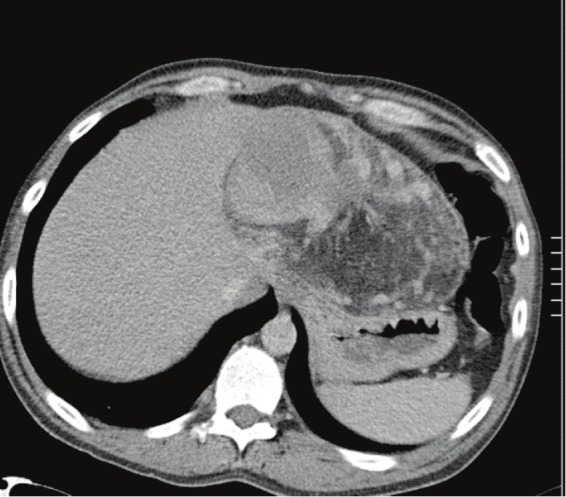
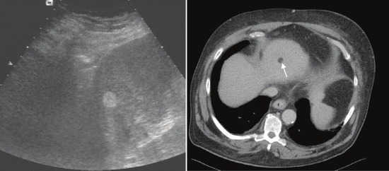
Hepatic angiomyolipoma is rare and may pose differential diagnostic difficulty, particularly if encountered in core needle biopsy. We studied 6 cases from 5 males and one female (median age, 48.6 yrs). All presented with non-specific symptoms or an incidentally discovered tumor mass. Two patients had a remote history of chemotherapy for hematological neoplasms (acute lymphoblastic leukemia and Hodgkin lymphoma respectively) and another had clear cell renal cell carcinoma and anaplastic pancreatic carcinoma diagnosed at autopsy without definable syndrome. None of the patients had evidence of the tuberous sclerosis complex or renal or other extra-renal angiomyolipoma. Three tumors were resected completely and three have been only biopsied and followed up. None of the resected cases recurred at a mean follow-up of 35 months. Histologically, tumors were classified as classical triphasic (1), lipomatous (2), epithelioid/oncocytoid (1), epithelioid trabecular (1) and myelolipoma-like (1). The adjacent liver parenchyma was normal in 3 cases, showed pigment cirrhosis in one case and mild fatty change in another case. One case had clinically diagnosed but histologically unverified cirrhosis. The initial diagnostic impression/frozen section was misleading in 5 of the cases and included vascular lesion, focal fatty change, myelolipoma, hepatocellular tumor and oncocytic neoplasm. All tumors expressed HMB45 and variably desmin. One epithelioid lesion expressed HMB45 and TFE3, but lacked desmin expression. In conclusion, hepatic angiomyolipomas are increasingly recognized as incidental findings during surveillance for cirrhosis or investigations for unrelated conditions. Awareness of their diverse morphological spectrum in liver biopsy is necessary to avoid misdiagnosis as hepatocellular carcinoma, metastatic melanoma or other malignant neoplasms.
Keywords: Angiomyolipoma; HMB45; PEComa; TFE3; liver; myelolipoma.
Figures


References
-
- Goodman ZD, Terracciano LM. Tumours and tumour-like lesions of the liver. In: Burt AD, Portmann BC, Ferrell LD, editors. MacSween’s Pathology Of The Liver. Churchill Livingstone; 2007. pp. 761–814.
-
- Lencioni R. Surveillance and early diagnosis of hepatocellular carcinoma. Dig Liver Dis. 2010 Jul;42(Suppl 3):S223–7. - PubMed
-
- Bonetti F, Pea M, Martignoni G, Doglioni C, Zamboni G, Capelli P, Rimondi P, Andrion A. Clear cell (“sugar”) tumor of the lung is a lesion strictly related to angiomyolipoma--the concept of a family of lesions characterized by the presence of the perivascular epithelioid cells (PEC) Pathology. 1994 Jul;26(3):230–6. - PubMed
-
- Ishak KG. Mesenchymal tumors of the liver. In: Okuda K, Peters RL, editors. Hepatocellular Carcinoma. New York: John Wiley & Sons; 1976.
Publication types
MeSH terms
LinkOut - more resources
Full Text Sources
Medical