Telmisartan counteracts TGF-β1 induced epithelial-to-mesenchymal transition via PPAR-γ in human proximal tubule epithelial cells
- PMID: 22949934
- PMCID: PMC3430109
Telmisartan counteracts TGF-β1 induced epithelial-to-mesenchymal transition via PPAR-γ in human proximal tubule epithelial cells
Abstract
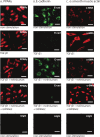
Chronic renal failure (CRF) mainly results from kidney fibrosis. Epithelial-to-mesenchymal transition (EMT) occurs in stressed tubular epithelial cells and contributes to renal fibrosis. Transforming growth factor-β1 (TGF-β1) has been shown to initiate and complete the whole EMT process. Peroxisome proliferators-activated receptor-γ (PPAR-γ) exerts anti-inflammatory, anti-fibrotic and vaculo-protective effects on different renal diseases. Telmisartan is a member of angiotensin II (Ang II) receptor blocker (ARB) family. Recent studies show that Telmisartan has a partial agonistic effect on PPAR-γ. Therefore, we tested the hypothesis that Telmisartan reverses the progression of induced EMT by TGF-β1 in cultured human renal proximal tubular epithelial (HK-2) cells. Cultured HK-2 cells were treated with TGF-β1 (3 ng/ml), a combination of TGF-β1 and Telmisartan (10-200 umol/L) and a combination of TGF-β1, Telmisartan and GW9662, a PPAR-γ antagonist for 48 hours. EMT was determined by quantitative real-time PCR analysis of E-cadherin (E-cad), Connective Tissue Growth Factor (CTGF) and PPAR-γ transcript expression and immunocytochemical analysis of E-cad, α-Smooth Muscle Actin (α-SMA) and PPAR-γ protein expression. TGF-β1 induced phenotypic EMT in cultured HK-2 cell line via significantly reduced E-cad expression and significantly increased CTGF, α-SMA expression in association with the loss of epithelial morphology. Telmisartan reversed all EMT markers in a dose-dependent manner which was inhibited by PPAR antagonist GW9662. In the present study, it was suggested that Telmisartan attenuated TGF-β1 induced EMT by agonistic activation of PPAR-γ.
Keywords: GW9662; PPAR-γ; TGF-β1; epithelial-to-mesenchymal transition; proximal tubular epithelial cells; telmisartan.
Figures


References
-
- Yoshino J, Monkawa T, Tsuji M, Inukai M, Itoh H, Hayashi M. Snail1 is involved in the renal epithelial-mesenchymal transition. Biochem Biophys Res Commun. 2007;362:63–68. - PubMed
-
- Ding Z, Chen Z, Chen X, Cai M, Guo H, Gong N. Adenovirus-mediated anti-sense ERK2 gene therapy inhibits tubular epithelial-mesenchymal transition and ameliorates renal allograft fibrosis. Transpl Immunol. 2011;25:34–41. - PubMed
-
- Carlisle RE, Heffernan A, Brimble E, Liu L, Jerome D, Collins CA, Mohammed-Ali Z, Margetts PJ, Austin RC, Dickhout JG. TDAG51 mediates epithelial-to-mesenchymal transition in human proximal tubular epithelium. Am J Physiol Renal Physio. 2012 - PubMed
Publication types
MeSH terms
Substances
LinkOut - more resources
Full Text Sources
Miscellaneous
