

Knockdown of apoptosis repressor with caspase recruitment domain (ARC) increases the sensitivity of human glioma cell line U251MG to VM-26
- PMID: 22949938
- PMCID: PMC3430098
Knockdown of apoptosis repressor with caspase recruitment domain (ARC) increases the sensitivity of human glioma cell line U251MG to VM-26
Abstract
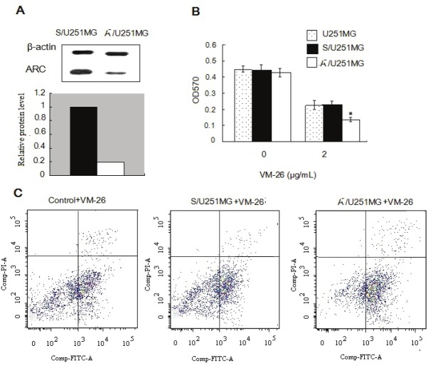
Previous studies have demonstrated that apoptosis repressor with caspase recruitment domain (ARC) is up-regulated in many forms of malignant tumors and low levels of ARC protein were expressed in normal human brain tissue. Little is known expression of ARC in glioma. Here, we found that ARC protein was highly expressed in primary human glioma when compared with normal brain tissues. A decrease in cell viability and an increase in apoptosis were observed in U251MG cells after ARC was knocked down. Knockdown of ARC was confirmed by western blotting. Knockdown of ARC promoted caspase-8, caspase-3 activation and Bax accumulation. These results indicate that ARC has a anti-apoptosis function in glioma.
Keywords: ARC; VM-26 sensitivity; apoptosis; glioma.
Figures


Similar articles
-
Role of apoptosis repressor with caspase recruitment domain in human health and chronic diseases.Ann Med. 2024 Dec;56(1):2409958. doi: 10.1080/07853890.2024.2409958. Epub 2024 Oct 1. Ann Med. 2024. PMID: 39351758 Free PMC article. Review.
-
A double-edged sword: role of apoptosis repressor with caspase recruitment domain (ARC) in tumorigenesis and ischaemia/reperfusion (I/R) injury.Apoptosis. 2023 Apr;28(3-4):313-325. doi: 10.1007/s10495-022-01802-4. Epub 2023 Jan 18. Apoptosis. 2023. PMID: 36652128 Review.
-
Curcumin suppressed anti-apoptotic signals and activated cysteine proteases for apoptosis in human malignant glioblastoma U87MG cells.Neurochem Res. 2007 Dec;32(12):2103-13. doi: 10.1007/s11064-007-9376-z. Epub 2007 Jun 12. Neurochem Res. 2007. PMID: 17562168
-
Curcumin differentially sensitizes malignant glioma cells to TRAIL/Apo2L-mediated apoptosis through activation of procaspases and release of cytochrome c from mitochondria.J Exp Ther Oncol. 2005;5(1):39-48. J Exp Ther Oncol. 2005. PMID: 16416600
-
Adenovirus-mediated transfer of siRNA against basic fibroblast growth factor mRNA enhances the sensitivity of glioblastoma cells to chemotherapy.Med Oncol. 2011 Mar;28(1):24-30. doi: 10.1007/s12032-010-9445-z. Epub 2010 Feb 24. Med Oncol. 2011. PMID: 20221717
Cited by
-
Role of apoptosis repressor with caspase recruitment domain in human health and chronic diseases.Ann Med. 2024 Dec;56(1):2409958. doi: 10.1080/07853890.2024.2409958. Epub 2024 Oct 1. Ann Med. 2024. PMID: 39351758 Free PMC article. Review.
-
The role of apoptosis repressor with a CARD domain (ARC) in the therapeutic resistance of renal cell carcinoma (RCC): the crucial role of ARC in the inhibition of extrinsic and intrinsic apoptotic signalling.Cell Commun Signal. 2017 May 2;15(1):16. doi: 10.1186/s12964-017-0170-5. Cell Commun Signal. 2017. PMID: 28464919 Free PMC article.
-
A double-edged sword: role of apoptosis repressor with caspase recruitment domain (ARC) in tumorigenesis and ischaemia/reperfusion (I/R) injury.Apoptosis. 2023 Apr;28(3-4):313-325. doi: 10.1007/s10495-022-01802-4. Epub 2023 Jan 18. Apoptosis. 2023. PMID: 36652128 Review.
-
Interplay of Phosphorylated Apoptosis Repressor with CARD, Casein Kinase-2 and Reactive Oxygen Species in Regulating Endothelin-1-Induced Cardiomyocyte Hypertrophy.Iran J Basic Med Sci. 2013 Aug;16(8):928-35. Iran J Basic Med Sci. 2013. PMID: 24106598 Free PMC article.
-
Cisplatin inhibits hippocampal cell proliferation and alters the expression of apoptotic genes.Neurotox Res. 2014 May;25(4):369-80. doi: 10.1007/s12640-013-9443-y. Epub 2013 Nov 26. Neurotox Res. 2014. PMID: 24277158 Free PMC article.
References
-
- Maher EA, Furnari FB, Bachoo RM, Rowitch DH, Louis DN, Cavenee WK, DePinho RA. Malignant glioma: genetics and biology of a grave matter. Genes Dev. 2001;15:1311–33. - PubMed
-
- Furnari FB, Fenton T, Bachoo RM, Mukasa A, Stommel JM, Stegh A, Hahn WC, Ligon KL, Louis DN, Brennan C, Chin L, DePinho RA, Cavenee WK. Malignant astrocytic glioma: genetics, biology, and paths to treatment. Genes Dev. 2007;21:2683–710. - PubMed
-
- Jakubowicz-Gil J, Langner E, Wertel I, Piersiak T, Rzeski W. Temozolomide, quercetin and cell death in the MOGGCCM astrocytoma cell line. Chem Biol Interact. 2010;188:190–203. - PubMed
-
- Li YC, Tzeng CC, Song JH, Tsia FJ, Hsieh LJ, Liao SJ, Tsai CH, Van Meir EG, Hao C, Lin CC. Genomic alterations in human malignant glioma cells associate with the cell resistance to the combination treatment with tumor necrosis factor-related apoptosis-inducing ligand and chemotherapy. Clin Cancer Res. 2006;12:2716–29. - PubMed
-
- Angileri FF, Aguennouz M, Conti A, La Torre D, Cardali S, Crupi R, Tomasello C, Germanò A, Vita G, Tomasello F. Nuclear factor-kappaB activation and differential expression of survivin and Bcl-2 in human grade 2-4 astrocytomas. Cancer. 2008;112:2258–66. - PubMed
Publication types
MeSH terms
Substances
LinkOut - more resources
Full Text Sources
Medical
Research Materials