A gated-7T MRI technique for tracking lung tumor development and progression in mice after exposure to low doses of ionizing radiation
- PMID: 22950352
- PMCID: PMC3478889
- DOI: 10.1667/rr2800.1
A gated-7T MRI technique for tracking lung tumor development and progression in mice after exposure to low doses of ionizing radiation
Abstract
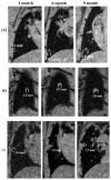
A gated-7T magnetic resonance imaging (MRI) application is described that can accurately and efficiently measure the size of in vivo mouse lung tumors from ∼0.1 mm(3) to >4 mm(3). This MRI approach fills a void in radiation research because the technique can be used to noninvasively measure the growth rate of lung tumors in large numbers of mice that have been irradiated with low doses (<50 mGy) without the additional radiation exposure associated with planar X ray, CT or PET imaging. High quality, high resolution, reproducible images of the mouse thorax were obtained in ∼20 min using: (1) a Bruker 7T micro-MRI scanner equipped with a 60 mm inner diameter gradient insert capable of generating a maximum gradient of 1000 mT/m; (2) a 35 mm inner diameter quadrature radiofrequency volume coil; and (3) an electrocardiogram and respiratory gated Fast Low Angle Shot (FLASH) pulse sequence. The images had an in-plane image resolution of 98 μm and a 0.5 mm slice thickness. Tumor diameter measured by MRI was highly correlated (R(2) = 0.97) with the tumor diameter measured by electronic calipers. Data generated with an initiation/promotion mouse model of lung carcinogenesis and this MRI technique demonstrated that mice exposed to 4 weekly fractions of 10, 30 or 50 mGy of CT radiation had the same lung tumor growth rate as that measured in sham-irradiated mice. In summary, this high-field, double-gated MRI approach is an efficient way of quantitatively tracking lung tumor development and progression after exposure to low doses of ionizing radiation.
Figures


References
-
- Division of Cancer Prevention and Control, National Center for Chronic Disease Prevention and Health Promotion. Centers for Disease Control and Prevention. ( http://www.cdc.gov/cancer/lung/statistics/index.htm) [Page last updated: November 23, 2010, cited: September 7, 2011]
-
- Jemal A, Siegel R, Xu J, Ward E. Cancer statistics, 2010. Cancer J Clin. 2010;60:277–300. - PubMed
-
- Pawaroo D, Cummings NM, Musonda P, Rintoul RC, Rassl D, Beadsmoore C. Non-small cell lung carcinoma: accuracy of PET/CT in determining the size of T1 and T2 primary tumors. AJR Am J Roentgenol. 2011;196:1176–81. - PubMed
-
- Fischer B, Lassen U, Mortensen J, Larsen S, Loft A, Bertelsen A, et al. Preoperative staging of lung cancer with combined PET-CT. N Engl J Med. 2009;361(1):32–9. - PubMed
Publication types
MeSH terms
Grants and funding
LinkOut - more resources
Full Text Sources
Medical

