Review
. 2012 Sep 1;2(9):a007765.
doi: 10.1101/cshperspect.a007765.

Do MHCII-presented neoantigens drive type 1 diabetes and other autoimmune diseases?

Affiliations
Review

Do MHCII-presented neoantigens drive type 1 diabetes and other autoimmune diseases?

Philippa Marrack et al. Cold Spring Harb Perspect Med. .

Abstract

The strong association between particular MHCII alleles and type 1 diabetes is not fully understood. Two ideas that have been considered for many years are that autoimmunity is driven by (1) low-affinity CD4(+) T cells that escape thymic negative selection and respond to certain autoantigen peptides that are particularly well presented by particular MHCII molecules, or (2) CD4(+) T cells responding to neoantigens that are absent in the thymus, but uniquely created in the target tissue in the periphery and presented by particular MHCII alleles. Here we discuss the recent structural data in favor of the second idea. We review studies suggesting that peptide antigens recognized by autoimmune T cells are uniquely proteolytically processed and/or posttranslationally modified in the target tissue, thus allowing these T cells to escape deletion in the thymus during T-cell development. We postulate that an encounter with these tissue-specific neoantigenic peptides presented by the particular susceptible MHCII alleles in the peripheral tissues when accompanied by the appropriate inflammatory milieu activates these T-cell escapees leading to the onset of autoimmune disease.


Figures

Figure 1.
Figure 1.
Schematic diagram of the various sources of peptides and the three main ways they are loaded into MHCII. (A) The major pathway involves newly synthesized MHCII trafficking to the endosomes from the Golgi complexed to invariant chain. After degradation of the invariant chain, DM/DO facilitates exchange of the residual invariant chain CLIP peptide for endosomally generated peptides. In the competition for binding among the peptides, those (colored) that best match the MHCII allelic binding motif are favored. The sources of these peptides are mostly proteins diverted from the secretory pathway or endocytosed from the cell membrane or extracellular cellular fluid, but in some antigen-presenting cells cytoplasmic proteins can enter this endosomal pathway. (B) Recycled cell-surface MHCII can reenter the endosome pathway and exchange its original peptide for a new internally generated one. (C) At the cell surface MHCII can lose its original peptide and take up one generated extracellularly.
Figure 2.
Figure 2.
Conventional peptide-MHCII presentation and recognition by CD4+ T-cell TCRs. (A) The conserved H-bonding network between MHCII amino acids and the bound peptide hold the peptide in a polyproline-like extended helix. The eight MHCII amino acids involved in the network are conserved among MHCII isotypes and alleles in all species. (B) The numbering scheme used for MHCII-bound peptides. (C) Using the mouse MHCII molecule IEk as an example, the pockets in the peptide-binding groove that accept the side chains of the p1 (yellow), p4 (green), p6 (red), and p9 (blue) amino acids of the peptide are shown occupied by amino acids from a self-peptide from hemoglobin (pHb) (PDB 1IEA). (D) The structures of two self-peptides, pHb and a peptide from heat shock protein 70 (pHsp70) (PDB 1IEB), and one foreign peptide from moth cytochrome c, pMCC (PDB 1KT2), are shown when bound to IEk, demonstrating the degeneracy allowed by the IEk p1-, p4-, p6-, and p9-binding pockets. (E) The conventional positions of TCR CDR loops are shown on surfaces of two MHCII-peptide ligands: a self-peptide from mutated triose phosphate isomerase (pTPI) bound to human HLA-DR1 and a foreign synthetic peptide, p3K, bound to mouse IAb. The tube representations of the CDR loops are colored as follows: Vα CDR1, green; CDR2, cyan; CDR3, blue; V CDR1, magenta; CDR2, orange; CDR3, red. The surfaces are colored as follows: MHCII strands, white; α helix of MHCIIα, light cyan; α helix of MHCII, light magenta; peptide, yellow.
Figure 3.
Figure 3.
Unconventional MHCII presentation and recognition of MBP to CD4+ T-cell autoimmunity. (A) The positions of the TCR CDR loops of a mouse MBP-specific autoreactive CD4+ T cell are shown on the surface of IAu bound to an MBP mimotope (PDB 1U3H). The CDR loops and surfaces are colored as in Figure 2E, except the portion of the peptide surface formed by amino acids not present in the natural MBP peptide is colored gray. (B) The positions of the TCR CDR loops of a human MBP-specific autoreactive CD4+ T cell are shown on the surface of HLA-DR2 bound to a different MBP peptide (PDB 1YMM). The CDR loops and surfaces are colored as in Figure 2E, except the portion of the peptide surface that is not within the TCR footprint is colored gray.
Figure 4.
Figure 4.
Unconventional IAg7 presentation of insulin to CD4+ T cells in NOD T1D. (A) The sequence of the insulin B9:23 peptide is shown along with the predicted positions that would occupy the IAg7-binding groove were the peptide to bind in register 3. Also shown are the maximal carboxy-terminal truncations, B:9–20 and B:9:21, tolerated by two types of B:9–23-reactive NOD diabetogenic CD4+ T cells, as well as versions of B:9–23 that mimic these truncations. (B) Models of the B:9–23 mimotopes for the type I (upper panel) and type II (lower panel) T cells shown in (A) were made based on the solved structure of IAg7 bound to a GAD peptide (PDB 1ES0) using Swiss PDB Viewer. The surface of IAg7 is shown colored as in Figure 2E. The surfaces of the portions of the mimotope peptides corresponding to the truncations in (A) are colored yellow. The rest of the peptide surface is colored gray. The p8, p9, and p10 amino acids are labeled.
Figure 5.
Figure 5.
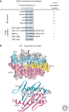
Unconventional IAg7 presentation of ChgA to CD4+ T cells in NOD T1D. (A) The activity of various ChgA peptides and mimotopes present by IAg7+ APC in stimulation ChgA-specific T cells. The peptide mimotopes are representative of those reported in three studies in which they were shown to stimulate strongly one or more ChgA-reactive T-cell clones or hybridomas (Judkowski et al. 2001; Yoshida et al. 2002; Stadinski et al. 2010b). The ChgA peptides are representative of those tested that contained the ChgA WSRMD sequence (Stadinski et al. 2010b). All sequences were aligned based on the WX(RK)M(DE) motif, which is highlighted. The putative positions of the peptide amino acids in the IAg7 groove are shown at the top, based on studies with the mimotopes. (B) A model of the ChgA WE14 peptide bound to IAg7 was created based on the structures of IAg7 bound to a GAD peptide (PDB 1ES0) and IEk bound to pHb (1IEA) using Swiss PDB Viewer. In the upper panel the predicted surface of the complex is shown colored as in Figure 2E. The lower panel shows a ribbon representation of the IAg7 α1 (cyan) and 1 (magenta) domains with a wire-frame representation of the first six amino acids of modeled WE14 peptide. The p5–p9 amino acids that make up the shared motif for ChgA-specific CD4+ T cells are labeled as in the p10 glutamine, which is the potential target of transglutaminase, treatment which greatly enhances the stimulatory activity of the WE14 peptide.

References

    1. Abadie V, Sollid LM, Barreiro LB, Jabri B 2011. Integration of genetic and immunological insights into a model of celiac disease pathogenesis. Annu Rev Immunol 29: 493–525 - PubMed
    1. Abiru N, Wegmann D, Kawasaki E, Gottlieb P, Simone E, Eisenbarth GS 2000. Dual overlapping peptides recognized by insulin peptide B:9-23 T cell receptor AV13S3 T cell clones of the NOD mouse. J Autoimmun 14: 231–237 - PubMed
    1. Anderton SM 2004. Post-translational modifications of self antigens: Implications for autoimmunity. Curr Opin Immunol 16: 753–758 - PubMed
    1. Bankovich AJ, Girvin AT, Moesta AK, Garcia KC 2004. Peptide register shifting within the MHC groove: Theory becomes reality. Mol Immunol 40: 1033–1039 - PubMed
    1. Burton AR, Vincent E, Arnold PY, Lennon GP, Smeltzer M, Li CS, Haskins K, Hutton J, Tisch RM, Sercarz EE, et al. 2008. On the pathogenicity of autoantigen-specific T-cell receptors. Diabetes 57: 1321–1330 - PubMed

Publication types

MeSH terms

Substances