. 2012 Oct;139(20):3827-37.
doi: 10.1242/dev.079178. Epub 2012 Sep 5.

Muscle length and myonuclear position are independently regulated by distinct Dynein pathways

Affiliations

Muscle length and myonuclear position are independently regulated by distinct Dynein pathways

Eric S Folker et al. Development. 2012 Oct.

Abstract

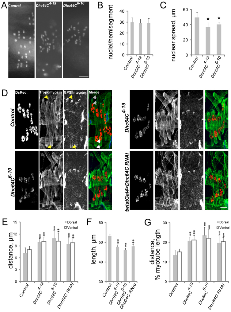
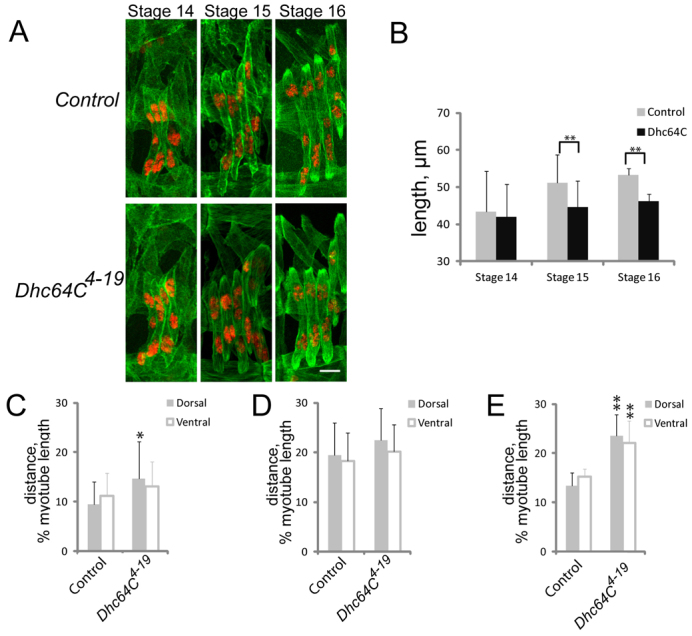
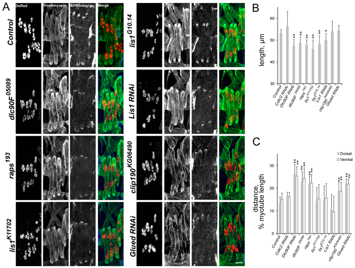
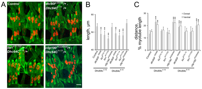
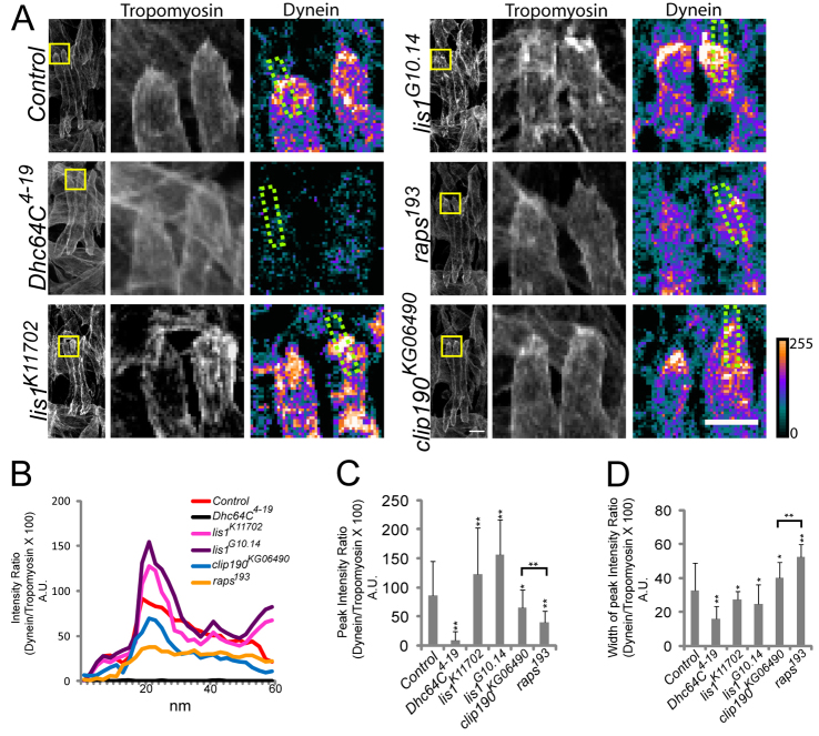
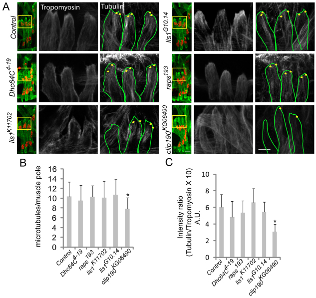
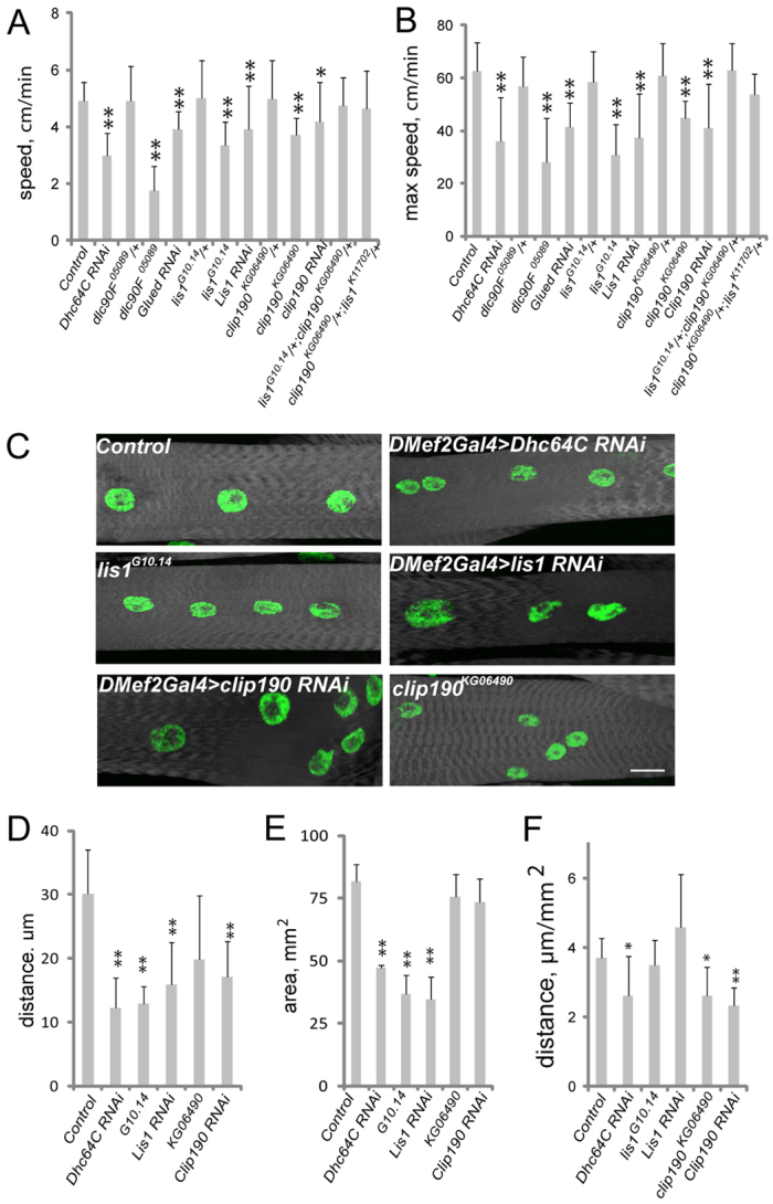
Various muscle diseases present with aberrant muscle cell morphologies characterized by smaller myofibers with mispositioned nuclei. The mechanisms that normally control these processes, whether they are linked, and their contribution to muscle weakness in disease, are not known. We examined the role of Dynein and Dynein-interacting proteins during Drosophila muscle development and found that several factors, including Dynein heavy chain, Dynein light chain and Partner of inscuteable, contribute to the regulation of both muscle length and myonuclear positioning. However, Lis1 contributes only to Dynein-dependent muscle length determination, whereas CLIP-190 and Glued contribute only to Dynein-dependent myonuclear positioning. Mechanistically, microtubule density at muscle poles is decreased in CLIP-190 mutants, suggesting that microtubule-cortex interactions facilitate myonuclear positioning. In Lis1 mutants, Dynein hyperaccumulates at the muscle poles with a sharper localization pattern, suggesting that retrograde trafficking contributes to muscle length. Both Lis1 and CLIP-190 act downstream of Dynein accumulation at the cortex, suggesting that they specify Dynein function within a single location. Finally, defects in muscle length or myonuclear positioning correlate with impaired muscle function in vivo, suggesting that both processes are essential for muscle function.


Figures

Fig. 1.
Fig. 1.
Cytoplasmic Dynein regulates muscle length and myonuclear position. (A) Fluorescence images of apRed nuclei in the lateral transverse (LT) muscles of live stage 17 Drosophila embryos (16.5-20 hours AEL) of the indicated genotypes. (B) The number of nuclei per hemisegment in stage 17 embryos of the indicated genotypes. (C) The distance between the dorsalmost and ventralmost nuclei in stage 17 embryos of the indicated genotypes. (D) Immunofluorescence images of stage 16 (16 hours AEL) embryos of the genotypes noted to the left and antigen listed at the top of the first image. Green, Tropomyosin (muscle); red, DsRed (nuclei); blue, β-PS-Integrin in merge. Arrows in the control panels denote points from which measurements were made for all genotypes. The distance between the points indicated by the yellow arrows in the Tropomyosin and β-PS-Integrin panels was used to determine muscle length. The distance between the pair of gray arrows at the top of the merged image was used to determine the distance between the nuclei and the dorsal pole. The distance between the pair of white arrows at the bottom of the merged image was used to determine the distance between the nuclei and the ventral pole. (E) The shortest distance between the indicated pole of the LT muscles (gray, dorsal; white, ventral) and the nearest cluster of nuclei in stage 16 embryos. (F) LT muscle length in stage 16 embryos. (G) The shortest distance between the indicated pole of the LT muscles (gray, dorsal; white, ventral) and the nearest cluster of nuclei normalized for muscle length in stage 16 embryos of the indicated genotypes. Error bars indicate s.d.; **P<0.01, *P<0.05. Scale bars: 10 μm.
Fig. 2.
Fig. 2.
Stage-dependent effects of Dynein on muscle length and myonuclear positioning. (A) Immunofluorescence images of the muscles (green, Tropomyosin) and nuclei (red, DsRed) at stage 14, 15 and 16 in control and Dhc64C4-19 Drosophila embryos. Scale bar: 10 μm. (B) The length of the muscles in control (gray) and Dhc64C4-19 (black) embryos at stage 14, 15 and 16. (C-E) The distance between the nearest nucleus and either the dorsal (gray) or ventral (white) pole of the muscle in stage 14 (C), 15 (D) and 16 (E) embryos. Error bars indicate s.d.; *P<0.05, **P<0.01.
Fig. 3.
Fig. 3.
Dynein-interacting proteins regulate myotube length and/or myonuclear position. (A) Immunofluorescence images of stage 16 Drosophila embryos of the indicated genotypes. Green, Tropomyosin; red, DsRed; blue, β-PS-Integrin in merge. The antigen for grayscale images is listed at the top of the first image. Scale bar: 10 μm. (B) The length of the LT muscles in stage 16 embryos. (C) The shortest distance between the indicated pole of the LT muscles (gray, dorsal; white, ventral) and the nearest cluster of nuclei normalized for muscle length. Error bars indicate s.d.; *P<0.05, **P<0.01.
Fig. 4.
Fig. 4.
Interactions between Dynein and associated genes during muscle development. (A) Immunofluorescence images of stage 16 Drosophila embryos that are doubly heterozygous for Dhc64C4-19 and the indicated allele. Green, Tropomyosin; red, DsRed. Scale bar: 10 μm. (B) LT muscle length in stage 16 embryos that are doubly heterozygous for the Dhc64C allele shown beneath the histogram and the listed alleles. (C) The shortest distance between the indicated pole of the LT muscles (gray, dorsal; white, ventral) and the nearest cluster of nuclei normalized for muscle length in stage 16 embryos that are doubly heterozygous for the Dhc64C allele indicated beneath the histogram and the listed alleles. Error bars indicate s.d.; **P<0.01.
Fig. 5.
Fig. 5.
The pathways controlling muscle length and myonuclear position do not genetically interact. (A) Immunofluorescence images of stage 16 Drosophila embryos that are doubly heterozygous for the indicated alleles. Green, Tropomyosin; red, DsRed. Scale bar: 10 μm. (B) LT muscle length in stage 16 embryos that are doubly heterozygous for the indicated alleles. (C) The shortest distance between the indicated pole of the LT muscles (gray, dorsal; white, ventral) and the nearest cluster of nuclei in stage 16 embryos normalized for muscle length. Error bars indicate s.d.; *P<0.05, **P<0.01.
Fig. 6.
Fig. 6.
Dynein hyperaccumulates in Lis1 mutant embryos. (A) Confocal projections of a single hemisegment from stage 16 Drosophila embryos immunostained for Tropomyosin (green) and Dynein heavy chain (red). The boxed regions are shown at higher magnification to the right, with Tropomyosin shown in grayscale and Dynein shown as a heat map (the scale indicates relative intensity). The regions outlined by the green dotted lines were used for linescan analysis. Scale bars: left-hand panel, 5 μm; right-hand panel, 10 μm. (B) Representative linescan analysis indicating the intensity of Dynein heavy chain immunofluorescence as a function of position across the muscle pole. (C) The peak intensity signal for Dynein heavy chain immunofluorescence in the indicated genotypes. (D) The width of the peak intensity of Dynein heavy chain immunofluorescence. Error bars indicate s.d.; *P<0.05, **P<0.01, compared with control.
Fig. 7.
Fig. 7.
Microtubule organization is disrupted in CLIP-190 mutant embryos. (A) Confocal projections of a single hemisegment from stage 16 Drosophila embryos immunostained for Tropomyosin (green) and Tubulin (grayscale). The boxed regions are shown at higher magnification to the right. Yellow arrows indicate individual and/or bundles of microtubules that reach the muscle pole. Scale bars: 10 μm. (B) The number of microtubules within 3 μm of the myofiber pole in the indicated genotypes. (C) The average intensity of Tubulin immunofluorescence in the 3 μm near the myotube pole in the indicated genotypes. Error bars represent s.d.; *P<0.05, compared with control.
Fig. 8.
Fig. 8.
Larval muscle organization and physiology are affected by Dynein and associated proteins. (A,B) The average (A) and maximum (B) speed of Drosophila larvae as they crawl towards a stimulus. (C) Fluorescence images of VL3 muscles from L3 larvae that were used in locomotion assays just prior to dissection. White, phalloidin; green, Hoechst. Scale bar: 20 μm. (D) The average distance between nuclei in larval muscles from the indicated genotypes. (E) The surface area of the muscles in larvae of the indicated genotypes. (F) The distance between nuclei in the larval muscles normalized for muscle size. Error bars indicate s.d. *P<0.05, **P<0.01.

References

    1. Bate M. (1990). The embryonic development of larval muscles in Drosophila. Development 110, 791-804 - PubMed
    1. Beckett K., Baylies M. K. (2007). 3D analysis of founder cell and fusion competent myoblast arrangements outlines a new model of myoblast fusion. Dev. Biol. 309, 113-125 - PMC - PubMed
    1. Bellen H. J., Levis R. W., Liao G., He Y., Carlson J. W., Tsang G., Evans-Holm M., Hiesinger P. R., Schulze K. L., Rubin G. M., et al. (2004). The BDGP gene disruption project: single transposon insertions associated with 40% of Drosophila genes. Genetics 167, 761-781 - PMC - PubMed
    1. Brent J. R., Werner K. M., McCabe B. D. (2009). Drosophila larval NMJ dissection. J. Vis. Exp. 24, 1107 - PMC - PubMed
    1. Caggese C., Moschetti R., Ragone G., Barsanti P., Caizzi R. (2001). dtctex-1, the Drosophila melanogaster homolog of a putative murine t-complex distorter encoding a dynein light chain, is required for production of functional sperm. Mol. Genet. Genomics 265, 436-444 - PubMed

Publication types

MeSH terms

LinkOut - more resources