. 2013 Jan;133(1):258-67.
doi: 10.1038/jid.2012.267. Epub 2012 Sep 6.

Toll-like receptor 4 has an essential role in early skin wound healing

Affiliations

Toll-like receptor 4 has an essential role in early skin wound healing

Lin Chen et al. J Invest Dermatol. 2013 Jan.

Erratum in

  • J Invest Dermatol. 2014 Feb;134(2):583

Abstract

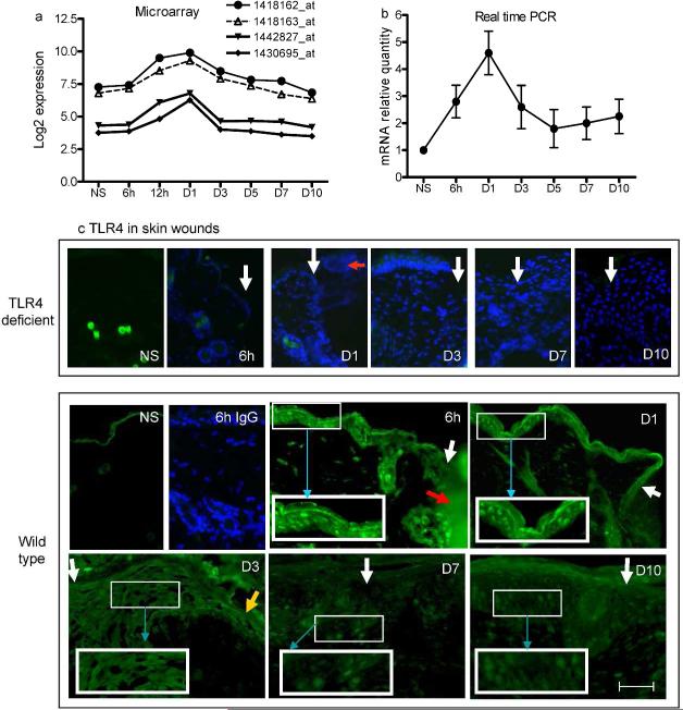
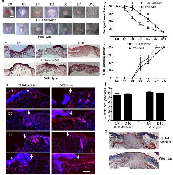
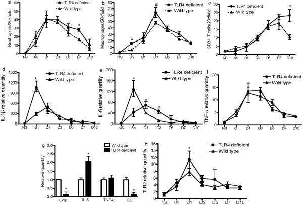
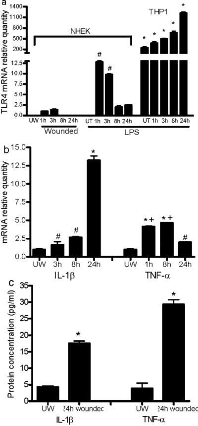
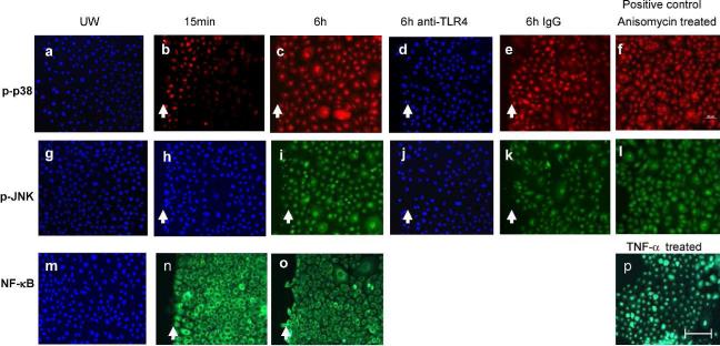
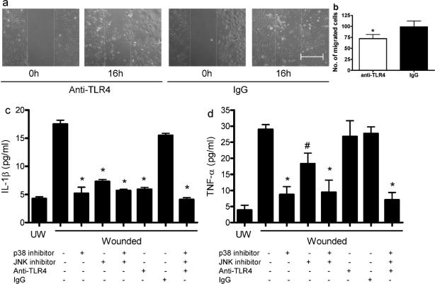
Toll-like receptor 4 (TLR4) has a key role in the initiation of innate immunity and in the regulation of adaptive immune responses. Using microarray analysis and PCR, TLR4 expression was observed to increase in murine skin wounds at the early stages. The cellular location of TLR4 was primarily in keratinocytes at the wound edges. The closure of excisional wounds was significantly delayed in TLR4-deficient (C3H/HeJ) as compared with wild-type mice, and both IL-1β and IL-6 production were significantly lower in the wounds of TLR4-deficient mice. EGF also markedly decreased in the wound edge of epidermis in TLR4-deficient mice. In vitro studies confirmed that a wound stimulus induces TLR4 mRNA expression in primary normal human epidermal keratinocytes (NHEK). In vitro injury also induced the phosphorylation of p38 and JNK MAPK (Jun N-terminal kinase mitogen-activated protein kinase) and the expression of IL-1β and tumor necrosis factor-α by NHEK. Blockade of TLR4 delayed NHEK migration and abolished the phosphorylation of p38 and JNK MAPK, and blockade of TLR4 and/or p38/JNK abolished IL-1β production. The results suggest that inflammatory cytokine production by injured NHEK is stimulated via the TLR4-p38 and JNK MAPK signaling pathway. Together, the results provide evidence for a role of TLR4 at sites of injury, and suggest that TLR4 is an important regulator of wound inflammation.


Figures

Figure 1
Figure 1
TLR4 gene and protein expression is upregulated in early skin wounds. (a) Microarray analysis shows that all four TLR4 probes in the array demonstrate a significant increase in expression, especially from 12h to 1 day (p<1.2×10⁻⁷, one-way ANOVA). (b) Real time PCR confirms that TLR4 mRNA is markedly increased in early stage wounds in TLR4 wild type mice (p=0.0003, one-way ANOVA). N=3 at each time point. (c) Photomicrographs of indirect immunofluorescence detection of TLR4 in skin wounds of TLR4 deficient and wild type mice. The control (rabbit IgG staining) histologic sections of wounds from TLR4 deficient mice, as well as the the 6h wound sample from wild type mice, were also stained with DAPI (blue). White arrows indicate wound edges; red arrow indicates fibrin clot, yellow arrow indicates advancing epidermal tip, and blue arrows show the higher magnification of the outlined areas. Bar=200μm.
Figure 2
Figure 2
Mice with a TLR4 mutation exhibit impaired early wound healing without vascularity changes. (a) Representative photomicrographs of wounds at 0 and 6h as well as 1, 3, 5, 7 and 10 days after injury. Bar=3mm. (b) Percent of original wound size, N=5, * p<0.01, # p<0.05 VS wild type. (c) Photomicrographs of HE stained histologic sections of wounds. Bar=500μm. (d) Rate of wound re-epithelialization measured by histomorphometric analysis of tissue sections. N=5, * p<0.01 VS wild type. (e) Photomicrograpahs of ki67+ (red) proliferating keratinocytes with nuclear DAPI counterstaining (blue). Arrows indicate wound edges. Bar=200μm. (f) Percent area occupied by CD31 stained vessels at day 7 and 10 after wounding. N=5 at each time point. (g) Photomicrographs of Masson's trichrome stained histologic sections of day 10 wounds. Bar=500μm.
Figure 3
Figure 3
Wounds of TLR4 deficient and wild type mice show differences in inflammatory cell and cytokine content. (a, b, and c) The number of neutrophils, macrophages, and CD3+ T cells, respectively, in the wounds, determined by immunohistochemistry. (d, e, f, and h) mRNA levels of IL1β, IL-6, TNF-α and TLR2 in wounds, as determined by real time PCR. * p<0.01, # P<0.05, N=5 at each time point. Expression in the normal skin of wild type mice was used as baseline. (g) mRNA levels of IL1β, IL-6, TNF-α and EGF in epithelial cells at the 6h wound edges determined by LCM and RT-PCR. Data are averages of triplicate wells, and are representative of two independent experiments. * p<0.01 VS wild type.
Figure 4
Figure 4
Scratch wound injury induces mRNA expression of TLR4 and production of inflammatory cytokines in NHEK cells. (a) LPS and injury-induced expression of TLR4 mRNA. # p<0.01 VS 1 and 3h expression in injured cells, and 8 and 24h LPS treated cells. THP1 treated with or without LPS were used as positive control. * p<0.0001 VS wounded and LPS treated NHEK. (b) Increased IL-1β and TNF-α mRNA expression in NHEK from 3 to 24h after wounding. # p<0.05 VS unwounded (IL-1β), * p<0.01 VS unwounded, 3h, and 8h (IL-1β), * p<0.01 and # p<0.05 VS unwounded (TNF-α), + p<0.01 VS 1h and 8h. Data in a&b represent the average of triplicate wells and are representative of two independent experiments. (c) Protein levels of IL-1β and TNF-α in the culture media 24h after injury. N=3, *p<0.01 VS unwounded cells. UW, unwounded; UT, LPS untreated.
Figure 5
Figure 5
Scratch wound injury induces expression of p-p38 and p-JNK MAPK, but not NF-κB nuclear translocation in NHEK. Nuclear NF-κB, p-p38, and p-JNK were detected using immunofluorescence 15 min and 6h after wounding. To test the effects of anti-TLR4 antibody on induction of p-p38 (red) and p-JNK (green), NHEK were treated with anti-TLR4 or mouse IgG prior to scratch injury. (a, g, and m) Unwounded NHEK; (b, h, and n) 15 min after injury; (c, d, e, i, j, and k) 6h after injury; (p) TNF-α treated NHEK as NF-κB positive control; (f and l) Anisomycin treated NHEK as positive control for p-p38 and p-JNK; (a, d, g, h, j, and m) were also stained by DAPI (blue). UW: unwounded. Arrows indicate wound edges. Bar=100μm.
Figure 6
Figure 6
Neutralization of TLR4 delays NHEK migration and abolishes IL-1β production in injured NHEK. (a and b) NHEK monolayers were pretreated with anti-TLR4 or normal mouse IgG (20μg/ml) followed by mitomycin C (10μg/ml) treatment, and then wounded by scratches. The defined areas were photographed at 0 and 16h after wounding. The numbers of cells migrated out of the initial areas were counted. N=4. * p<0.05 compared to IgG control. (c and d) Cultured NHEK were incubated with anti-TLR4 and/or p38 inhibitor SB203580, and/or JNK inhibitor SP600125 and then cells were injured by scratching. The supernatants were collected 24h later for IL-1β and TNF-α ELISA analyses. N=3,*p<0.01, # p<0.05 compared to wounded untreated cells. UW, unwounded. Bar=100μm.

References

    1. Akira S, Takeda K. Toll-like receptor signalling. Nat Rev Immunol. 2004;4:499–511. - PubMed
    1. Asea A, Rehli M, Kabingu E, Boch JA, Bare O, Auron PE, et al. Novel signal transduction pathway utilized by extracellular HSP70: role of toll-like receptor (TLR) 2 and TLR4. J Biol Chem. 2002;277:15028–34. - PubMed
    1. Baker BS, Ovigne JM, Powles AV, Corcoran S, Fry L. Normal keratinocytes express Toll-like receptors (TLRs) 1, 2 and 5: modulation of TLR expression in chronic plaque psoriasis. Br J Dermatol. 2003;148:670–9. - PubMed
    1. Becker CE, O'Neill LA. Inflammasomes in inflammatory disorders: the role of TLRs and their interactions with NLRs. Semin Immunopathol. 2007;29:239–48. - PubMed
    1. Bettinger DA, Pellicane JV, Tarry WC, Yager DR, Diegelmann RF, Lee R, et al. The role of inflammatory cytokines in wound healing: accelerated healing in endotoxin-resistant mice. J Trauma. 1994;36:810–3. discussion 3-4. - PubMed

Publication types

MeSH terms

LinkOut - more resources