. 2012 Dec;82(12):1284-96.
doi: 10.1038/ki.2012.192. Epub 2012 Sep 5.

The IgA1 immune complex-mediated activation of the MAPK/ERK kinase pathway in mesangial cells is associated with glomerular damage in IgA nephropathy

Affiliations

The IgA1 immune complex-mediated activation of the MAPK/ERK kinase pathway in mesangial cells is associated with glomerular damage in IgA nephropathy

Houda Tamouza et al. Kidney Int. 2012 Dec.

Abstract

IgA nephropathy (IgAN), the most common primary glomerulonephritis worldwide, has significant morbidity and mortality as 20-40% of patients progress to end-stage renal disease within 20 years of onset. In order to gain insight into the molecular mechanisms involved in the progression of IgAN, we systematically evaluated renal biopsies from such patients. This showed that the MAPK/ERK signaling pathway was activated in the mesangium of patients presenting with over 1 g/day proteinuria and elevated blood pressure, but absent in biopsy specimens of patients with IgAN and modest proteinuria (<1 g/day). ERK activation was not associated with elevated galactose-deficient IgA1 or IgG specific for galactose-deficient IgA1 in the serum. In human mesangial cells in vitro, ERK activation through mesangial IgA1 receptor (CD71) controlled pro-inflammatory cytokine secretion and was induced by large-molecular-mass IgA1-containing circulating immune complexes purified from patient sera. Moreover, IgA1-dependent ERK activation required renin-angiotensin system as its blockade was efficient in reducing proteinuria in those patients exhibiting substantial mesangial activation of ERK. Thus, ERK activation alters mesangial cell-podocyte crosstalk, leading to renal dysfunction in IgAN. Assessment of MAPK/ERK activation in diagnostic renal biopsies may predict the therapeutic efficacy of renin-angiotensin system blockers in IgAN.


Conflict of interest statement

The authors declare no competing financial interests.

Figures
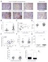
Figure 1
Figure 1. Mesangial activation of MAPK/ERK pathway correlates with increased proteinuria and MBP
(A) Immunostaining of p-ERK1/2 in renal biopsy tissues from IgAN patients and healthy controls. Patients were divided into two groups according to the p-ERK1/2 mesangial score (low <1.1 and high >1.1). (B) Proteinuria in patients presenting a high or low mesangial score for p-ERK1/2 staining. (C) Correlation between p-ERK1/2 mesangial staining score and proteinuria. (D) MBP distribution in patients presenting a high or low mesangial score for p-ERK1/2 staining. (E) Correlation between p-ERK1/2 mesangial staining score and MBP. (F) Age distribution in patients presenting a high or low mesangial score for p-ERK1/2 staining. (G) Hematuria in patients presenting a high or low mesangial score for p-ERK1/2 staining. (H) eGFRMDRD values in patients presenting a high or low mesangial score for p-ERK1/2 staining. (I) Sex distribution (males in filled histograms and females in open histograms) in patients presenting a high or low mesangial score for p-ERK1/2 staining. All data are mean ± s.e.m. *P < 0.05, **P < 0.01, *** P < 0.001
Figure 2
Figure 2. MAPK/ERK pathway of mesangial activation is not associated with histological variables of the MEST score from the Oxford Classification
Distribution of mesangial proliferation (M0/M1) (A), endocapillary proliferation (E0/E1) (B), glomerulosclerosis (S0/S1) (C) and tubular atrophy and interstitial fibrosis (T0/T1/T2) (D) scores in IgAN patients presenting low or high-mesangial score for p-ERK1/2 labeling.
Figure 3
Figure 3. MAPK/ERK pathway of mesangial activation in other proteinuric glomerulonephritis
Immunostaining of p-ERK1/2 in renal biopsy tissues from patients with systemic lupus erythematosus or MGN showing that proteinuria can be observed without p-ERK1/2 expression in mesangial cells in patients with MGN.
Figure 4
Figure 4. Activation of the MAPK/ERK pathway in mesangial cells is not associated with serum levels of aberrantly glycosylated IgA1, IgG-IgA immune complex, or anti-Gal-deficient-IgA1 IgG antibodies
(A) Serum levels of Gal-deficient IgA1 (evaluated by ELISA using HAA lectin) in healthy subjects, in all IgAN patients, and in IgAN patients presenting high or low levels of p-ERK1/2 mesangial score. (B) Detection of Gal-deficient IgA1 following neuraminidase treatment. (C–D) Detection of IgG-IgA complexes and IgG anti-Gal-deficient IgA1 in the serum of patients presenting a high or low p-ERK1/2 mesangial score.
Figure 5
Figure 5. Activation of human mesangial cells (HMC) by pIgA1 induced MAPK/ERK and PI3K/Akt/mTOR signaling pathways
(A) Circulating immune complexes (CIC) from an IgAN patient and uncomplexed pIgA1 myeloma protein activate MAPK/ERK1/2 pathway in HMC. Serum-starved HMC were stimulated for 15 min with PDGF-BB, IgA1-containing circulating immune complexes (CIC) from an IgAN patient (CIC-S, large-molecular-mass stimulatory CIC; CIC-I, small-molecular-mass inhibitory CIC), and uncomplexed naturally Gal-deficient pIgA1 myeloma protein (pIgA1 Mce, IgA1). Cell lysates were immunoblotted with anti-p-ERK1/2 and re-probed for actin as control for equal protein loading. (B, C) Time-course stimulation of HMC with pIgA1 and PDGF-BB. Serum-starved HMC were treated with or without 1 μM wortmannin (W) or 25 nM rapamycin (R) for 30 min and then activated with pIgA1 (Dou) (pIgA1) (B) or PDGF-BB (C) for the indicated lengths of time. Cell lysates were immunoblotted with anti-p-Akt, anti-p-mTOR and anti-p-ERK1/2 antibodies as described in Methods section. Anti-ERK1/2 antibody was used as control for equal protein loading. Data are representative of at least three independent experiments. (D) Circulating immune complexes (CIC) from an IgAN patient and uncomplexed pIgA1 myeloma protein activated Akt1/FKHRL1 pathway in HMC. Serum-starved HMC were stimulated for 15 or 60 min with PDGF-BB, IgA1-containing CIC from an IgAN patient (CIC-S, large-molecular-mass stimulatory CIC; CIC-I, small-molecular-mass inhibitory CIC) and uncomplexed Gal-deficient pIgA1 myeloma protein (pIgA1 Mce, IgA1). Cell lysates were immunoblotted with anti-p-FKHRL1 antibodies (p-FKHRL1) and re-probed with actin as control for equal protein loading.
Figure 6
Figure 6. Activation of MAPK/ERK and PI3K/Akt/mTOR signaling pathways are involved in IL-6 secretion and HMC proliferation, respectively
(A) Activation of PI3K/Akt/mTOR pathway regulates IgA1-dependent HMC proliferation. Serum-starved HMC were pre-incubated with inhibitors of PI3 kinase (wortmannin; 1 μM), mTOR (rapamycin; 25 nM) or ERK (PD98059, 10 μM) for 2 h and then activated with pIgA1 (Dou; 0.5 mg/ml) for another 48 h before an 18-h incorporation of [3H]-thymidine. Non-stimulated (NS) HMC served as control. Data are representative of three independent experiments (error bars, SD). * p < 0.05, ** p < 0.01, *** p < 0.001 (two-tailed t test). (B) Blocking of MAPK/ERK pathway inhibits IL-6 secretion by pIgA1-stimulated HMC. Serum-starved HMC were pre-incubated with inhibitors for 2 h and then activated with or without pIgA1 (0.5 mg/ml) overnight. Non-stimulated (NS) HMC served as control. Supernatants were then collected and IL-6 secreted in cultures was quantified by ELISA. Data are means +/− SD of three independent experiments. * p < 0.05, ** p < 0.01, *** p < 0.001 (two-tailed t test).
Figure 7
Figure 7. pIgA1-dependent HMC stimulation is abrogated by TfR1 blocking
(A and B) The anti-TfR1 mAb A24 inhibited IL-6 secretion (A) and cellular proliferation (B) in pIgA1-stimulated HMC. HMC were pre-incubated for 2 h with mAb A24 (50 μg/ml) before activation with pIgA1 (1 mg/ml). Cytokine secretion and cell proliferation were assessed after overnight and 48-h incubation, respectively, as described in Methods section. Data are the means +/− SD from three independent experiments. * p < 0.05, ** p < 0.01, *** p < 0.001 (two-tailed t test). (C) HMC were pretreated for 30 min with wortmannin (1 μM), rapamycin (25 nM), or soluble TfR1 (sTfR, 200 μg/ml) before stimulation with pIgA1 (0.5 mg/ml) for 10 min. Cell lysates were immunoblotted with anti-p-Akt1 (p-Akt1) and equal loading of proteins was determined by blotting with anti-Akt1 antibody. (D) PDGF-BB-dependent HMC stimulation was not abrogated by soluble TfR1. HMC were pretreated for 30 min with wortmannin (1 μM), rapamycin (25 nM), or soluble TfR1 (200 μg/ml) before stimulation with PDGF-BB (10 ng/ml) for 10 min. Cell lysates were immunoblotted with anti-p-Akt1 (p-Akt1) and equal loading of proteins was determined by blotting with anti-Akt1 antibody.
Figure 8
Figure 8. Calcium signaling is triggered by pIgA1 (but not by other TfR1 ligands) in HMC
The cells grown on glass coverslips were loaded with Fura-2/AM and intracellular calcium was measured as the ratio of Fura-2/AM intensities (excitation, 340 and 380 nm; emission, 515 nm; F/F0) before and after exposure to pIgA1, Fe-Tf, Apo-Tf (1: 0.5 μg/ml, 2: 5 μg/ml, 3: 50 μg/ml, 4: 250 μg/ml, 5: 500 μg/ml) or PDGF-BB (6: 10 ng/ml). The maximal calcium mobilization was measured after stimulation of the cells with ionomycin (i: 10 ng/ml) as an internal control.
Figure 9
Figure 9. pIgA1-induced HMC activation is dependent on ATR1 signaling
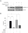
Serum-starved HMC were pre-incubated with AT1R antagonist losartan (10 μM) overnight and then activated overnight with or without pIgA1 (1 mg/ml) in the presence or absence of angiotensin II (ANGII, 10 nM). (A) The presence of p-ERK1/2 (p-ERK1/2) was examined by immunoblotting of cell lysates. Blotting with anti-actin was used to control for equal protein loading. (B) IL-6 in cell culture supernatants was quantified by ELISA (data are the mean +/− SD from three independent experiments. * p < 0.05, ** p < 0.01, *** p < 0.001; two-tailed t test).
Figure 10
Figure 10. Mesangial score for p-ERK1/2 predicts decrease in proteinuria (but not MBP) induced by RAS blockers therapy
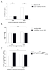
(A) Baseline and last follow-up visit values of proteinuria after beginning RAS blockers therapy in patients with low or high scores of p-ERK1/2 staining. (B) Baseline and last follow-up visit values of MBP after beginning RAS blockers therapy.

References

    1. Berger J, Hinglais N. Intercapillary deposits of IgA-IgG. J Urol Nephrol (Paris) 1968;74:694–695. - PubMed
    1. Monteiro RC, Moura IC, Launay P, et al. Pathogenic significance of IgA receptor interactions in IgA nephropathy. Trends Mol Med. 2002;8:464–468. - PubMed
    1. Coppo R, D’Amico G. Factors predicting progression of IgA nephropathies. J Nephrol. 2005;18:503–512. - PubMed
    1. Locatelli F, Marcelli D, Comelli M, et al. Proteinuria and blood pressure as causal components of progression to end-stage renal failure. Northern Italian Cooperative Study Group. Nephrol Dial Transplant. 1996;11:461–467. - PubMed
    1. Stoycheff N, Pandya K, Okparavero A, et al. Early change in proteinuria as a surrogate outcome in kidney disease progression: a systematic review of previous analyses and creation of a patient-level pooled dataset. Nephrol Dial Transplant. 2011;26:848–857. - PMC - PubMed

Publication types

MeSH terms