Review
. 2012 Nov 4;48(85):10472-80.
doi: 10.1039/c2cc35042d.

Nucleic acid aptamers: an emerging frontier in cancer therapy

Affiliations
Review

Nucleic acid aptamers: an emerging frontier in cancer therapy

Guizhi Zhu et al. Chem Commun (Camb). .

Abstract

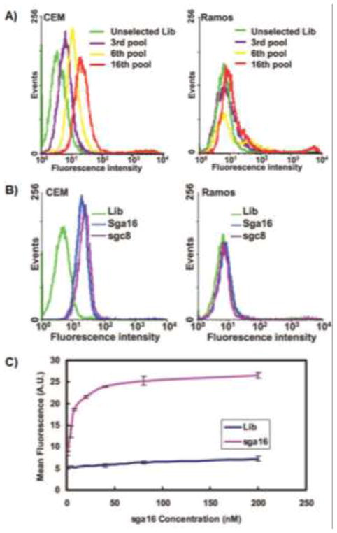
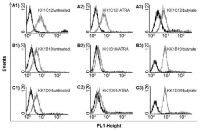
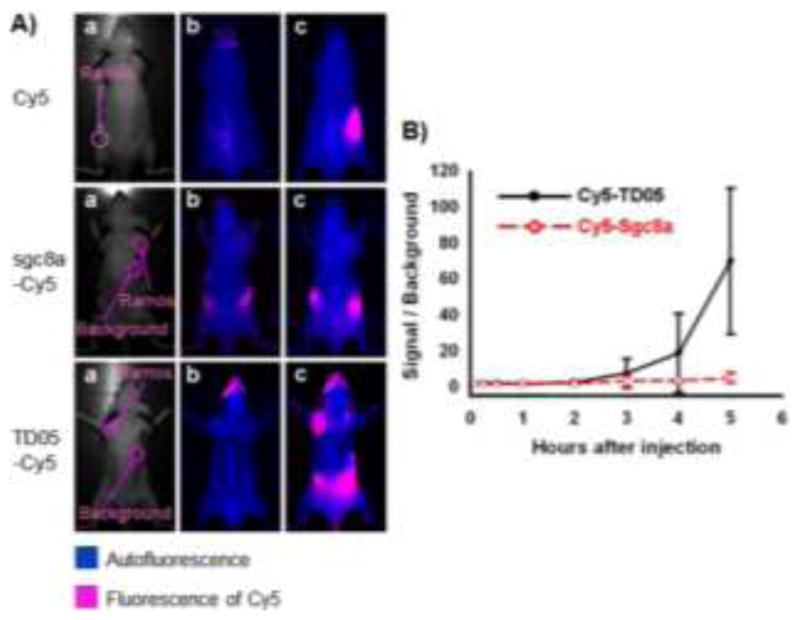
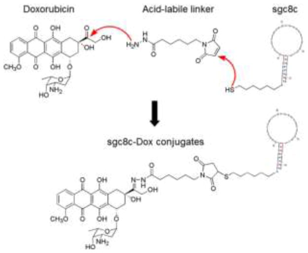
The last two decades have witnessed the development and application of nucleic acid aptamers in a variety of fields, including target analysis, disease therapy, and molecular and cellular engineering. The efficient and widely applicable aptamer selection, reproducible chemical synthesis and modification, generally impressive target binding selectivity and affinity, relatively rapid tissue penetration, low immunogenicity, and rapid systemic clearance make aptamers ideal recognition elements for use as therapeutics or for in vivo delivery of therapeutics. In this feature article, we discuss the development and biomedical application of nucleic acid aptamers, with emphasis on cancer cell aptamer isolation, targeted cancer therapy, oncology biomarker identification and drug discovery.


Figures

Fig. 1
Fig. 1
Monitoring of cell-SELEX and characterization of selected aptamers. (A) Flow cytometry assay to monitor the binding of selected pools and enrichment of aptamers with CEM cells (target) and Ramos cells (control). For target CEM cells, there was an increase in binding ability of the pool as the selection was progressing, whereas there was little enhancement for the control cells. (B) Flow cytometry assay for the binding of the FITC-labeled sequences sga16 and sgc8 obtained from the above selection. (C) Flow cytometry assay to determine the binding affinity of FITC-labeled sga16 to CEM cells. The nonspecific binding was measured using an FITC-labeled random DNA sequence library. Adapted from ref. (Copyright (2006) National Academy of Sciences, U.S.A.)
Fig. 2
Fig. 2
Flow cytometry assay showing the binding pro les of aptamers KH1C12 (A1-A3), KK1B10 (B1–B3) and KK1D04 (C1–C3) to the untreated, ATRA-treated and sodium butyrate-treated HL60 cells. The dark curves show fluorescence background using the random DNA sequence library. Adapted from ref.
Fig. 3
Fig. 3
An aptamer identified through cell-SELEX for in vivo imaging of Ramos subcutaneous xenograft. (A) Optical and fluorescent imaging of tumors in mice using Cy5 dye and Cy5-labeled aptamers. Target Ramos cells were injected subcutaneously on the back of BALB/c nude mice. Cy5 dye, Cy5-labeled sgc8a (control), or Cy5-labeled TD05 was subsequently injected intravenously, and images of the dorsal side of live mice were taken at various time points after injection. Images a, b and c represent the optical photographs, the fluorescent images before injection, and 3.5 h after injection, respectively. (B) Quantification of the signal-to-background ratio for Cy5-labeled TD05 and Cy5-labeled sgc8a. The number of mice used to derive statistical information for each probe is 5. Data represent mean ± standard error. Adapted from ref.
Fig. 4
Fig. 4
Schematic illustration of conjugation of a chemotherapeutic drug, Doxorubicin (Dox), with an DNA aptamer sgc8c for targeted drug delivery to cancer cells. Adapted from ref.
Fig. 5
Fig. 5
Schematic illustration of the self-assembled aptamer-based drug carrier SD for bi-specific cytotoxicity. A bi-specific aptamer-based drug carrer, SD, was self-assembled from molecularly engineered aptamers sgc8c (S) and sgc5a (D), simultaneously forming Dox intercalation sites in the dsDNA linker. Subsequently, Dox was loaded onto SD through site-specific Dox intercalation and bi-specifically delivered into target cells in a mixed cell environment, where it displayed bi-specific cytotoxicity. Adapted from ref.
Fig. 6
Fig. 6
Aptamer-micelles for targeted delivery of doped dye. (A) Schematic illustration of aptamer micelle formation. Bright field and fluorescent images of Ramos cells after incubation with free CellTracker Green BODIPY for 2 h (B) and 12 h (C), or incubation with biotin-TD05-micelle doped with CellTracker Green BODIPY for 2 h (D). Image E is the enlarged fluorescent image after postlabeling the biotinylated TD05 aptamer with QD705 streptavidin. The inset in image C is the enlarged individual cell image. The inset in image E is the fluorescent image of the dead cell. (F) Real-time monitoring of doped CellTracker released from the core of the micelles. The numbers indicated the time (minutes) of incubation at room temperature. Adapted from ref.
Fig. 7
Fig. 7
Schematic illustration of polymeric aptamer construction. Polymeric aptamers, which work as flexible molecular probes, were engineered from multiple dye-labeled reporting elements and aptamer-labeled targeting elements. Adapted from ref.

References

    1. Ellington AD, Szostak JW. Nature. 1990;346:818–822. - PubMed
    1. Tuerk C, Gold L. Science. 1990;249:505–510. - PubMed
    1. Huizenga DE, Szostak JW. Biochemistry. 1995;34:656–665. - PubMed
    1. Hermann T, Patel DJ. Science. 2000;287:820–825. - PubMed
    1. Mallikaratchy P, Stahelin RV, Cao Z, Cho W, Tan W. Chem Commun. 2006:3229–3231. - PubMed

Publication types