Review
. 2013 Sep-Oct;31(5):722-35.
doi: 10.1016/j.biotechadv.2012.08.006. Epub 2012 Aug 24.

Generation of tissue constructs for cardiovascular regenerative medicine: from cell procurement to scaffold design

Affiliations
Review

Generation of tissue constructs for cardiovascular regenerative medicine: from cell procurement to scaffold design

Vishal Tandon et al. Biotechnol Adv. 2013 Sep-Oct.

Abstract

The ability of the human body to naturally recover from coronary heart disease is limited because cardiac cells are terminally differentiated, have low proliferation rates, and low turn-over rates. Cardiovascular tissue engineering offers the potential for production of cardiac tissue ex vivo, but is currently limited by several challenges: (i) Tissue engineering constructs require pure populations of seed cells, (ii) Fabrication of 3-D geometrical structures with features of the same length scales that exist in native tissue is non-trivial, and (iii) Cells require stimulation from the appropriate biological, electrical and mechanical factors. In this review, we summarize the current state of microfluidic techniques for enrichment of subpopulations of cells required for cardiovascular tissue engineering, which offer unique advantages over traditional plating and FACS/MACS-based enrichment. We then summarize modern techniques for producing tissue engineering scaffolds that mimic native cardiac tissue.


Figures

Figure 1
Figure 1
Scheme of Hele-Shaw flow chamber geometry and shear stress profile for the device geometry designed by Usami et al. (1993) and developed by Murthy et al. (2004). Figure adapted from Murthy et al. (2004).
Figure 2
Figure 2
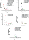
Cell adhesion on a variety of functionalized surfaces as a function of shear stress for (a) endothelial cells, (b) endothelial progenitor cells, (c) fibroblasts, (d) smooth muscle cells, and (e) stem cells. Open symbols denote antibodies as the capture molecules, and close symbols represent peptides or other proteins.
Figure 3
Figure 3
Two-stage microfluidic system for separation of cells with different densities of surface receptors. Each device stage has a different surface density of capture molecules, resulting in enrichment of a particular cell type. Figure from Vickers et al. (2012) (Permission Pending).
Figure 4
Figure 4
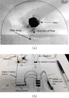
(a) Micrograph of the inlet of a microfluidic device for selective capture and release of cells using degradable hydrogels. The pillar-array geometry increases the likelihood of collisions between the cells and the hydrogel coating. The device is symmetrical, where the outlet is similar to the inlet. (b) Sequence of devices for adhesion-based microfluidic separation of cells against multiple surface markers. Following capture and release from device (i), cells expressing marker 1 enter device (ii) where a calcium chloride solution is co-injected to neutralize the EDTA present in the cell suspension. Device (iii) mixes the calcium chloride solution and cell suspension. Device (iv) captures cells against marker 2, which can then be eluted out using an injection of EDTA solution. Figure from Hatch et al. (2012) (Permission Pending).
Figure 5
Figure 5
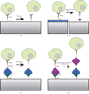
Schemes of potential cell capture/release mechanisms. (a) Shear stress is applied to remove cells from capture antibodies. (b) Capture molecules are functionalized to a degradable surface. When the surface is removed, cells are released. (c) A photo- or electrodegradable linker between antibodies and other proteins is used to tag specific cells with biotin. Cells are released when the linker molecule is destroyed. (d) Avidin-desthiobiotin binding is disrupted by competitive binding from biotin, releasing captured cells.
Figure 6
Figure 6
Cardiovascular tissue engineering. (a) Accordion-like honeycomb scaffolds for anisotropic cardiac tissue engineering (Engelmayr et al., 2008). (b) Microfluidic vessel network (Zheng et al., 2012). (c) Microfluidic tissue gauges used to measure temporal contractility response of micro-tissue (Legant et al., 2009).
Figure 7
Figure 7
Characterization of functionalized collagen scaffolds with immobilized VEGF+Ang1 growth factors. (a) SEM image of collagen sponge. (b) Tube formation H5V cells on collagen scaffold immobilized with VEGF+Ang1. (c) and (d) show cell morphology and organization after 7 days of cultivation in vitro, using (c) hemotoxylin and eosin staining, and (d) Von Willebrand factor staining. Figure adapted from Chiu and Radisic (2010a).

Similar articles

Cited by

References

    1. Au HT, Cheng I, Chowdhury MF, Radisic M. Interactive effects of surface topography and pulsatile electrical field stimulation on orientation and elongation of fibroblasts and cardiomyocytes. Biomaterials. 2007;28(29):4277–4293. - PMC - PubMed
    1. Beltrami AP, Barlucchi L, Torella D, Baker M, Limana F, Chimenti S, Kasahara H, Rota M, Musso E, Urbanek K, Leri A, Kajstura J, Nadal-Ginard B, Anversa P. Adult cardiac stem cells are multipotent and support myocardial regeneration. Cell. 2003;114(6):763–776. - PubMed
    1. Bergmann O, Mhardwaj RD, Bernard S, Zdunek S, Barnabe-Heider F, Walsh S, Zupicich J, Alkass K, Buchholz BA, Druid H, Jovinge S, Frisen J. Evidence for cardiomyocyte renewal in humans. Science. 2009;324:98–102. - PMC - PubMed
    1. Black LDr, Meyers JD, Weinbaum JS, Shvelidze YA, Tranquillo RT. Cell-induced alignment augments twitch force in fibrin gel-based engineered myocardium via gap junction modification. Tissue Eng Part A. 2009;15(10):3099–3108. - PMC - PubMed
    1. Boudou T, Legant WR, Mu A, Borochin MA, Thavandiran N, Radisic M, Zandstra PW, Epstein JA, Margulies KB, Chen CS. A microfabricated platform to measure and manipulate the mechanics of engineered cardiac microtissues. Tissue engineering. Part A. 2012 - PMC - PubMed

Publication types

LinkOut - more resources