. 2012 Nov 15;72(22):6013-23.
doi: 10.1158/0008-5472.CAN-12-0895. Epub 2012 Sep 4.

A synthetic matrix with independently tunable biochemistry and mechanical properties to study epithelial morphogenesis and EMT in a lung adenocarcinoma model

Affiliations

A synthetic matrix with independently tunable biochemistry and mechanical properties to study epithelial morphogenesis and EMT in a lung adenocarcinoma model

Bartley J Gill et al. Cancer Res. .

Abstract

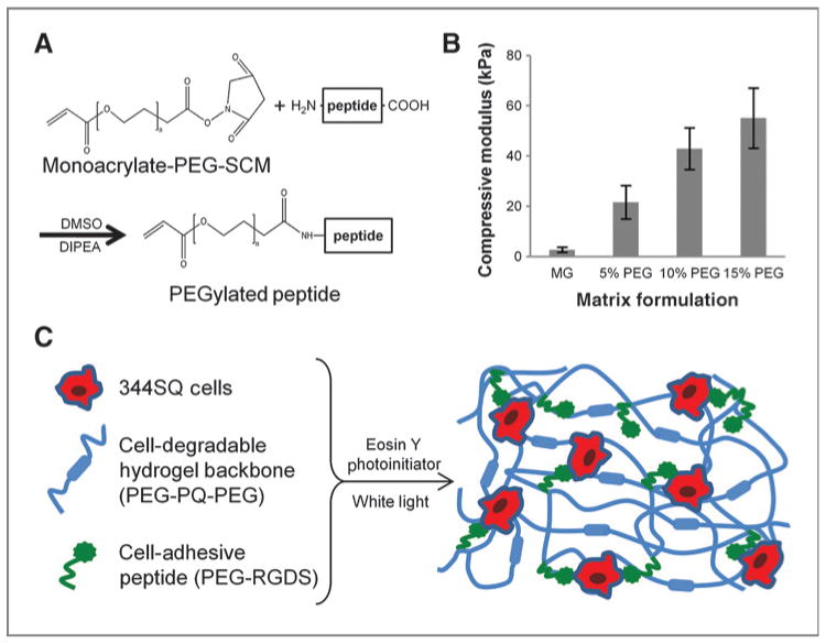
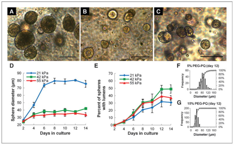
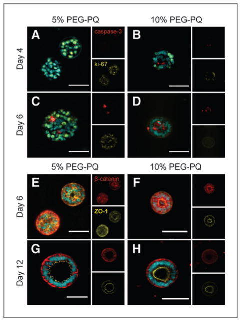
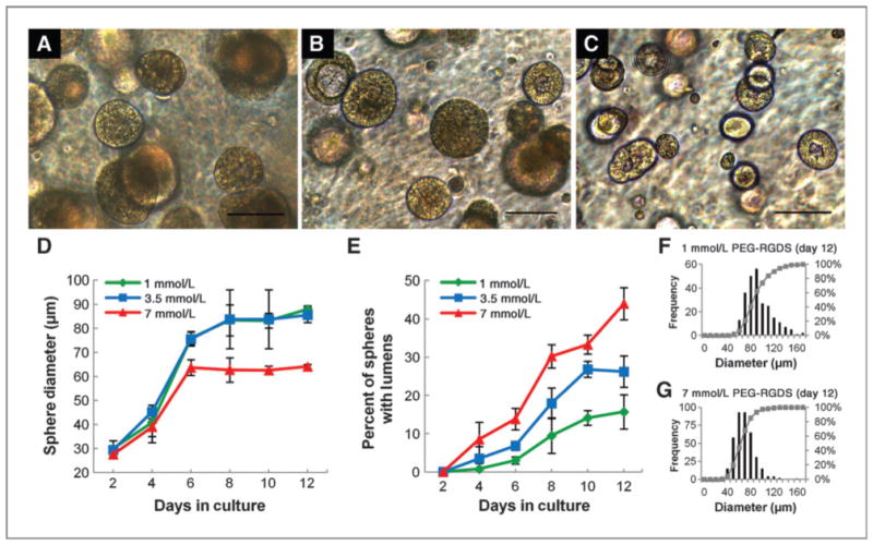
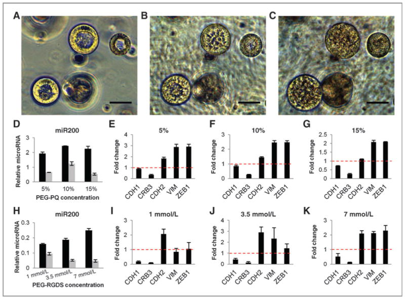
Better understanding of the biophysical and biochemical cues of the tumor extracellular matrix environment that influence metastasis may have important implications for new cancer therapeutics. Initial exploration into this question has used naturally derived protein matrices that suffer from variability, poor control over matrix biochemistry, and inability to modify the matrix biochemistry and mechanics. Here, we report the use of a synthetic polymer-based scaffold composed primarily of poly(ethylene glycol), or PEG, modified with bioactive peptides to study murine models of lung adenocarcinoma. In this study, we focus on matrix-derived influences on epithelial morphogenesis of a metastatic cell line (344SQ) that harbors mutations in Kras and p53 (trp53) and is prone to a microRNA-200 (miR-200)-dependent epithelial-mesenchymal transition (EMT) and metastasis. The modified PEG hydrogels feature biospecific cell adhesion and cell-mediated proteolytic degradation with independently adjustable matrix stiffness. 344SQ encapsulated in bioactive peptide-modified, matrix metalloproteinase-degradable PEG hydrogels formed lumenized epithelial spheres comparable to that seen with three-dimensional culture in Matrigel. Altering both matrix stiffness and the concentration of cell-adhesive ligand significantly influenced epithelial morphogenesis as manifest by differences in the extent of lumenization, in patterns of intrasphere apoptosis and proliferation, and in expression of epithelial polarity markers. Regardless of matrix composition, exposure to TGF-β induced a loss of epithelial morphologic features, shift in expression of EMT marker genes, and decrease in mir-200 levels consistent with EMT. Our findings help illuminate matrix-derived cues that influence epithelial morphogenesis and highlight the potential utility that this synthetic matrix-mimetic tool has for cancer biology.


Conflict of interest statement

Disclosure of Potential Conflicts of Interest: No potential conflicts of interest were disclosed.

Figures

Figure 1
Figure 1
Bioactive PEG matrix with tunable mechanical properties. A, bioactive peptides are reacted with monoacrylate-PEG-SCM to form pegylated peptides: PEG-RGDS to permit cell adhesion and, with a 2-molar excess of PEG-SCM, PEG-PQ-PEG that serves as a matrix backbone susceptible to cell-mediated degradation via cleavage by MMP-2 and 9. B, elastic moduli derived from uniaxial compressive testing show matrix stiffness can be tuned by altering weight percent PEG incorporated in the polymer mixture. All PEG matrices are stiffer than Matrigel (MG; P < 0.01). C, matrix components are mixed with 344SQ cells and a photoinitiator and polymerized to form a PEG hydrogel featuring independently tunable matrix stiffness and adhesive ligand concentration with encapsulated cells interacting with bioactive components.
Figure 2
Figure 2
Culture in PEG matrix recapitulates3D cultureinMatrigel. A and B, bright-field images of 344SQ WT cells encapsulated in Matrigel (A) or a 10% PEG-PQ/3.5 mmol/L PEG-RGDS matrix (B) after 10 days in culture show sphere lumenization. C and D, staining for basolateral marker α6-integrin (red) and apical marker ZO-1 (green) in 344SQ cells encapsulated in Matrigel (C) and PEG (D) indicate that spheres have adopted organized epithelial polarity. E to G, staining for polarity markers β-catenin (red) and ZO-1 (green) of 344P (E), 429 (F), and 393P (G) cells encapsulated in PEG-PQ matrices recapitulate morphology and polarity seen in 3D Matrigel culture (13). DAPI, blue. Scale bar, 50 μm.
Figure 3
Figure 3
Matrix stiffness influences epithelial morphogenesis. A–C, bright-field images of spheres at 12 days in culture in 5% PEG-PQ (A; 21 kPa),10% PEG-PQ (B; 42 kPa), and 15% PEG-PQ (C; 55 kPa) matrices with 3.5 mmol/L PEG-RGDS show differences in sphere size and lumenization. D, significantly larger spheres form in the softer hydrogel (day 12, 21 kPa vs. others, P < 0.01), whereas in E, a higher degree of lumenization occurred in stiffer matrices (day 12, 42 kPa compared with 21 kPa, P < 0.05). F and G, histograms of pooled size data for all spheres in all samples show much greater heterogeneity in sphere development in softer hydrogels. Scale bar, 100 μm.
Figure 4
Figure 4
Matrix stiffness influences apoptosis and proliferation in developing spheres and degree of epithelial polarity. A–D, staining for cleaved caspsase-3 (red) and ki-67 (yellow) at day 4 (A and B) and day 6 (C and D) in 5% (A and C) and 10% (B and D) hydrogels show earlier localization of proliferation at sphere periphery (B) in the stiffer hydrogel and high core apoptosis activity to induce more rapid lumenization (D) compared with diffuse proliferation activity (A) and unlocalized apoptosis (C) in softer hydrogels. E-H, staining for polarity markers b-catenin (red) and ZO-1 (yellow) at day 6 (E and F) and day 12 (G and H) in 5% (E and G) and 10% (F and H) PEG-PQ hydrogels shows polar organization in spheres in only stiff hydrogels (F) at early time points compared with disorganization in softer hydrogels (E), whereas at later time points, lumenized spheres in both softer and stiffer matrices show a high degree of organized polarity (G and H) with a notable absence of basolateral β-catenin on the apical edge. DAPI, cyan. Scale bar, 50 μm.
Figure 5
Figure 5
Adhesive ligand concentration influences epithelial morphogenesis. A–C, bright-field images of spheres at 12 days in culture in 5% PEG-PQ gels with PEG-RGDS at a concentration of 1 mmol/L (A), 3.5 mmol/L (B), or 7 mmol/L (C) show differences in sphere size and lumenization. D, smaller spheres formed in matrices with a high PEG-RGDS concentration (7 mmol/L vs. others, P < 0.01 at day 12), whereas in E, lumenization occurred more rapidly and completely in spheres encapsulated in matrices with higher PEG-RGDS concentrations (day 12, 7 mmol/L vs. others, P < 0.01 and 3.5 mmol/L vs. 1 mmol/L, P < 0.05). F and G, histograms of pooled size data show greater heterogeneity in sphere development in hydrogels with little adhesive ligand. Scale bar, 100 μm.
Figure 6
Figure 6
Adhesive ligand concentration influences apoptosis and proliferation in developing spheres and degree of epithelial polarity. A–D, representative images stained for cleaved caspsase-3 (red) and ki-67 (yellow) at day 4 (A and B) and day 6 (C and D) in 5% PEG-PQ hydrogels with 1 mmol/L (A and C) or 7 mmol/L (B and D) PEG-RGDS. Early peripheral proliferation and central apoptosis localization is evident with higher adhesive ligand concentrations (B) followed by rapid and organized lumenization (D), whereas spheres in matrices with low adhesive ligand concentration show a high degree of proliferation (A) and unlocalized apoptotic activity (C). E–H, representative images stained for polarity markers β-catenin (red) and ZO-1 (yellow) at day 6 (E and F) and day 12 (G and H) in 5% matrices with PEG-RGDS at 1 mmol/L (E and G) or 7 mmol/L (F and H) show the appearance of spheres with polar organization at early time points in matrices with high adhesive ligand concentration (F) with completely polar structures widespread at later time points (H). Spheres in hydrogels with low adhesive ligand concentration do not show organized polarity at early time points (E), and, at later time points, spheres that do display lumenization and polarity (G) have adopted less organized, more irregular morphologies. DAPI, cyan. Scale bar, 50 μm.
Figure 7
Figure 7
PEG-encapsulated structures show EMT-related morphologic, epigenetic, and gene expression changes with exposure to TGF-β. A–C, bright-field images of lumenized spheres encapsulated in a 5% PEG-PQ, 7 mmol/L PEG-RGDS hydrogel at 12 days in culture before 5 ng/mL TGF-β exposure (A) show breakdown of lumen organization after 1 day of treatment (B) and complete lumen filling and loss of organization after 4 days (C). Scale bar, 50 μm. D–K, q-PCR data for miR-200b before (black bars) and after (gray bars) TGF-β and fold change in mRNA of EMT-related genes with TGF-β treatment in matrices with 3.5 mmol/L PEG-RGDS and varied stiffness (D–G) and matrices with fixed 5% PEG-PQ stiffness and varied PEG-RGDS concentration (H–K) show a decrease in miR-200, decreased expression of epithelial genes (CDH1 and CRB3), and increased expression of mesenchymal genes (CHD2, VIM, and ZEB1) in all matrix formulations following exposure consistent with EMT-related genetic and epigenetic changes seen in 344SQ in Matrigel cultures (for every formulation, miR-200b before vs. after TGF-β, P < 0.01). miR-200 is expressed relative to miR16 levels and fold change mRNA relative to L32 mRNA levels.

References

    1. Thiery JP, Acloque H, Huang RYJ, Nieto MA. Epithelial-mesenchymal transitions in development and disease. Cell. 2009;139:871–90. - PubMed
    1. Provenzano PP, Inman DR, Eliceiri KW, Trier SM, Keely PJ. Contact guidance mediated three-dimensional cell migration is regulated by Rho/ROCK-dependent matrix reorganization. Biophys J. 2008;95:5374–84. - PMC - PubMed
    1. Provenzano PP, Inman DR, Eliceiri KW, Knittel JG, Yan L, Rueden CT, et al. Collagen density promotes mammary tumor initiation and progression. BMC Med. 2008;6:11. - PMC - PubMed
    1. Provenzano PP, Eliceiri KW, Campbell JM, Inman DR, White JG, Keely PJ. Collagen reorganization at the tumor-stromal interface facilitates local invasion. BMC Med. 2006;4:38. - PMC - PubMed
    1. Levental KR, Yu H, Kass L, Lakins JN, Egeblad M, Erler JT, et al. Matrix crosslinking forces tumor progression by enhancing integrin signaling. Cell. 2009;139:891–906. - PMC - PubMed

Publication types

MeSH terms