. 2012 Oct 19;287(43):36322-30.
doi: 10.1074/jbc.M112.402149. Epub 2012 Sep 5.

Hallmarks of processivity in glycoside hydrolases from crystallographic and computational studies of the Serratia marcescens chitinases

Affiliations

Hallmarks of processivity in glycoside hydrolases from crystallographic and computational studies of the Serratia marcescens chitinases

Christina M Payne et al. J Biol Chem. .

Abstract

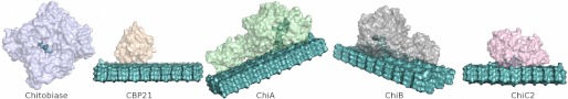
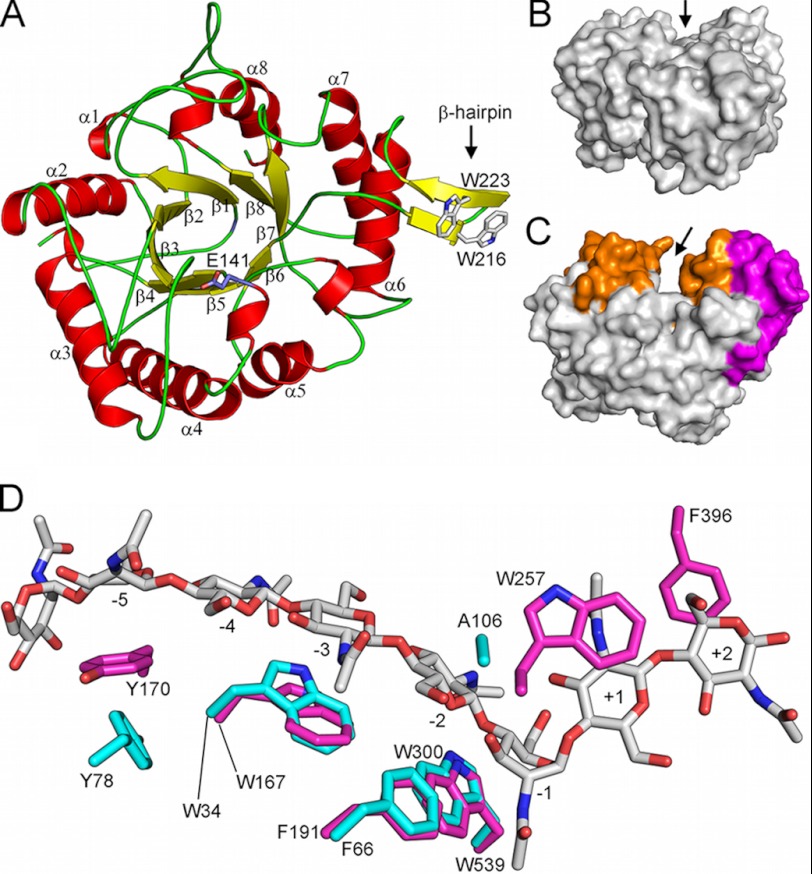
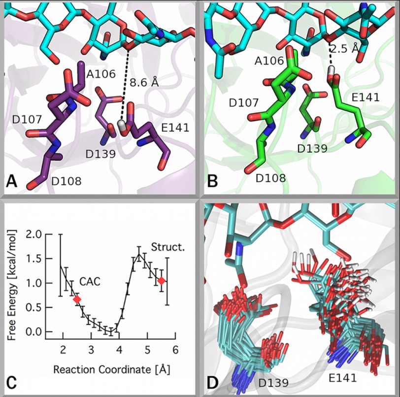
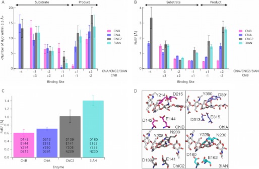
Degradation of recalcitrant polysaccharides in nature is typically accomplished by mixtures of processive and nonprocessive glycoside hydrolases (GHs), which exhibit synergistic activity wherein nonprocessive enzymes provide new sites for productive attachment of processive enzymes. GH processivity is typically attributed to active site geometry, but previous work has demonstrated that processivity can be tuned by point mutations or removal of single loops. To gain additional insights into the differences between processive and nonprocessive enzymes that give rise to their synergistic activities, this study reports the crystal structure of the catalytic domain of the GH family 18 nonprocessive endochitinase, ChiC, from Serratia marcescens. This completes the structural characterization of the co-evolved chitinolytic enzymes from this bacterium and enables structural analysis of their complementary functions. The ChiC catalytic module reveals a shallow substrate-binding cleft that lacks aromatic residues vital for processivity, a calcium-binding site not previously seen in GH18 chitinases, and, importantly, a displaced catalytic acid (Glu-141), suggesting flexibility in the catalytic center. Molecular dynamics simulations of two processive chitinases (ChiA and ChiB), the ChiC catalytic module, and an endochitinase from Lactococcus lactis show that the nonprocessive enzymes have more flexible catalytic machineries and that their bound ligands are more solvated and flexible. These three features, which relate to the more dynamic on-off ligand binding processes associated with nonprocessive action, correlate to experimentally measured differences in processivity of the S. marcescens chitinases. These newly defined hallmarks thus appear to be key dynamic metrics in determining processivity in GH enzymes complementing structural insights.


Figures

FIGURE 1.
FIGURE 1.
The chitinolytic system from S. marcescens. The enzyme system comprises three family GH18 chitinases, processive ChiA and ChiB and nonprocessive ChiC, a family GH20 chitobiase that cleaves chitobiose and CBP21, a lytic polysaccharide monooxygenase. The structures of all enzymes, except ChiC, have been reported previously (, , , , –35). The enzymes are shown with their substrates in light teal. An SDS-PAGE gel of the entire system has been included in supplemental Fig. S1.
FIGURE 2.
FIGURE 2.
Overall structure of the catalytic module of ChiC, ChiC2. A, ChiC2 exhibits an (a/b)8 TIM barrel fold with the catalytic glutamate, Glu-141, positioned between β-strand 4 and α-helix 4 (side chain shown in stick representation). Note the β-hairpin subdomain containing two exposed tryptophans. B and C, comparison of ChiC2 (nonprocessive) with ChiA (processive, PDB ID: 1EHN (29)) shows the difference in depth of the substrate-binding clefts that are suggestive of the nonprocessive and processive properties of these enzymes, respectively. The “walls” of the ChiA substrate-binding cleft are constituted from several insertions (colored orange) in addition to a large a+b subdomain insertion (colored magenta). Arrows indicate the binding clefts. D, aromatic amino acids lining the substrate-binding cleft of ChiA (magenta sticks) and ChiC2 (cyan sticks). The substrate (GlcNAc)8 bound to the 1EHN ChiA structure is shown in gray stick representation, and subsites to which the sugars are bound are indicated. Nitrogen and oxygen atoms are colored blue and red, respectively. Note the lack of aromatic amino acids in the +1 and +2 subsites of the ChiC2 substrate-binding cleft; Tyr-275 of the +1 subsite in ChiA is replaced with an Ala in ChiC2, and Phe-396 in the +2 subsite of ChiA is part of a loop that does not exist in ChiC.
FIGURE 3.
FIGURE 3.
The catalytic center of ChiC. The catalytic glutamate of ChiC exhibits a conformation inconsistent with the putative catalytically active conformation (CAC) (19). A and B, illustrations of these conformations are given showing the conformation found in the ChiC2 crystal structure (A) and showing the putative catalytically active conformation from an MD simulation (B). C, the free energy difference of the catalytic glutamate between the solvent-exposed conformation and the catalytically active complex. A reaction coordinate value of ∼2.5 Å corresponds to the catalytically active complex (19), and a value near 5.5 Å corresponds to the Glu-141 conformation observed in the crystal structure (4AXN). Error bars were obtained by bootstrapping in WHAM. D, superposition of frames from MD simulation of ChiC2 with a GlcNAc7 ligand bound (see supplemental text for details) illustrating the significant flexibility of Glu-141.
FIGURE 4.
FIGURE 4.
MD simulations of two processive chitinases, ChiA and ChiB, and two nonprocessive chitinases, ChiC2 and 3IAN. All simulations were done on enzymes in which an oligomer of GlcNAc (GlcNAc5 in ChiB, GlcNAc6 in ChiA and 3IAN, and GlcNAc7 in ChiC2) had been docked to the −4 to +2 subsites of the reducing end enzymes and the +3 to −2 subsites of ChiB (see supplemental text for additional details). In panels A and B, only subsites appearing in all four enzymes are shown. The product and substrate subsite definitions are as shown above panel A. The panels show the average solvation of the ligand by binding site (A), the root mean square fluctuations (RMSF) of the ligand by the binding site (B), and the root mean square fluctuations of the tetrad of catalytic residues, listed for each enzyme (C). Error bars in panel A represent one standard deviation. Error bars in panels B and C were obtained with block averaging. D, the catalytic tetrad of each enzyme examined here with simulation with the residues labeled. These residues represent the set for analysis in panel C. The enzyme structures used for the simulation input are described in detail in the supplemental text.

References

    1. Himmel M. E., Ding S. Y., Johnson D. K., Adney W. S., Nimlos M. R., Brady J. W., Foust T. D. (2007) Biomass recalcitrance: engineering plants and enzymes for biofuels production. Science 315, 804–807 - PubMed
    1. Vaaje-Kolstad G., Westereng B., Horn S. J., Liu Z., Zhai H., Sørlie M., Eijsink V. G. (2010) An oxidative enzyme boosting the enzymatic conversion of recalcitrant polysaccharides. Science 330, 219–222 - PubMed
    1. Mba Medie F., Davies G. J., Drancourt M., Henrissat B. (2012) Genome analyses highlight the different biological roles of cellulases. Nat. Rev. Microbiol. 10, 227–234 - PubMed
    1. Bayer E. A., Belaich J. P., Shoham Y., Lamed R. (2004) The cellulosomes: multienzyme machines for degradation of plant cell wall polysaccharides. Annu. Rev. Microbiol. 58, 521–554 - PubMed
    1. Demain A. L., Newcomb M., Wu J. H. (2005) Cellulase, Clostridia, and ethanol. Microbiol. Mol. Biol. Rev. 69, 124–154 - PMC - PubMed

Publication types

MeSH terms

Associated data

LinkOut - more resources