. 2012 Oct 19;287(43):36509-17.
doi: 10.1074/jbc.M112.376111. Epub 2012 Sep 5.

Suppressor of cytokine signaling 6 (SOCS6) negatively regulates Flt3 signal transduction through direct binding to phosphorylated tyrosines 591 and 919 of Flt3

Affiliations

Suppressor of cytokine signaling 6 (SOCS6) negatively regulates Flt3 signal transduction through direct binding to phosphorylated tyrosines 591 and 919 of Flt3

Julhash U Kazi et al. J Biol Chem. .

Abstract

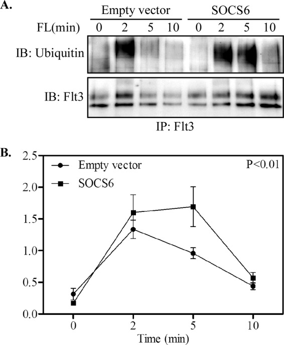
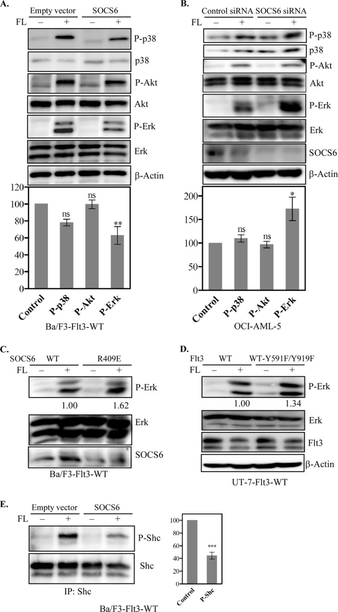
The receptor tyrosine kinase Flt3 is an important growth factor receptor in hematopoiesis, and gain-of-function mutations of the receptor contribute to the transformation of acute myeloid leukemia. SOCS6 (suppressor of cytokine signaling 6) is a member of the SOCS family of E3 ubiquitin ligases that can regulate receptor tyrosine kinase signal transduction. In this study, we analyzed the role of SOCS6 in Flt3 signal transduction. The results show that ligand stimulation of Flt3 can induce association of SOCS6 and Flt3 and tyrosine phosphorylation of SOCS6. Phosphopeptide fishing indicated that SOCS6 binds directly to phosphotyrosines 591 and 919 of Flt3. By using stably transfected Ba/F3 cells with Flt3 and/or SOCS6, we show that the presence of SOCS6 can enhance ubiquitination of Flt3, as well as internalization and degradation of the receptor. The presence of SOCS6 also induces weaker activation of Erk1/2, but not Akt, in transfected Ba/F3 and UT-7 cells and in OCI-AML-5 cells. The absence of SOCS6 promotes Ba/F3 and UT-7 cell proliferation induced by oncogenic internal tandem duplications of Flt3. Taken together, these results suggest that SOCS6 negatively regulates Flt3 activation, the downstream Erk signaling pathway, and cell proliferation.


Figures

FIGURE 1.
FIGURE 1.
SOCS6 interacts with Flt3 in response to FL-stimulation. A, COS-1 cells were cotransfected with SOCS6 and the WT Flt3 expression vector. Cells were serum-starved overnight and stimulated by FL or not for 5 min before lysis. Cell lysates were subjected to immunoprecipitation (IP) using either anti-Flt3 or anti-SOCS6 antibody and analyzed by Western blotting. B, COS-1 cells were cotransfected with SOCS6 and Flt3 ITD. Cells were then processed as described for A. C, OCI-AML-5 cells were starved overnight and stimulated with FL for 5 min. Cells were lysed and processed for immunoprecipitation and Western blotting. D, Ba/F3 cells transfected with Flt3 ITD and SOCS6 were starved for 4 h, followed by 5 min of FL stimulation. Cells were lysed and processed for immunoprecipitation and Western blotting. E, transfected COS-1 cells were serum-starved overnight and then treated with 100 ng/ml FL for the indicated times before lysis and immunoprecipitated with anti-SOCS6 antibody. F, COS-1 cells were transfected with Flt3 and SOCS6 mutants. After overnight starvation, cells were treated with 100 ng/ml FL for 5 min before lysis and immunoprecipitated with anti-SOCS6 antibody. TCL, total cell lysate.
FIGURE 2.
FIGURE 2.
Identification of Tyr-591 and Tyr-919 as SOCS6-binding sites. A, phosphopeptides corresponding to 12 known tyrosine phosphorylation sites in Flt3 were synthesized and immobilized on UltraLink. Peptide-bound slurry was incubated with SOCS6-transfected COS-1 cell lysates, and pulled down proteins were then processed for Western blotting using anti-SOCS6 antibody to screen the SOCS6-binding sites. The c-Kit Tyr-568 peptide was used as a positive control. pY, phospho-Tyr; IB, immunoblot. B, immobilized phospho- and non-phosphopeptides were used to pull down SOCS6 proteins and processed as described for A to determine whether the binding was dependent on tyrosine phosphorylation. C, COS-1 cells were cotransfected with SOCS6 and WT Flt3 or Flt3(Y591F/Y919F). Cells were serum-starved and stimulated with FL for 5 min before lysis. Lysates were immunoprecipitated (IP) with anti-Flt3 or anti-SOCS6 antibody and analyzed by Western blotting to show that Tyr-591 and Tyr-919 are the SOCS6-binding sites in vivo.
FIGURE 3.
FIGURE 3.
Expression of Flt3 and SOCS6 in Ba/F3 cells. A, stably transfected Ba/F3 cells were labeled with an isotype control or phycoerythrin-conjugated anti-Flt3 antibody and analyzed by flow cytometry. The gray line indicates cells labeled with the isotype control, and the black line indicates cells labeled with anti-Flt3 antibody. EV, empty vector. B, lysates of stably transfected Ba/F3-Flt3 cells with or without SOCS6 were immunoprecipitated (IP) with anti-SOCS6 antibody and analyzed by Western blotting. IB, immunoblot. C, endogenous SOCS6 and Flt3 expression was detected using specific antibodies in different AML cell lines.
FIGURE 4.
FIGURE 4.
FL induces SOCS6 gene expression in Ba/F3-Flt3 cells. A, Ba/F3-Flt3 cells were serum-starved and stimulated with FL for the indicated times. Cells were lysed, and total RNAs were isolated. Expression of SOCS6 mRNA was analyzed by qPCR as described under “Experimental Procedures.” B, SOCS6 expression in acute promyelocytic leukemia patients carrying either WT Flt3 or the Flt3 ITD mutation. C, SOCS6 expression in an AML patient and in corresponding normal cells. D, SOCS6 expression in AML patients with complete remission or relapse and in corresponding normal cells. ns, not significant. *, p < 0.05; ***, p < 0.001.
FIGURE 5.
FIGURE 5.
SOCS6 expression increases ubiquitination of the Flt3 receptor. A, Ba/F3-Flt3 cells with or without SOCS6 were serum-starved and stimulated with 100 ng/ml FL for the indicated times. Cells were lysed, and lysates were immunoprecipitated (IP) with anti-Flt3 antibody, followed by Western blotting. IB, immunoblot. B, signal intensities were quantified using Multi-Gauge software to calculate the receptor ubiquitination.
FIGURE 6.
FIGURE 6.
SOCS6 expression increases Flt3 receptor internalization and degradation. A, Ba/F3-Flt3 cells with or without SOCS6 were serum-starved and stimulated with 100 ng/ml FL for the indicated times. Cells were transferred to ice, immediately followed by incubation with phycoerythrin-conjugated anti-Flt3 antibody. The Flt3 surface expression level was analyzed by flow cytometry. Internalization of Flt3 was quantified compared with unstimulated cells. B, Ba/F3-Flt3 cells with or without SOCS6 were serum-starved and preincubated with cycloheximide for 4 h. Cells were then stimulated with 100 ng/ml FL for 30 min before lysis. Cell lysates were immunoprecipitated with anti-Flt3 antibody and analyzed by Western blotting. Signal intensities were quantified using Multi-Gauge software to calculate the receptor degradation. **, p < 0.01.
FIGURE 7.
FIGURE 7.
Effects of SOCS6 expression on FL-induced activation of Erk, p38, Akt, and Shc. A, Ba/F3-Flt3 cells with or without SOCS6 were treated with 100 ng/ml FL or not for 5 min before lysis. Total cell lysates were separated by SDS-PAGE, and membranes were probed with phospho-specific antibodies. Membranes were then stripped and reprobed with the respective antibodies to show loading. Signal intensities from three independent experiments were quantified using Multi-Gauge software to calculate the inhibition. ns, not significant. **, p < 0.01. B, OCI-AML-5 cells were transfected with control or SOCS6 siRNA using electroporation. One day after transfection, cells were starved overnight and stimulated with FL, followed by Western blot analysis. *, p < 0.05. C, Ba/F3-Flt3 cells transfected with WT SOCS6 or the SOCS6(R409E) mutant were starved for 4 h before a 5-min FL stimulation. Cells were then processed for Western blotting. D, UT-7 cells transfected with WT Flt3 or Flt3(Y591F/Y919F) were starved overnight before a 5-min FL stimulation. Cells were then processed for Western blotting. E, cell lysates were immunoprecipitated (IP) with anti-Shc antibody and analyzed by Western blotting. Signal intensities were quantified using Multi-Gauge software to calculate the inhibition. ***, p < 0.001.
FIGURE 8.
FIGURE 8.
Presence of SOCS6 down-regulates cell proliferation of Ba/F3-Flt3-ITD cells. A, Ba/F3-Flt3-ITD cells with or without SOCS6 (Empty vector) were grown for 48 h in the presence or absence of ligand. Viable cells were counted by the trypan blue exclusion method. ns, not significant. B, cells were labeled with annexin V and 7-aminoactinomycin D, and living cells were measured by flow cytometry. IL-3 was used as a positive control. Ba/F3-Flt3-ITD/empty vector cells or Ba/F3-Flt3-ITD/SOCS6-WT cells (C), Ba/F3-Flt3-ITD/SOCS6-WT cells or Ba/F3-Flt3-ITD/SOCS6-R409E (SOCS6-RE) cells (D), and UT-7-Flt3-ITD cells or UT-7-Flt3-ITD-Y591F/Y919F (Flt3-ITD-YYFF) cells (E) were washed to remove IL-3 and seeded in 96-well plates. Cells were then treated with either ligand or not for 48 h, followed by PrestoBlue cell viability assays. *, p < 0.05; **, p < 0.01; ***, p < 0.001.

Similar articles

Cited by

References

    1. Masson K., Rönnstrand L. (2009) Oncogenic signaling from the hematopoietic growth factor receptors c-Kit and Flt3. Cell. Signal. 21, 1717–1726 - PubMed
    1. Pawson T. (2004) Specificity in signal transduction: from phosphotyrosine-SH2 domain interactions to complex cellular systems. Cell 116, 191–203 - PubMed
    1. Levis M., Small D. (2003) FLT3: ITDoes matter in leukemia. Leukemia 17, 1738–1752 - PubMed
    1. Stirewalt D. L., Kopecky K. J., Meshinchi S., Engel J. H., Pogosova-Agadjanyan E. L., Linsley J., Slovak M. L., Willman C. L., Radich J. P. (2006) Size of FLT3 internal tandem duplication has prognostic significance in patients with acute myeloid leukemia. Blood 107, 3724–3726 - PMC - PubMed
    1. Meshinchi S., Alonzo T. A., Stirewalt D. L., Zwaan M., Zimmerman M., Reinhardt D., Kaspers G. J., Heerema N. A., Gerbing R., Lange B. J., Radich J. P. (2006) Clinical implications of FLT3 mutations in pediatric AML. Blood 108, 3654–3661 - PMC - PubMed

Publication types

MeSH terms

Substances