. 2012 Nov 15;303(10):F1454-9.
doi: 10.1152/ajprenal.00206.2011. Epub 2012 Sep 5.

Intrarenal localization of the plasma membrane ATP channel pannexin1

Affiliations

Intrarenal localization of the plasma membrane ATP channel pannexin1

Fiona Hanner et al. Am J Physiol Renal Physiol. .

Abstract

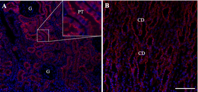
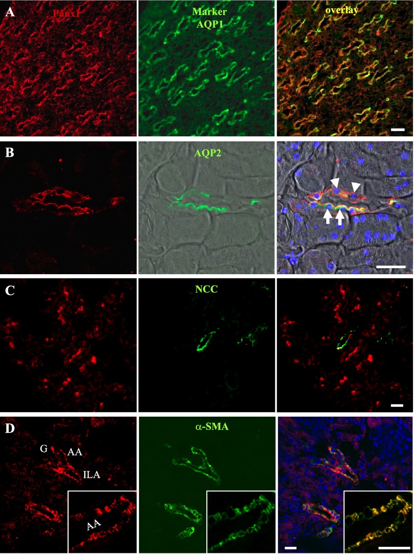
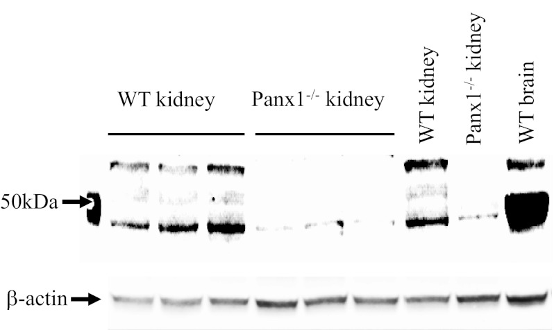
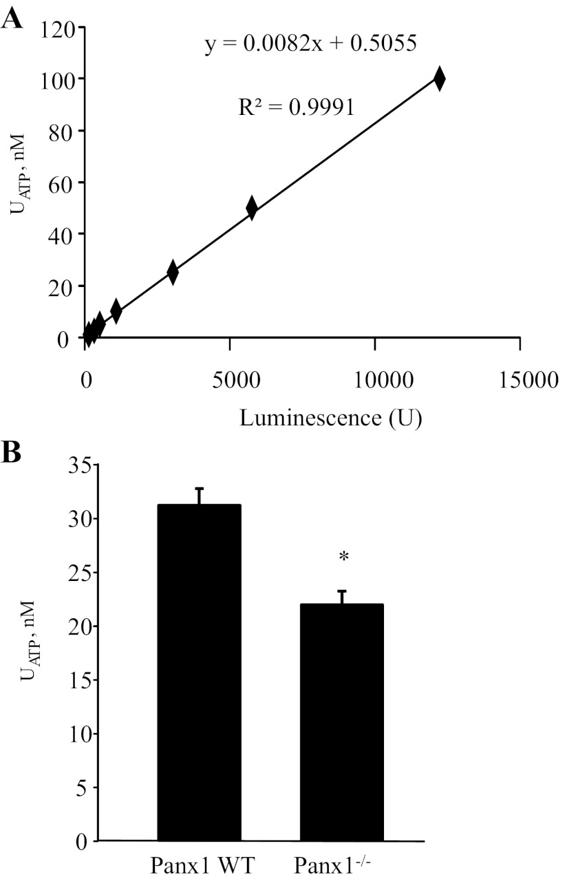
In the renal tubules, ATP released from epithelial cells stimulates purinergic receptors, regulating salt and water reabsorption. However, the mechanisms by which ATP is released into the tubular lumen are multifaceted. Pannexin1 (Panx1) is a newly identified. ubiquitously expressed protein that forms connexin-like channels in the plasma membrane, which have been demonstrated to function as a mechanosensitive ATP conduit. Here, we report on the localization of Panx1 in the mouse kidney. Using immunofluorescence, strong Panx1 expression was observed in renal tubules, including proximal tubules, thin descending limbs, and collecting ducts, along their apical cell membranes. In the renal vasculature, Panx1 expression was localized to vascular smooth muscle cells in renal arteries, including the afferent and efferent arterioles. Additionally, we tested whether Panx1 channels expressed in renal epithelial cells facilitate luminal ATP release by measuring the ATP content of urine samples freshly collected from wild-type and Panx1(-/-) mice. Urinary ATP levels were reduced by 30% in Panx1(-/-) compared with wild-type mice. These results suggest that Panx1 channels in the kidney may regulate ATP release and via purinergic signaling may participate in the control of renal epithelial fluid and electrolyte transport and vascular functions.


Figures

Fig. 1.
Fig. 1.
Overview of pannexin1 (Panx1) immunofluorescence in the mouse kidney. A: among cortical tubules, Panx1 labeling was strongest in proximal tubules (PT) in the apical brush border (inset). There was no signal in the glomerulus (G). B: in the medulla, intense Panx1 immunofluorescence was observed in the merging collecting duct (CD) system. In addition, thin-walled tubular structures showed weaker labeling. Nuclei were stained with 4,6-diamidino-2-phenylindole (DAPI; blue). Bar = 50 μm.
Fig. 2.
Fig. 2.
Immunofluorescence colabeling of Panx1 (red, left) with cell-specific markers (green, middle) in the mouse kidney and their merged images (overlay, right). A: in the loop of Henle, Panx1 (red) and aquaporin-1 (AQP1; green) colocalized (yellow) in descending thin limbs. B: Panx1 (red) was coexpressed with aquaporin-2 (AQP2; green), a marker of the principal cells in collecting ducts (arrows), mainly at the apical cell membranes. Labeling was also observed in the AQP2-negative intercalated cells (arrowheads). DIC background was added and merged with fluorescence. Nuclei were stained with DAPI (blue). C: colabeling of the distal convoluted tubule marker the sodium-chloride cotransporter (NCC; green) and Panx1 (red) failed to show any overlap in signal. D: immunofluorescence colabeling of Panx1 (red) and α-smooth muscle actin (α-SMA; green), a marker of vascular smooth muscle cells. Panx1 expression was found in vascular smooth muscle cells of large and small renal arteries, including interlobular arteries (ILA) and afferent arterioles (AA). A single AA is shown magnified in the inset. G, glomerulus. Bar = 20 μm.
Fig. 3.
Fig. 3.
Confirmation of kidney tissue Panx1 deficiency in Panx1−/− mice. Whole kidney homogenates collected from wild-type (WT) and Panx1−/− mice (3 animals each, 1 pair loaded twice as indicated) were immunoblotted and probed with Panx1 antibodies. Strong bands characteristic of Panx1 at ∼50 kDa were present in WT but not in Panx1−/− tissues. A very weak lower band was still detectable in Panx1−/− kidneys. A brain tissue homogenate served as a positive control. β-Actin was used as a loading control.
Fig. 4.
Fig. 4.
Panx1 is necessary for ATP release into the urine. A: calibration curve for ATP measurements. Changes in luminescence intensity were detected in response to serial dilutions of ATP (at concentrations of 1, 2.5, 5, 10, 25, 50, and 100 nM) using a luciferin- and luciferase-based ATP bioluminescence assay. The equation and R2 value of linear regression analysis are displayed. B: summary graph of ATP concentration in freshly harvested urine in WT and Panx1−/− mice (n = 10 each). Values are means ± SE. *P < 0.05 vs. Panx1 wild-type.

References

    1. Ambrosi C, Gassmann O, Pranskevich JN, Boassa D, Smock A, Wang J, Dahl G, Steinem C, Sosinsky GE. Pannexin1 and pannexin2 channels show quaternary similarities to connexons and different oligomerization numbers from each other. J Biol Chem 285: 24420–24431, 2010 - PMC - PubMed
    1. Bailey MA. Inhibition of bicarbonate reabsorption in the rat proximal tubule by activation of luminal P2Y1 receptors. Am J Physiol Renal Physiol 287: F789–F796, 2004 - PubMed
    1. Bao L, Locovei S, Dahl G. Pannexin membrane channels are mechanosensitive conduits for ATP. FEBS Lett 572: 65–68, 2004 - PubMed
    1. Baranova A, Ivanov D, Petrash N, Pestova A, Skoblov M, Kelmanson I, Shagin D, Nazarenko S, Geraymovych E, Litvin O, Tiunova A, Born TL, Usman N, Staroverov D, Lukyanov S, Panchin Y. The mammalian pannexin family is homologous to the invertebrate innexin gap junction proteins. Genomics 83: 706–716, 2004 - PubMed
    1. Barbe MT, Monyer H, Bruzzone R. Cell-cell communication beyond connexins: the pannexin channels. Physiology 21: 103–114, 2006 - PubMed

Publication types

LinkOut - more resources