. 2012 Aug;14(8):678-89.
doi: 10.1593/neo.12922.

Integrin-associated CD151 drives ErbB2-evoked mammary tumor onset and metastasis

Affiliations

Integrin-associated CD151 drives ErbB2-evoked mammary tumor onset and metastasis

Xinyu Deng et al. Neoplasia. 2012 Aug.

Abstract

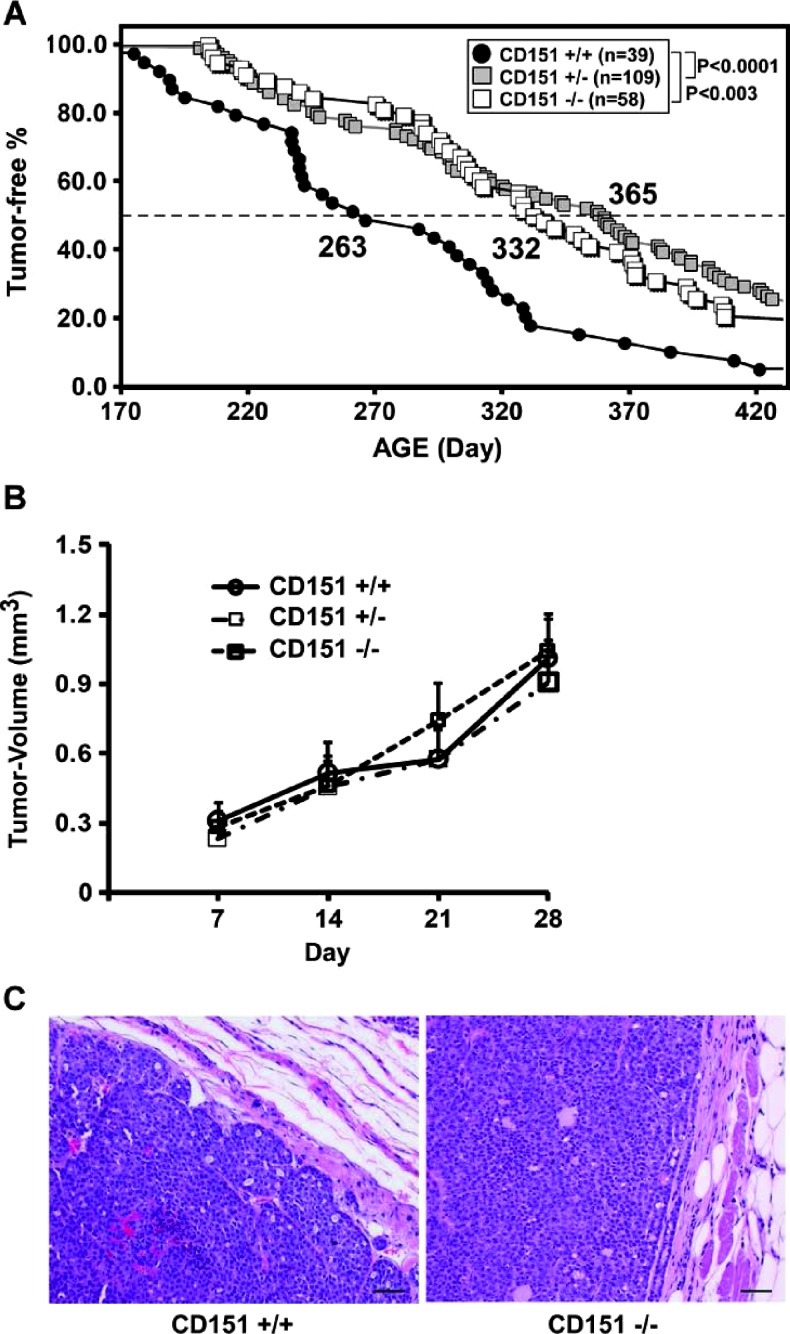
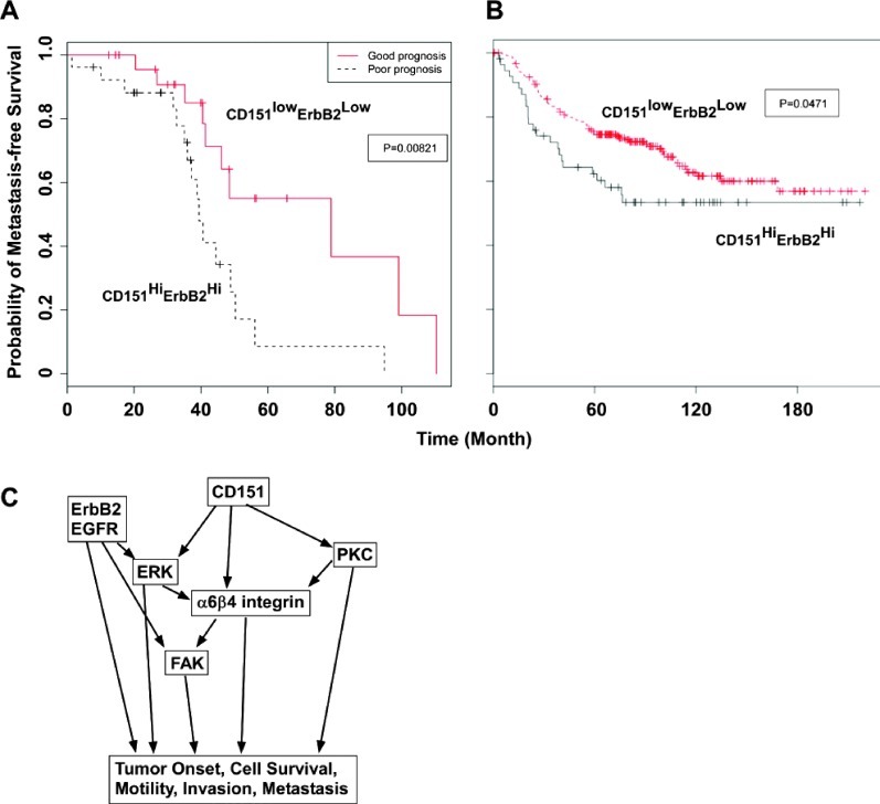
ErbB2+ human breast cancer is a major clinical problem. Prior results have suggested that tetraspanin CD151 might contribute to ErbB2-driven breast cancer growth, survival, and metastasis. In other cancer types, CD151 sometimes supports tumor growth and metastasis. However, a definitive test of CD151 effects on de novo breast cancer initiation, growth, and metastasis has not previously been done. We used CD151 gene-deleted mice expressing the MMTV-ErbB2 transgene to show that CD151 strongly supports ErbB2+ mammary tumor initiation and metastasis. Delayed tumor onset (by 70-100 days) in the absence of CD151 was accompanied by reduced survival of mammary epithelial cells and impaired activation of FAK- and MAPK-dependent pathways. Both primary tumors and metastatic nodules showed smooth, regular borders, consistent with a less invasive phenotype. Furthermore, consistent with impaired oncogenesis and decreased metastasis, CD151-targeted MCF-10A/ErbB2 cells showed substantial decreases in three-dimensional colony formation, EGF-stimulated tumor cell motility, invasion, and transendothelial migration. These CD151-dependent functions were largely mediated through α6β4 integrin. Moreover, CD151 ablation substantially prevented PKC- and EGFR/ERK-dependent α6β4 integrin phosphorylation, consistent with retention of epithelial cell polarity and intermediate filament cytoskeletal connections, which helps to explain diminished metastasis. Finally, clinical data analyses revealed a strong correlation between CD151 and ErbB2 expression and metastasis-free survival of breast cancer patients. In conclusion, we provide strong evidence that CD151 collaborates with LB integrins (particularly α6β4 and ErbB2 (and EGFR) receptors to regulate multiple signaling pathways, thereby driving mammary tumor onset, survival, and metastasis. Consequently, CD151 is a useful therapeutic target in malignant ErbB2+ breast cancer.


Figures

Figure 1
Figure 1
Impact of CD151 removal on ErbB2-induced mammary tumorigenesis. (A) Kaplan-Meier plots for tumor-free survival comparing mice with varying CD151 genotypes. The median time to tumor onset (in days) is shown. (B) Tumor volume (mean ± SEM) over time, starting with initial tumor detection, is indicated for tumor-bearing mice (for each group, n = 15). (C) Representative images of hematoxylin and eosin-stained primary tumor sections prepared from a total of 20 to 25 mice per CD151 genotype. Scale bar, 100 µm.
Figure 2
Figure 2
CD151 deletion and pulmonary metastasis. (A) Numbers of pulmonary lesions per mouse, plus average values, are indicated. Differences were assessed using nonparametric Kruskal-Wallis test; P values are indicated. Numbers of mice with pulmonary metastasis are indicated for each CD151 genotype. (B) Typical hematoxylin and eosin-stained sections of pulmonary lesions from wild-type and null tumor-bearing mice. Scale bar, 100 µm.
Figure 3
Figure 3
Effects of CD151 deletion on primary tumor proliferation and survival. (A) IHC analyses of sections of primary tumors from CD151 wild-type (WT) and null (KO) mice: (a and c) staining for Ki-67; (b and d) for caspase 3. Arrowheads indicate caspase 3-positive cells. (B) Quantitation of Ki-67 and caspase 3-positive cells (mean ± SEM). Numbers of positive cells per area were calculated for n = 5 mice for each CD151 genotype. Cleaved caspase 3-positive cells represent 1.5% and 7.5% of total cells in areas counted for WT and KO mice, respectively. Scale bar, 100 µm.
Figure 4
Figure 4
Loss of CD151 impairs signaling in mouse mammary tumors. (A) Lysates were prepared from four representative individual tumors from CD151 +/+ and -/- mice and blotted with indicated antibodies. The numbers below ERK1/2 and FAK represent phosphorylated/total protein ratios from densitometry measurements. Activation of ErbB2 and EGFR and their major downstream effectors was determined by blot analysis with antibodies recognizing Y1221/1222 of ErbB2, S473 of Akt, and T202/Y204 of ERK. (B) Changes in integrin-mediated tyrosine phosphorylation cascade. Phosphorylation of FAK and Src was detected using antibodies recognizing Y397 of FAK, Y416 of Src, and p130Cas, respectively. Results are representative of multiple independent analyses.
Figure 5
Figure 5
Ablation of CD151 decreases survival of MCF-10A/ErbB2 cells cultured in three dimensions. (A) Images of acinar-like structures are shown for cells expressing control short hairpin RNA (shRNA) (a, c) or CD151 shRNA (b, d) after 5 days of three-dimensional culture. Top panel: Representative fields were visualized using a phase-contrast microscope (a, b) and by confocal immunofluorescence imaging of antibody-stained colonies (c, d). Blue indicates DAPI staining of nuclei; green, antibody staining for smooth muscle actin (SMA). Scale bar, 50 µm. Right panel: Average colony areas in multiple random fields (mean ± SEM, n = 3). (B) Inhibition of acinar-like colony formation (mean ± SEM, n = 3). Three days after treatment with 0.5 µM lapatinib, 1.0 µM TAE266, 2.0 µM U0126, or 0.5 µM doxorubicin, colonies were imaged and quantitated. (C) Immunofluorescence analyses were carried out with indicated antibodies or DAPI for nuclear visualization. Confocal images of stained colonies: cells expressing control shRNA (a, c, e); CD151 shRNA (b, d, f). Staining for Ki-67 (a and b); caspase 3 (c and d); ERK1/2 (e and f). Blue indicates DAPI staining; red, antibody staining. Scale bar, 50 µm. Bottom panel: Percentages of positively stained cells (mean ± SEM; n = 3). All data are representative of multiple experiments. *P < .05, **P < .01, ***P < .001.
Figure 6
Figure 6
Ablation of CD151 inhibits tumor cell motility, invasion, and signaling. (A) To assess random motility, MCF-10A/ErbB2 cells (with or without CD151 knockdown) were treated with or without 10 ng/ml EGF or 5 ng/ml TGF-β1 for 12 hours. Twenty individual cells per treatment were tracked, and average distances traveled were calculated (mean ± SEM). (B) Differences in transendothelial migration (mean ± SEM, n = 6). EGF (10 ng/ml) was added to the bottom chamber as chemoattractant. (C) Invasive capabilities of MCF-10A/ErbB2 cells ± CD151 ablation (mean ± SEM, n = 3) and ±10 ng/ml EGF stimulation. **P < .01, ***P < .001. (D) Activation of FAK and ERK in response to EGF stimulation. The ratios of phosphorylated versus total proteins were calculated by densitometry analyses.
Figure 7
Figure 7
CD151-mediated tumorigenesis involves α6β4 integrin. (A) Mouse stable cell lines were established from primary mouse ErbB2 tumors, labeled with [3H]-palmitate and immunoprecipitated with integrin-specific monoclonal antibodies as described [10,29]. (B) Human MCF-10A/ErbB2 cells were treated with nonimmune IgG or GöH3 (anti-integrin α6) or 1A5 (anti-CD151) antibodies for 5 days and photographed. Mean sizes of colonies are indicated. (C) Human MCF-10A/ErbB2 cells were infected with virus expressing intact human CD151 or EC mutant (defective in α6β4 integrin association) as previously described [30]. The EC mutant was generated by replacing a stretch of amino acids located in the large extracellular loop of CD151 protein with a corresponding portion from the non-integrin-binding tetraspanin CD231/A15.
Figure 8
Figure 8
Impact of CD151 removal on α6β4 integrin phosphorylation. (A) Tissue extracts prepared from four representative tumors from CD151 +/+ and -/- mice were blotted with the indicated antibodies to show β4 integrin phosphorylation. (B) Phosphorylation of β4 integrin in cultured MCF-10A/ErB2 cells was analyzed. Control shRNA- and CD151 shRNA-expressing cells were stimulated with EGF (10 ng/ml) for indicated times (minutes) and lysed in RIPA buffer. Antibodies used to assess phosphorylation of β4 integrin were described [27,28]. Numbers under blots indicate phosphorylated/total protein ratios from densitometry analyses. All samples were run on the same gels and blotted simultaneously with the same nitrocellulose membranes.
Figure 9
Figure 9
Correlation of CD151 gene expression with metastasis-free survival of breast cancer patients. (A) Kaplan-Meier metastasis-free survival curves were constructed from data sets containing CD151 and ErbB2 gene expression information from 132 human primary tumor samples [44]. (B) Similar curves are constructed from data sets containing CD151 and ErbB2 information from 295 human samples [45]. Statistical significance (in A and B) is evaluated using the log-rank test. (C) A working model is shown for CD151 function during ErbB2-evoked mammary tumor onset and metastasis.

References

    1. Lohrisch C, Piccart M. An overview of HER2. Semin Oncol. 2001;28:3–11. - PubMed
    1. Eccles SA. The role of c-erbB-2/HER2/neu in breast cancer progression and metastasis. J Mammary Gland Biol Neoplasia. 2001;6:393–406. - PubMed
    1. Esteva FJ, Cheli CD, Fritsche H, Fornier M, Slamon D, Thiel RP, Luftner D, Ghani F. Clinical utility of serum HER2/neu in monitoring and prediction of progression-free survival in metastatic breast cancer patients treated with trastuzumab-based therapies. Breast Cancer Res. 2005;7:R436–R443. - PMC - PubMed
    1. Esteva FJ, Valero V, Booser D, Guerra LT, Murray JL, Pusztai L, Cristofanilli M, Arun B, Esmaeli B, Fritsche HA, et al. Phase II study of weekly docetaxel and trastuzumab for patients with HER-2-overexpressing metastatic breast cancer. J Clin Oncol. 2002;20:1800–1808. - PubMed
    1. Hemler ME. Specific tetraspanin functions. J Cell Biol. 2001;155:1103–1107. - PMC - PubMed

Publication types

MeSH terms

LinkOut - more resources