. 2012;8(8):e1002937.
doi: 10.1371/journal.pgen.1002937. Epub 2012 Aug 30.

Reduced prostasin (CAP1/PRSS8) activity eliminates HAI-1 and HAI-2 deficiency-associated developmental defects by preventing matriptase activation

Affiliations

Reduced prostasin (CAP1/PRSS8) activity eliminates HAI-1 and HAI-2 deficiency-associated developmental defects by preventing matriptase activation

Roman Szabo et al. PLoS Genet. 2012.

Abstract

Loss of either hepatocyte growth factor activator inhibitor (HAI)-1 or -2 is associated with embryonic lethality in mice, which can be rescued by the simultaneous inactivation of the membrane-anchored serine protease, matriptase, thereby demonstrating that a matriptase-dependent proteolytic pathway is a critical developmental target for both protease inhibitors. Here, we performed a genetic epistasis analysis to identify additional components of this pathway by generating mice with combined deficiency in either HAI-1 or HAI-2, along with genes encoding developmentally co-expressed candidate matriptase targets, and screening for the rescue of embryonic development. Hypomorphic mutations in Prss8, encoding the GPI-anchored serine protease, prostasin (CAP1, PRSS8), restored placentation and normal development of HAI-1-deficient embryos and prevented early embryonic lethality, mid-gestation lethality due to placental labyrinth failure, and neural tube defects in HAI-2-deficient embryos. Inactivation of genes encoding c-Met, protease-activated receptor-2 (PAR-2), or the epithelial sodium channel (ENaC) alpha subunit all failed to rescue embryonic lethality, suggesting that deregulated matriptase-prostasin activity causes developmental failure independent of aberrant c-Met and PAR-2 signaling or impaired epithelial sodium transport. Furthermore, phenotypic analysis of PAR-1 and matriptase double-deficient embryos suggests that the protease may not be critical for focal proteolytic activation of PAR-2 during neural tube closure. Paradoxically, although matriptase auto-activates and is a well-established upstream epidermal activator of prostasin, biochemical analysis of matriptase- and prostasin-deficient placental tissues revealed a requirement of prostasin for conversion of the matriptase zymogen to active matriptase, whereas prostasin zymogen activation was matriptase-independent.


Conflict of interest statement

The authors have declared that no competing interests exist.

Figures

Figure 1
Figure 1. Effect of St14 gene dosage and c-Met activity on embryonic development in HAI-1– and HAI-2–deficient mice.
(A) Matriptase haploinsufficiency partially restores early embryonic development of HAI-2 deficient mice. Relative frequency of Spint2−/−;St14+/+ (blue diamonds and trend line), Spint2−/−;St14+/− (red squares and trendline), and Spint2−/−;St14−/− (green triangles and trendline) embryos in offspring from interbred Spint2 +/− ;St14+/− mice at E8.5–E15.5. The expected 25% Mendelian frequency is shown with the dotted trend line. 59–250 embryos were genotyped at each stage. (B) Matriptase haploinsufficiency does not rescue development of HAI-1-deficient mice. Genotype distribution of E8.5–E11.5 embryos and newborn (P1) offspring from interbred Spint1 +/− ;St14+/− mice. No living St14+/+;Spint1 −/− or St14+/−;Spint1 −/− embryos are observed after E9.5. (C) Distribution of Spint1 genotypes in c-Met-expressing (Hgfr+/+ or Hgfr+/−, blue bars) and c-Met-deficient (Hgfr−/−, green bars) embryos from interbred Spint1 +/− ;Hgfr+/− mice at E11.5–13.5. Loss of c-Met activity does not improve embryonic survival of HAI-1-deficient mice. (D and E) Distribution of Spint2 genotypes in c-Met-expressing (Hgfr+/+ or Hgfr+/−, blue bars) and c-Met-deficient (Hgfr−/−, green bars) embryos from Spint2 +/− ;Hgfr+/−×Spint2 +/− ;Hgfr+/− (D) or Spint2 +/− ;Hgfr+/−×Spint2 +/− ;Hgfr+/−;St14+/− (E) breeding pairs at E9.5–10.5. Only St14+/− embryos are shown in (E). Loss of c-Met does not improve survival of HAI-2-deficient embryos. (F) Frequency of exencephaly observed in 153 control (Spint2+;Hgfr+), 53 c-Met- (Spint2+;Hgfr−/−), 24 HAI-2- (Spint2−/−,Hgfr+), and 6 c-Met and HAI-2 double- (Spint2−/−;Hgfr−/−) deficient embryos at E9.5. Loss of c-Met activity fails to correct neural tube defects in HAI-2-deficient mice.
Figure 2
Figure 2. Prostasin expression in embryonic and extraembryonic tissues.
(A–C) Immunohistochemical detection of prostasin at E8.5 in epithelial cells of surface ectoderm (examples with arrows in A and B) overlying the cranial neural tube region. Specificity of staining is shown by the absence of staining of Prss8−/− surface ectoderm (arrow in C). Filled arrowhead shows non-specific staining of yolk sac. No expression was observed in the neuroepithelium (A and B, open arrowheads). (D–F) Immunohistochemical detection of prostasin in the chorionic ectoderm (examples with arrows) of mouse placenta at E8.5. Specificity of staining is shown by the absence of staining of Prss8−/− chorionic ectoderm (F). Filled arrowheads in D and F shows non-specific staining of trophoblast giant cells. No expression was detected in the trophoblast stem cell-containing chorionic epithelium (open arrowhead in E). (G–I) Immunohistochemical detection of prostasin in the placental labyrinth (examples with arrows in G and H) of mouse placenta at E12.5. Specificity of staining is shown by the absence of staining of the Prss8−/− labyrinth (I). No expression was detected in the trophoblast stem cell-containing chorionic epithelium (open arrowhead in H). Scale bars: A, C, D, F, G, and I, 100 µm; B, E, and H, 25 µm. (J) Enzymatic activity of wildtype (red), V170D (blue), S238A (grey), and zymogen (black) forms of prostasin. Prostasin variants were incubated with 50 µM pERTKR-AMC fluorogenic peptide at 37°C. V170D prostasin exhibited about 6% of the amidolytic activity of wildtype prostasin. No activity of catalytically inactive prostasin or prostasin zymogen was detected. (K) Western blot detection of SDS-stable complexes between prostasin and protein nexin-1 (PN-1). Wildtype zymogen (lanes 1 and 2), activated wildtype (lanes 3 and 4), V170D (frizzy) zymogen (lanes 5 and 6), activated V170D (lanes 7 and 8), S238A zymogen (lanes 9 and 10), and activated S238A (lanes 11 and 12) prostasin variants were incubated with (lanes 2, 4, 6, 8, 10, and 12) or without (lanes 1, 3, 5, 7, 9, and 11) 250 ng of recombinant human PN-1. Wildtype, but not V170D or S238A variants of prostasin formed SDS-stable complexes with PN-1. Positions of pro-prostasin, activated prostasin (migrating slightly faster than the zymogen due to removal of the 12 aa propeptide that is not detected after 4–12% SDS/PAGE with anti-prostasin antibody), and prostasin/PN-1 complexes are indicated. Positions of molecular weight markers (kDa) are shown on left.
Figure 3
Figure 3. Reduced prostasin activity restores placental development and embryonic survival of HAI-1–deficient mice.
(A) Distribution of genotypes of born offspring of intercrossed Spint1 +/− ;Prss8fr/− mice. No Spint1−/− mice expressing one or two wildtype prostasin alleles (Prss8+/+ or Prss8+/fr, blue bars) were identified, while Spint1−/− embryos carrying two mutant prostasin alleles (Prss8fr/fr, green bars) were found in near-expected frequency. (B–G) Representative low (B–D) and high (E–G) magnification images showing the histological appearance of H&E-stained placental tissues of (Spint1+;Prss8+) (B and E), (Spint1−/−;Prss8+) (C and F), and (Spint1−/−;Prss8fr/fr) (D and G) embryos at E11.5. The thickness of the placental labyrinth (two-sided arrows between the dotted lines in B–D), as well as the number of fetal vessels (E–G, arrows) and lacunae filled with maternal blood (E–G, arrowheads) within the labyrinth is markedly reduced in prostasin-sufficient (C and F), but not in prostasin-deficient (D and G) Spint1−/− embryos, when compared to the controls (B and E). (H, I) Quantification of the maximum thickness of the labyrinth layer (H) and the number of fetal vessels in the placental labyrinth (I) of Spint1+;Prss8+, Spint1+;Prss8fr/fr, Spint1−/−;Psrr8+, and Spint1−/−;Psrr8fr/fr embryos at E11.5. The thickness of the labyrinth and fetal vessel density were strongly diminished in HAI-1-deficient mice but completely restored in HAI-1-deficient mice with low prostasin activity. (J) Outward appearance of one-year-old Spint1−/−;Prss8fr/fr and littermate Spint1+;Prss8+ mice. ***, p<0.0001, Student's t-Test, two tailed. Scale bars: B–D, 100 µm; E–G, 25 µm.
Figure 4
Figure 4. Reduced prostasin activity restores placental differentiation, embryonic survival, and neural tube closure in HAI-2–deficient mice.
(A) Distribution of Spint2 genotypes in prostasin-sufficient (Prss8+/+ or Prss8+/fr) E9.5–11.5 offspring from interbred Spint2 +/− ;Prss8fr/+ mice. No Spint2−/− embryos were observed (P<0.025, Chi-square test). (B) Distribution of Spint2 genotypes in prostasin-sufficient (Prss8+/+ or Prss8+/fr, blue bars) and prostasin-deficient (Prss8fr/fr, green bars) mouse embryos from interbred Spint2 +/− ;Prss8fr/+ mice at E13.5–15.5. No prostasin-expressing Spint2−/− embryos were observed (P<0.001, Chi-square test), while survival of prostasin-deficient Spint2−/− embryos was restored. (C) Distribution of Spint2 genotypes in newborn prostasin-sufficient, matriptase wildtype (Prss8+/+ or Prss8+/fr;St14+/+, blue bars), prostasin-deficient, matriptase wildtype (Prss8fr/fr;St14+/+, green bars), and prostasin-deficient, matriptase haploinsufficient (Prss8fr/fr;St14+/−, red bars) offspring from Spint2 +/− ;Prss8fr/−×Spint2 +/− ;Prss8fr/+;St14+/− breeding pairs. Reduced prostasin activity restored embryonic survival of Spint2−/− mice partially in matriptase wildtype and completely in matriptase haploinsufficient mice. (D–G) Macroscopic (D and E) and histological (H&E staining) (F and G) appearance of the HAI-2-deficient, matriptase- and prostasin-sufficient (Spint2−/−;Prss8+/+ or Prss8+/fr, St14+/−, D and F) or HAI-2- and prostasin-deficient, matriptase-sufficient (Spint2−/−;Prss8fr/fr, St14+/+ or St14+/−) (E and G) embryos at E9.5. HAI-2 deficiency prevents convergence of neural folds in the cranial region of neural tube (D and F, arrows) leading to exencephaly. Convergence and fusion of neural folds are restored in HAI-2-deficient mice with low prostasin activity (E and G, arrows). Presence of medial (F, open arrowhead) and absence of dorsolateral (F, arrowheads) hinge points. (H) Frequency of exencephaly in E9.5–18.5 Spint2−/− embryos with different levels of prostasin activity (Prss8+/+, Prss8fr/+ or Psrr8fr/fr) and matriptase (St14+/+, St14+/− or St14−/−). The frequency of neural tube defects is inversely correlated with the combined number of wildtype Prss8 and St14 alleles. A total of 524 embryos were analyzed. (I–L) Histological appearance (H&E staining) (I and J), thickness of placental labyrinth (K), and number of fetal vessels within the labyrinth (L) in the placentas of HAI-2 and prostasin-sufficient (Spint2+;Prss8+) and HAI-2 and prostasin double-deficient (Spint2−/−;Prss8fr/fr) embryos at E12.5. Reduced prostasin activity restores differentiation of placental labyrinth in Spint2−/− mice to levels not significantly (N.S.) different from wildtype littermate controls. Arrows in I and J show examples of fetal vessels. Scale bars: F, 50 µm G, I, and J, 100 µm.
Figure 5
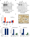
Figure 5. Prostasin is required for the activation of matriptase during placental differentiation.
(A and B) Expression of prostasin (A) and matriptase (B) in placental tissues of wildtype mice at E11.5. Both proteins were expressed in the chorionic (arrows) and labyrinthine (arrowheads) trophoblasts. (C) Western blot detection of active prostasin in the fetal part of the placenta of wildtype (Prss8+/+ and St14+/+, lanes 2, 4, and 6), prostasin-deficient (St14+/+;Prss8−/−) (lane 1), and matriptase-deficient (St14−/−;Prss8+/+) (lanes 3 and 5) embryos at E11.5 after immunoprecipitation with anti-mouse HAI-1 antibodies. Immunoprecipitated proteins in lanes 5 and 6 were acid-exposed to dissociate prostasin-HAI-1 complexes, and then incubated with PN-1 prior to western blot analysis. Positions of bands corresponding to active prostasin, prostasin/PN-1 complex, as well as non-specific signals of IgG heavy and light chains are indicated on the right. Positions of molecular weight markers (kDa) are shown on the left. (D) Omission of anti-HAI-1 antibody resulted in loss of detectable prostasin (compare lanes 1 and 2), indicating that the detected prostasin formed complexes with HAI-1. (E) Quantification of the relative amount of active prostasin in wildtype and matriptase placentae by densitometric scanning of prostasin western blots of HAI-1 immunoprecipitated material from (Prss8−/−;St14+/+, N = 3, Prss8+/+;St14−/−, N = 3, and Prss8+/+;St14+/+, N = 6). Data are shown as mean ± standard deviation (N.S., not significant). (F and G) Western blot detection of active matriptase in the fetal part of the placenta at E11.5 (F) after anti-HAI-1 immunoprecipitation, and in the epidermis of newborn skin (G) of wildtype (Prss8+/+ and St14+/+) (F, lanes 1, and 3, and G, lane 2), prostasin-deficient (St14+/+;Prss8−/−) (F, lane 4 and G, lane 1), and matriptase-deficient (St14−/−;Prss8+/+) (F, lane 2, and G, lane 3) embryos. A 30 kDa band representing the active serine protease domain of matriptase (Mat SPD) was present in extracts from wildtype (lanes 1 and 3 in F), but not in matriptase- (lane 2 in F) or prostasin-deficient (lane 4 in F) placenta. Zymogen (Mat FL) and active (Mat SPD) forms of matriptase were detected in extracts from both wildtype and prostasin-deficient, but not matriptase-deficient epidermis. (H–H″) Immunohistochemical staining of matriptase in control Prss8+ (H) and prostasin-deficient Prss8−/− (H′) placenta at E11.5. Specificity of staining of chorionic and labyrinthine trophoblasts (examples with arrows) is shown by the absence of staining of corresponding cells in St14−/− placenta (H″). Insets in H and H′ are parallel sections stained with prostasin antibodies. Open arrowheads in H–H″ show examples of non-specific staining. Scale bars: A, B, H, H′, and H″, 50 µm.
Figure 6
Figure 6. Prostasin activates matriptase on the surface of HEK293 cells.
(A and B) Western blot detection of matriptase in cell lysates (A) and in the conditioned medium (B) from HEK293 cells transiently transfected with wildtype recombinant human matriptase and HAI-1 expression vectors (lanes 1 and 2), catalytically inactive (S805A) matriptase and HAI-1 (lanes 3 and 4), HAI-1 alone (lanes 5 and 6), and cells transfected with a control empty vector (lanes 7 and 8) that were incubated with (lanes 2, 4, 6, and 8) or without (lanes 1, 3, 5, and 7) 100 nM soluble recombinant human prostasin. Addition of prostasin promoted conversion of the matriptase zymogen to its activated two-chain form. Positions of bands corresponding to full length matriptase (Mat FL), matriptase pro-enzyme processed by autocatalytic cleavage within the SEA domain (Mat SEA), and activated matriptase serine protease domain (Mat SPD) are indicated on the right. Positions of molecular weight markers (kDa) are shown on left. (C) Quantification of the activation of PAR-2 in HEK293 cells expressing recombinant human PAR-2 in combination with wildtype (WT) or inactive (S805A) variants of matriptase and HAI-1, HAI-1 alone, or transfected with an empty vector, incubated without (blue bars) or with (red bars) 100 nM soluble recombinant human prostasin. Prostasin induced matriptase activity-dependent activation of PAR-2. (D) Immunohistochemical analysis of the expression of the gamma subunit of the epithelial sodium channel (ENaC) in placenta of control mice at E11.5. The expression was detected in the populations of chorionic (arrow) and labyrinthine (arrowhead) trophoblasts. Scale bar: 50 µm. (E) Frequency of exencephaly in amiloride-treated wildtype (Spint2+/+, N = 56), untreated HAI-2-deficient (Spint2−/−, N = 12) and amiloride-treated HAI-2-deficient (Spint2−/−, N = 7) embryos at E9.5. Amiloride treatment failed to rescue neural tube defects in Spint2−/−; St14+/− embryos. (F) Distribution of Spint2 genotypes in ENaC-expressing (Scnn1a+/+ or Scnn1a+/−, blue bars) and ENaC-deficient (Scnn1a−/−, green bars) offspring from Spint2 +/− ;Scnn1a+/−×Spint2 +/− ;Scnn1a+/− breeding pairs at E9.5. Loss of ENaC expression did not rescue early embryonic lethality in Spint2−/− mice. (G) Distribution of Spint2 genotypes in matriptase-haploinsufficient ENaC-expressing (St14+/−;Scnn1a+/+ or Scnn1a+/−, blue bars) and ENaC-deficient (St14+/−; Scnn1a−/−, green bars) offspring from Spint2 +/− ;Scnn1a+/−, St14+/−×Spint2 +/− ;Scnn1a+/−;St14+/+ breeding pairs at birth. Loss of ENaC expression did not rescue overall embryonic survival in Spint2−/−; St14+/− mice.
Figure 7
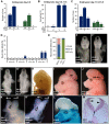
Figure 7. Neural tube defects and embryonic lethality in HAI-2–deficient mice are not dependent on PAR-2, and combined PAR-1 and matriptase deficiency does not phenocopy combined PAR-1 and PAR-2 deficiency.
(A) Distribution of Spint2 genotypes at E9.5 in PAR-2-expressing (F2rl1+/+ or F2rl1+/−, blue bars) and PAR-2-deficient (F2rl1−/−, green bars) offspring from interbred Spint2+/−,F2rl1+/− mice. No Spint2−/− embryos were detected irrespective of PAR-2 expression. (B) Frequency of exencephaly observed in HAI-2 and PAR-2-sufficient (Spint2+;F2rl1+ N = 366), PAR-2-deficient (Spint2+;F2rl1−/−, N = 164), HAI-2-deficient (Spint2−/−,F2rl1+, N = 18), and PAR-2 and HAI-2 double- (Spint2−/−;F2rl1−/−, N = 12) deficient embryos extracted at E9.5–E11.5. Loss of PAR-2 activity fails to correct neural tube defects in HAI-2-deficient embryos. (C) Distribution of St14 alleles at E11.5–15.5 in PAR-1-expressing (F2r+/+ or F2r+/−, blue bars) and PAR-1-deficient (F2r−/−, green bars) embryos from interbred St14 +/− ;F2r+/− mice. Loss of PAR-1 activity does not affect embryonic survival of matriptase-deficient mice. (D) Frequency of exencephaly (Ex), spina bifida (SB), and curly tail (CT) in E9.5–18.5 embryos with different levels of expression of PAR-1 (F2r+ or F2r−/−) and matriptase (St14+or St14−/−). A total of 326 embryos were analyzed. Loss of matriptase does not significantly increase the incidence of neural tube defects in PAR-1-deficient embryos. (E) Comparison of the severity of exencephaly in HAI-2-deficient (Spint2−/−, N = 29) and PAR-1 and PAR-2 double-deficient (F2r−/−;F2rl1−/−, N = 39) embryos. 95% of affected F2r−/−;F2rl1−/− embryos exhibited exencephaly that was confined to hindbrain region of the cranium (HB only, green bars), with the remaining 5% extending to the midbrain region (MB-HB, blue bars). In contrast, only 10% of exencephalies observed in Spint2−/−-deficient mice were confined to the hindbrain, with 59% extended to midbrain, and 31% to forebrain region (FB-HB, red bars). (F–G′) Ventral (F and G) and dorsal (F′ and G′) view of non-affected control (F and F′) and affected PAR-1 and PAR-2 double-deficient (F2r−/−;F2rl1−/−) (G and G′) embryos at E9.5. The initial stages of neural tube closure all appear to be unaffected by the combined absence of PAR-1 and PAR-2. (H–J) Appearance of control (H) and PAR-1 and PAR-2 double-deficient embryos with exencephaly (I and J) at E14.5. Exencephaly in 95% of the affected PAR-1 and PAR-2 double-deficient embryos was restricted to hindbrain region (HB, two-sided arrow in I) and extended to midbrain (MB-HB, two-sided arrow in J) in only 5% of the cases. (K and K′) Ventral (K) and dorsal (K′) view of the macroscopic appearance of HAI-2-deficient (Spint2−/−) embryos at E9.5. Divergence of neural folds (arrows) and defects in neural tube closure extending from forebrain region to cervix are obvious. Open arrowheads show normal formation of medial hinge points. (L) Macroscopic appearance of a HAI-2-deficient embryo with exencephaly at E14.5. 90% of embryos presented with exencephaly that included at least midbrain and hindbrain regions of the developing cranium. (M) Histological appearance (nuclear fast red staining) of PAR-1 and PAR-2 double-deficient embryo with exencephaly at E9.5. Defined medial (arrow) and dorsolateral (arrowheads) hinge points are clearly visible. Scale bar: 150 µm.

References

    1. Szabo R, Bugge TH (2011) Membrane anchored serine proteases in cell and developmental biology. Annu Rev Cell and Developmental Biology 27: 213–235. - PMC - PubMed
    1. Bugge TH, Antalis TM, Wu Q (2009) Type II transmembrane serine proteases. J Biol Chem 284: 23177–23181. - PMC - PubMed
    1. Antalis TM, Buzza MS, Hodge KM, Hooper JD, Netzel-Arnett S (2010) The cutting edge: membrane-anchored serine protease activities in the pericellular microenvironment. Biochem J 428: 325–346. - PMC - PubMed
    1. Guipponi M, Tan J, Cannon PZ, Donley L, Crewther P, et al. (2007) Mice deficient for the type II transmembrane serine protease, TMPRSS1/hepsin, exhibit profound hearing loss. Am J Pathol 171: 608–616. - PMC - PubMed
    1. Scott HS, Kudoh J, Wattenhofer M, Shibuya K, Berry A, et al. (2001) Insertion of beta-satellite repeats identifies a transmembrane protease causing both congenital and childhood onset autosomal recessive deafness. Nat Genet 27: 59–63. - PubMed

Publication types

MeSH terms