. 2012;7(8):e43019.
doi: 10.1371/journal.pone.0043019. Epub 2012 Aug 29.

Structures of human DPP7 reveal the molecular basis of specific inhibition and the architectural diversity of proline-specific peptidases

Affiliations

Structures of human DPP7 reveal the molecular basis of specific inhibition and the architectural diversity of proline-specific peptidases

Gustavo Arruda Bezerra et al. PLoS One. 2012.

Abstract

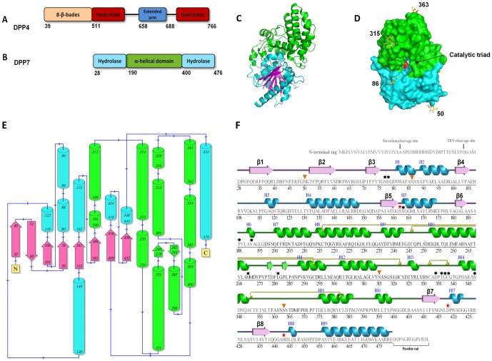
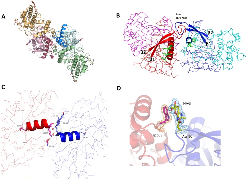
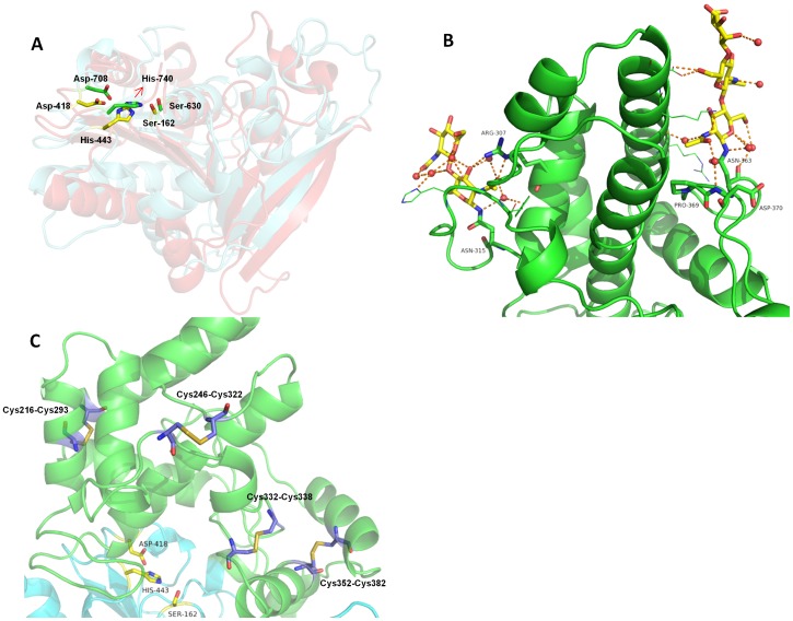
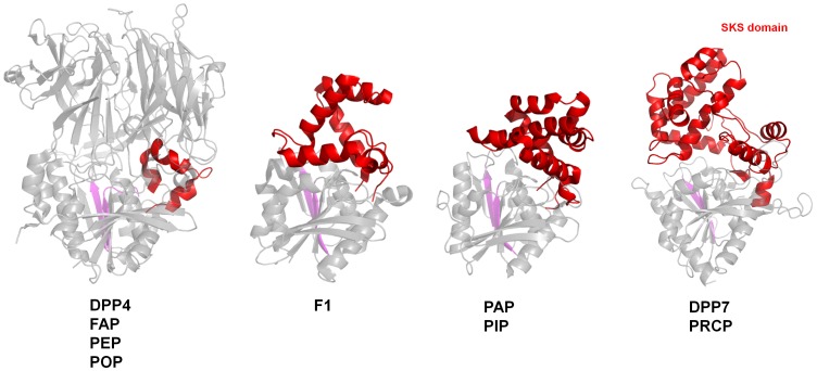
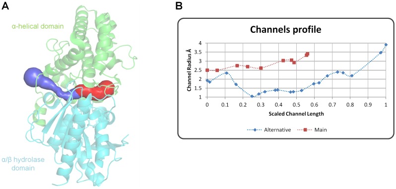
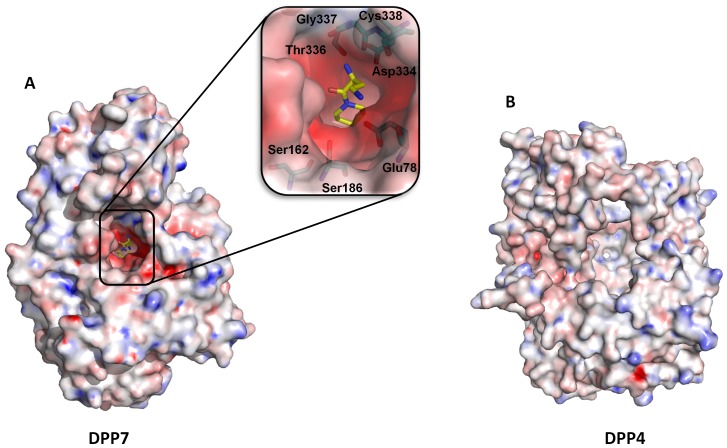
Proline-specific dipeptidyl peptidases (DPPs) are emerging targets for drug development. DPP4 inhibitors are approved in many countries, and other dipeptidyl peptidases are often referred to as DPP4 activity- and/or structure-homologues (DASH). Members of the DASH family have overlapping substrate specificities, and, even though they share low sequence identity, therapeutic or clinical cross-reactivity is a concern. Here, we report the structure of human DPP7 and its complex with a selective inhibitor Dab-Pip (L-2,4-diaminobutyryl-piperidinamide) and compare it with that of DPP4. Both enzymes share a common catalytic domain (α/β-hydrolase). The catalytic pocket is located in the interior of DPP7, deep inside the cleft between the two domains. Substrates might access the active site via a narrow tunnel. The DPP7 catalytic triad is completely conserved and comprises Ser162, Asp418 and His443 (corresponding to Ser630, Asp708 and His740 in DPP4), while other residues lining the catalytic pockets differ considerably. The "specificity domains" are structurally also completely different exhibiting a β-propeller fold in DPP4 compared to a rare, completely helical fold in DPP7. Comparing the structures of DPP7 and DPP4 allows the design of specific inhibitors and thus the development of less cross-reactive drugs. Furthermore, the reported DPP7 structures shed some light onto the evolutionary relationship of prolyl-specific peptidases through the analysis of the architectural organization of their domains.


Conflict of interest statement

Competing Interests: The work was funded in part by the companies GlaxoSmithKline and Merck & Co, Inc. In addition, co-authors LMS, AMH, SMS, TDS, PJM, KOJ, and KMK-W are employees of the commercial funder GlaxoSmithKline. This does not alter the authors' adherence to all the PLOS ONE policies on sharing data and materials.

Figures

Figure 1
Figure 1. Structure of DPP7.
(A) Schematic showing the DPP4 domain structure. The domains are represented as boxes and their borders are indicated. The propeller domain is in yellow, the hydrolase domain in dark red and the extended arm in blue. (B) Schematic showing the DPP7 domain structure aligned with that of DPP4. The hydrolase domain is in aquamarine and the big α-helical (SKS) domain in green. (C) Ribbon presentation of the DPP7 protomer structure. The domains are colored as in (B) with the β-strands characteristic for the hydrolase fold presented in magenta. (D) Surface representation of the DPP7 protomer. The domain is colored as in (B) and (C). The catalytic triad (Ser162, Asp418 and His443) is shown in red. The carbohydrates identified in the molecule are represented as sticks and colored ‘per atom’ (yellow, blue and red for C, N, and O, respectively). The corresponding amino acid numbers are shown in black. (E) Topology diagram evidencing how the new fold is positioned in relation to the catalytic fold. Color code is the same of Figure 1. (F) Expressed sequence information. Secondary structure of DPP7 was aligned to the amino acid sequence. Residues without secondary structure are not observed and presumed flexible. The color code is the same as in the previous figures. The catalytic triad (Ser162, Asp418 and His443) is indicated by a “red star”. The strands are represented by arrows and helices by bars. The glycosylated residues identified in the electron density map are marked with orange triangles and the suggested inhibitor interacting residues with asterisks. Disulfide bonds are indicated by yellow circles linked to the corresponding partners by yellow bars. Items C and D were prepared using the program PyMOL (http://www.pymol.org/).
Figure 2
Figure 2. The DPP7 dimerization interface.
(A) Cartoon representation of DPP7 overall structure highlighting the two protomers: helical domains are shown in orange and green, the α/β-hydrolase domains in red and blue. Carbohydrates are shown as sticks. (B) Dimerization mediated by the loop Arg39 – Asn50. Ribbon representation with two strands of the central β-sheet and the loop Arg39 – Asn50 represented as cartoon. The protomers are colored in blue and red, respectively. The supposed leucine zipper motifs are highlighted as cartoon and shown in green. (C) Dimerization mediated by helix α5. The catalytic Ser162 and residues participating in hydrogen bonds at the other of the helix are represented as sticks. Two water molecules are shown as red spheres. Helix α5 is represented as cartoon and the protomers are represented as ribbons in red and blue. (D) Stacking interaction between N-acetylglycosamine (NAG) linked to Asn50 and Trp389. A 2Fo-Fc electron density map of NAG and Trp389 is shown contoured at 1 σ. The figure was prepared using the program PyMOL (http://www.pymol.org/).
Figure 3
Figure 3. The catalytic domain and SKS domain.
SKS domain shown in green cartoon and the catalytic domain shown in aquamarine cartoon. The catalytic triad is represented as yellow sticks. (A) Conservation of the catalytic triad evidenced by the structural superposition. The catalytic fold of DPP4 is shown as red cartoon. DPP7 catalytic residues (Ser-162, Asp-418 and His-443) are colored in yellow, while DPP4 catalytic residues (Asp-708, His-740 and Ser-630) in green. The corresponding amino acid numbers are shown in black. (B) The SKS domain is shown in green cartoon, while the glycans linked to Asn363 and Asn315 are shown as sticks. Both glycans seem to have a scaffold function by holding the loop Glu354-Asp375 to the helix α8 and the loop Asp324-Tyr314 to the helix α12, respectively. (C) Four disulfide bonds present in the SKS domain. Cys216-Cys293 and Cys246-Cys322 seem to have a structure stabilization role. Cys332-Cys338 is very likely involved in the activity regulation of the enzyme, while Cys352-Cys382 could play either regulatory and structural role. The figure was prepared using the program PyMOL (http://www.pymol.org/).
Figure 4
Figure 4. Representation of the evolution of the SKS domain.
The α/β-hydrolase domain in all enzymes and the β-propeller domain in DPP4 are shown as grey cartoons. The cap domain in all enzymes are shown as red cartoon. DPP4, FAP, PEP and POP, share the same extended arm (red cartoon) which increases in complexity to become the cap domain in the F1 aminopeptidase. The degree of complexity is further increased in PAP and PIP, until it reaches its maximum convolution in the form of SKS domain in DPP7 and PRCP. For clarity purpose, only DPP4, F1, PAP and DPP7 are displayed in the Figure. β-strands 6 and 7 are shown in magenta. The figure was prepared using the program PyMOL (http://www.pymol.org/).
Figure 5
Figure 5. The channel system in DPP7.
(A) The main channel is represented in red, and should be the route for the substrates to access the active site. It can be also visualized in Figure 1D. The products should be released either through the main channel or by an alternative channel (blue), identified by the Caver algorithm . (B) Channels profile indicating the radius in Å vs. the scaled length, starting from the inhibitor position towards the protein surface. The figure was prepared using the program PyMOL (http://www.pymol.org/).
Figure 6
Figure 6. Inhibitor Dap-Pip in the DPP7 active site.
(A) Omit and 2Fo-Fc electron density maps for the bound inhibitor are shown at 1 and 1.5σ, respectively. (B) General overview of the inhibitor in the binding pocket. The α/β–hydrolase domain is shown in aquamarine and the SKS–domain in green. The inhibitor is shown in magenta. (C) Stereo view of the residues involved in inhibitor binding. The coloring scheme is the same as above. Dashed yellow lines denote hydrogen bonding interactions (D) Representation of the interaction of the inhibitor with surrounding residues prepared using LigPlot+ . (E) Superimposition of the DPP7 Dab-Pip complex with the structure of DPP4 complexed with Diprotin A . The coloring scheme for DPP7 is the same as above. The Diprotin A is shown using thinner, yellow sticks. The figure was prepared using the program PyMOL (http://www.pymol.org/).
Figure 7
Figure 7. Poisson-Boltzmann electrostatic potential.
(A) Surface representation of DPP7 showing the negatively charged substrate binding pocket. Inset: Zoom in the binding pocket depicting the amino acid residues that contributes to the negative charge. (B) Surface representation of the electrostatic potential of DPP4 evidencing the more neutral to positive charge in the binding pocket. The calculations were done with the chain A of DPP7 (PDB code 3JYH) and DPP4 (PDB code: 3EIO) using the software APBS . The figure was prepared using the program PyMOL (http://www.pymol.org/).

References

    1. Yaron A, Naider F (1993) Proline-dependent structural and biological properties of peptides and proteins. Crit Rev Biochem Mol Biol 28: 31–81. - PubMed
    1. Rosenblum JS, Kozarich JW (2003) Prolyl peptidases: a serine protease subfamily with high potential for drug discovery. Curr Opin Chem Biol 7: 496–504. - PubMed
    1. Yazbeck R, Howarth GS, Abbott CA (2009) Dipeptidyl peptidase inhibitors, an emerging drug class for inflammatory disease? Trends Pharmacol Sci 30: 600–607. - PubMed
    1. Busek P, Malik R, Sedo A (2004) Dipeptidyl peptidase IV activity and/or structure homologues (DASH) and their substrates in cancer. Int J Biochem Cell Biol 36: 408–421. - PubMed
    1. Gorrell MD, Gysbers V, McCaughan GW (2001) CD26: a multifunctional integral membrane and secreted protein of activated lymphocytes. Scand J Immunol 54: 249–264. - PubMed

Publication types

MeSH terms