. 2012;7(8):e43196.
doi: 10.1371/journal.pone.0043196. Epub 2012 Aug 29.

Characterization of omental immune aggregates during establishment of a latent gammaherpesvirus infection

Affiliations

Characterization of omental immune aggregates during establishment of a latent gammaherpesvirus infection

Kathleen S Gray et al. PLoS One. 2012.

Abstract

Herpesviruses are characterized by their ability to establish lifelong latent infection. The gammaherpesvirus subfamily is distinguished by lymphotropism, establishing and maintaining latent infection predominantly in B lymphocytes. Consequently, gammaherpesvirus pathogenesis is closely linked to normal B cell physiology. Murine gammaherpesvirus 68 (MHV68) pathogenesis in laboratory mice has been extensively studied as a model system to gain insights into the nature of gammaherpesvirus infection in B cells and their associated lymphoid compartments. In addition to B cells, MHV68 infection of macrophages contributes significantly to the frequency of viral genome-positive cells in the peritoneal cavity throughout latency. The omentum, a sheet of richly-vascularized adipose tissue, resides in the peritoneal cavity and contains clusters of immune cell aggregates termed milky spots. Although the value of the omentum in surgical wound-healing has long been appreciated, the unique properties of this tissue and its contribution to both innate and adaptive immunity have only recently been recognized. To determine whether the omentum plays a role in gammaherpesvirus pathogenesis we examined this site during early MHV68 infection and long-term latency. Following intraperitoneal infection, immune aggregates within the omentum expanded in size and number and contained virus-infected cells. Notably, a germinal-center B cell population appeared in the omentum of infected animals with earlier kinetics and greater magnitude than that observed in the spleen. Furthermore, the omentum harbored a stable frequency of viral genome-positive cells through early and into long-term latency, while removal of the omentum prior to infection resulted in a slight decrease in the establishment of splenic latency following intraperitoneal infection. These data provide the first evidence that the omentum is a site of chronic MHV68 infection that may contribute to the maintenance of chronic infection.


Conflict of interest statement

Competing Interests: The authors have declared that no competing interests exist.

Figures

Figure 1
Figure 1. Increased size and frequency of omental immune aggregates in MHV68-infected mice.
Omenta from mice inoculated intraperitoneally (i.p.) with 1000 plaque forming units (pfu) virus in complete media (cDMEM) or cDMEM alone (mock) and harvested at the indicated times post infection. Omenta were cryosectioned and stained using DAPI-containing mounting media. Dense DAPI-staining in immune aggregates, “milky spots,” is outlined by dashed ovals.
Figure 2
Figure 2. B cell populations in omentum and peritoneal exudate following MHV68 infection.
Cells from the omentum and peritoneal cavity (PECs) were isolated at the indicated times post-intraperitoneal infection and analyzed by flow cytometry. Live cells were gated on A) CD3 and total B cells determined by CD19 expression or B) CD3CD19+ cells to determine CXCR5 expression.
Figure 3
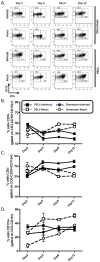
Figure 3. Alterations in B1-B cell populations in the omentum and peritoneal exudate following MHV68 infection.
Cells from the omentum and peritoneal cavity (PECs) were isolated at the indicated times post-intraperitoneal infection and analyzed by flow cytometry. Live cells were gated on CD3CD19+CD11b+ cells and B1-a and B1-b populations discriminated by CD5 expression. A) Representative flow plots and B–D) compiled data from experimental groups of mice represented as line graphs for B1a B cells (B), B1b B cells (C), and total B1 B cell (D) frequencies. Squares, [cells from] peritoneal cavity; Circles, [cells from] omentum; Open symbols, mock-infected; filled symbols, MHV68-infected.
Figure 4
Figure 4. Germinal center B cell expansion in omentum precedes expansion in spleen.
Cells from the omentum and spleen were isolated at the indicated times post-intraperitoneal infection and analyzed by flow cytometry. Live cells were gated on CD3CD19+ cells and germinal center populations identified by Fas (CD95) and GL7 expression. A) Representative flow plots and B) data from individual mice represented as line graph. Squares, [cells from] spleen; Circles, [cells from] omentum; Open symbols, mock-infected; filled symbols, MHV68-infected.
Figure 5
Figure 5. Visualization of YFP+ cells in the omentum.
Omenta from mice inoculated intraperitoneally (i.p.) with 1000 plaque forming units (pfu) virus in complete media (cDMEM) or cDMEM alone (mock) and harvested at day 9 (omentum) and day 12 (spleen). Omenta (A–D) and spleen (E) were cryosectioned and stained with the antibodies to detect YFP, CD11b or B220 as described in Materials and Methods. a) YFP+ (virus-infected) cells within the B220+ area of omental immune aggregate; boxed area magnified in (b). c) CD11b+ YFP+ cells; boxed area magnified in (d). e) YFP+ splenic B cell follicles with surrounding CD11b+ extrafollicular region.
Figure 6
Figure 6. The omentum supports early latent infection.
Mice were inoculated i.p. with 1000 pfu virus and PECs, spleen and omenta harvested at day 18 post-infection. Single cell suspensions were used for limiting dilution PCR analyses (omentum) (A) or prepared (see Materials and Methods) and plated immediately for limiting dilution reactivation analyses (spleen, peritoneum, and omentum) (B). A) Frequency of positive PCR reactions after limiting-dilution plating and DNA-quantity controlled PK digest/PCR. B) Frequency of CPE in MEF monolayers after limiting-dilution plating of intact cells as evidence of virus reactivation. Mechanically-disrupted cells were plated in parallel to control for the presence of preformed infectious virus (data not shown).
Figure 7
Figure 7. Reduced establishment in omentectomized mice during early latency.
Mice underwent omentectomy or sham surgery and allowed to recover for four weeks, then were inoculated i.p. with 1000 pfu virus. Spleen and omenta were harvested at day 18 post-infection and single cell suspensions analyzed by limiting dilution reactivation analyses (A), or used for limiting dilution PCR analyses (B). A) Frequency of CPE in MEF monolayers after limiting-dilution plating of intact cells as evidence of virus reactivation. Mechanically-disrupted cells were plated in parallel to control for the presence of preformed infectious virus (data not shown). B) Frequency of positive PCR reactions after limiting-dilution plating and DNA-quantity-controlled PK digest/PCR.
Figure 8
Figure 8. The omentum harbors MHV68 infected cells during long-term latency.
Mice were inoculated i.p. with 1000 pfu virus and PECs, spleen, and omenta harvested at day 90 post-infection. Single cell suspensions were prepared used for limiting dilution PCR analyses to determine the frequency of viral genome positive cells.

References

    1. Barton E, Mandal P, Speck SH (2011) Pathogenesis and host control of gammaherpesviruses: lessons from the mouse. Annu Rev Immunol 29: 351–397. - PubMed
    1. Speck SH, Ganem D (2010) Viral latency and its regulation: lessons from the gamma-herpesviruses. Cell Host Microbe 8: 100–115. - PMC - PubMed
    1. Willer DO, Speck SH (2003) Long-term latent murine Gammaherpesvirus 68 infection is preferentially found within the surface immunoglobulin D-negative subset of splenic B cells in vivo. J Virol 77: 8310–8321. - PMC - PubMed
    1. Hwang S, Wu TT, Tong LM, Kim KS, Martinez-Guzman D, et al. (2008) Persistent gammaherpesvirus replication and dynamic interaction with the host in vivo. J Virol 82: 12498–12509. - PMC - PubMed
    1. Peacock JW, Bost KL (2000) Infection of intestinal epithelial cells and development of systemic disease following gastric instillation of murine gammaherpesvirus-68. J Gen Virol 81: 421–429. - PubMed

Publication types

LinkOut - more resources