. 2012;7(8):e43924.
doi: 10.1371/journal.pone.0043924. Epub 2012 Aug 31.

Involvement of microglia activation in the lead induced long-term potentiation impairment

Affiliations

Involvement of microglia activation in the lead induced long-term potentiation impairment

Ming-Chao Liu et al. PLoS One. 2012.

Abstract

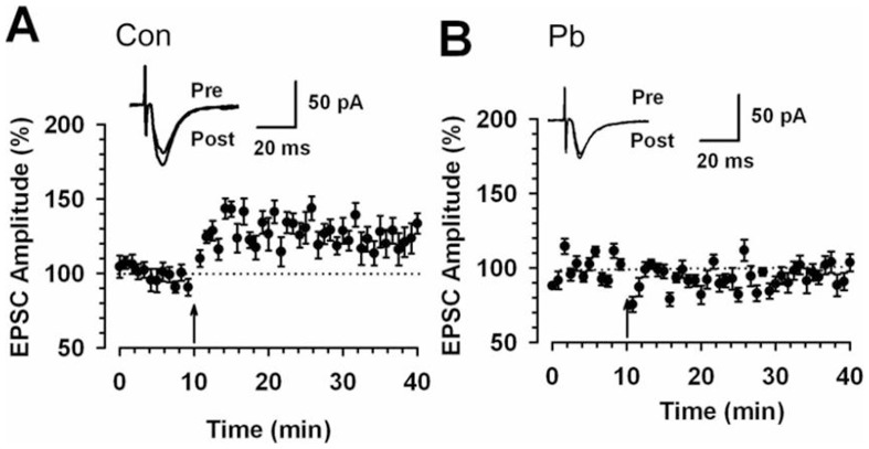
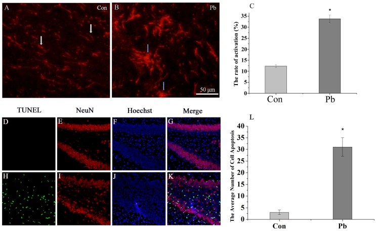
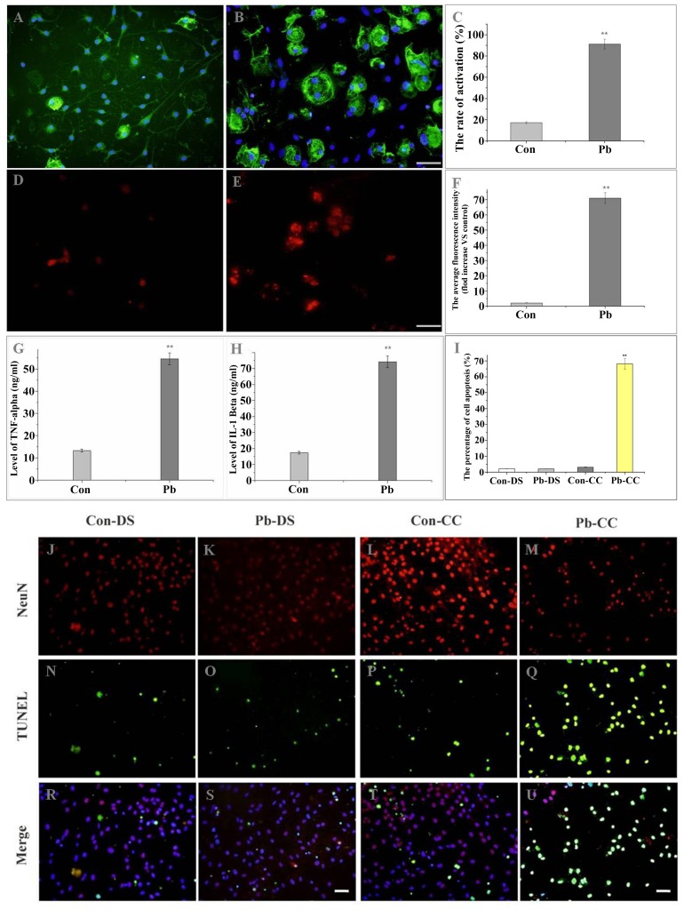
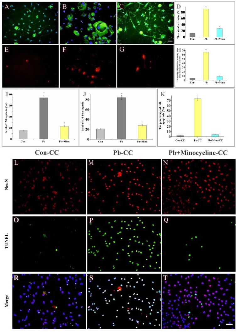
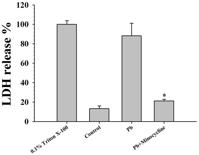
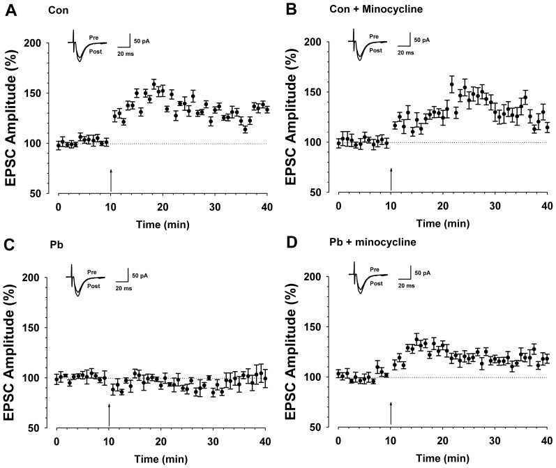
Exposure of Lead (Pb), a known neurotoxicant, can impair spatial learning and memory probably via impairing the hippocampal long-term potentiation (LTP) as well as hippocampal neuronal injury. Activation of hippocampal microglia also impairs spatial learning and memory. Thus, we raised the hypothesis that activation of microglia is involved in the Pb exposure induced hippocampal LTP impairment and neuronal injury. To test this hypothesis and clarify its underlying mechanisms, we investigated the Pb-exposure on the microglia activation, cytokine release, hippocampal LTP level as well as neuronal injury in in vivo or in vitro model. The changes of these parameters were also observed after pretreatment with minocycline, a microglia activation inhibitor. Long-term low dose Pb exposure (100 ppm for 8 weeks) caused significant reduction of LTP in acute slice preparations, meanwhile, such treatment also significantly increased hippocampal microglia activation as well as neuronal injury. In vitro Pb-exposure also induced significantly increase of microglia activation, up-regulate the release of cytokines including tumor necrosis factor-alpha (TNF-α), interleukin-1β (IL-1β) and inducible nitric oxide synthase (iNOS) in microglia culture alone as well as neuronal injury in the co-culture with hippocampal neurons. Inhibiting the microglia activation with minocycline significantly reversed the above-mentioned Pb-exposure induced changes. Our results showed that Pb can cause microglia activation, which can up-regulate the level of IL-1β, TNF-α and iNOS, these proinflammatory factors may cause hippocampal neuronal injury as well as LTP deficits.


Conflict of interest statement

Competing Interests: The authors have declared that no competing interests exist.

Figures

Figure 1
Figure 1. Abolishment of hippocampus synaptic potentiation in 100 ppm Pb treated rats.
(A) LTP was induced in hippocampal pyramidal neurons in control rats (n = 13 slices/6 rats, t-test; P<0.001 compared with baseline). (B) LTP was lost in hippocampal pyramidal neurons in Pb treated rats (n = 8 slices/6 rats, t-test; P>0.05 compared with baseline). Pairing training is indicated by an arrow. The dashed line indicates the mean basal synaptic responses.
Figure 2
Figure 2. Effects of hippocampal neuronal injury and microglia activation induced by Pb in the hippocampus.
Microglia activation was detected by immunocytochemistry (OX42). (A) control; (B) 100 ppm Pb for 8 weeks. Activation of microglia was evaluated by counting the number of activated microglial cells (B blue arrow); White arrows (A) indicate resting microglia. (C) The results were quantified and are expressed as the mean ± S.D. of average activated cell rate in random fields (n = 20). * P<0.05 compared with control groups. Scale bar indicates 50 µm. Hippocampal neuronal injury was detected with in situ TUNEL (green fluorescence). (K) The apoptotic neurons in the Pb group were significantly higher than the control group (G). Hippocampal neuronal injury was evaluated by analyze the number of apoptotic neurons of each group. (L) The results were quantified and are expressed as the mean ± S.D. of apoptotic neurons in DG zone of hippocampus (n = 8). * P<0.05 compared with control groups. Scale bar indicates 50 µm.
Figure 3
Figure 3. Pb activated microglia, enhanced TNF-α and IL-1β secretes, iNOS expression, and induces hippocampal neuronal injury.
Purified microglia cells were seeded in 12-well culture plates. Cells were treated with vehicle (control) (A) or Pb (50 µmol) (B) for the indicated times. Microglia activation was detected by immunocytochemistry (OX42). (A) control; (B) 50 µmol Pb for 48 h. (C) The results were quantified. Results (A and B) are expressed as the mean ± S.D. of average activated cell rate in random fields (n = 20). **P<0.001 compared with control groups. Scale bar indicates 50 µm. iNOS was detected by immunofluorescence staining. Activation of iNOS was evaluated by measure the average fluorescence of iNOS-immunoreactive cells (D, E). (F) The results were quantified and are expressed as the mean ± S.D. of average fluorescence of iNOS-immunoreactive cells in random fields (n = 20). ** P<0.001 compared with control groups. Scale bar indicates 50 µm. TNF-α (G) and IL-1β (H) were determined by ELISA. Purified microglia were cultured with vehicle (control) or Pb (50 µmol) for 48 h for the TNF-α and IL-1β assay, respectively. ** P<0.001 compared with control groups. The purified microglia were co-cultured with hippocampal neurons for 48 h after culturing with vehicle (control) (L, P, T) or Pb (50 µmol) (M, Q, U) for 48 h. Apoptosis of hippocampal neurons was detected with in situ TUNEL (green fluorescence). Degeneration of hippocampal neurons was evaluated by counting the number of apoptotic neurons. As compared with the co-culture method, hippocampal neurons were also treated with vehicle (control) (J, N, R) or Pb (50 µmol) (K, O, S) directly for 48 h. (I) The results were quantified and are expressed as the mean ± SD of apoptotic neurons percentage in random fields (n = 20). ** P<0.001 compared with control groups. Scale bar indicates 50 µm. CC in figure means co-culture method; DS in figure means direct stimulation method.
Figure 4
Figure 4. Minocycline blocks the activation of microglia; attenuates Pb-induced secrete of TNF-α, IL-1β and expression of iNOS; and protects co-cultured hippocampal neurons.
Purified microglia were treated with vehicle (control) (A), Pb (50 µmol) (B), or Pb (50 µmol)+minocycline (45 µmol) (C) for 48 h, and were co-cultured with hippocampal neurons for another 48 h. (A, B, C) Microglia activation was detected by OX-42 antibody. (D) The results were quantified. Results (A, B and C) are expressed as the mean ± S.D. of average activated cell rate in random fields (n = 20). **P<0.001 compared with control groups. *P<0.01 compared with Pb-treated groups. iNOS was detected by anti-iNOS antibody (E, F, G. red fluorescence). Activation of iNOS was evaluated by measure the average fluorescence of iNOS-immunoreactive cells. (H) The results were quantified. Results (E, F and G) are expressed as the mean ± S.D. of average fluorescence of iNOS-immunoreactive cell number in random fields (n = 20). **P<0.001 compared with control groups. *P<0.01 compared with Pb-treated groups. TNF-α (I) and IL-1β (J) were determined by ELISA. Purified microglia were treated with vehicle (control) or Pb (50 µmol), or Pb (50 µmol)+minocycline (45 µmol) for 48 h for the TNF-α and IL-1β assay, respectively. **P<0.001 compared with control groups. *P<0.01 compared with Pb-treated groups. Apoptotic hippocampal neurons were detected and compared using TUNEL (green fluorescence) (L–T). Degeneration of hippocampal neurons was evaluated by counting the number of apoptotic neurons. (K) The results were quantified and are expressed as the mean ± S.D. of apoptotic neurons percentage in random fields (n = 20). **P<0.001 compared with control groups. *P<0.01 compared with Pb-treated groups. All scale bar indicates 50 µm.
Figure 5
Figure 5. Effect of minocycline on LDH-release after Pb exposure.
Purified microglia were treated with vehicle (control), Pb (50 µmol) or Pb (50 µmol)+minocycline (45 µmol) for 48 h, and were co-cultured with hippocampal neurons for another 48 h. LDH-release were measured from co-culture supernatant by LDH-cytotoxicity assay kit. The mean value of LDH-release of the positive control group (0.1% triton X-100) was considered as 100%. n = 6. Mean ± S.D. *P<0.01 compared with Pb-treated groups.
Figure 6
Figure 6. Minocycline reversed the induction of LTP in hippocampus after 100 ppm Pb treated.
(A) LTP was induced in hippocampal pyramidal neurons in control rats (n = 11 slices/6 rats, t-test; P<0.001 compared with baseline). (B) LTP was induced in hippocampal pyramidal neurons in minocycline treated rats (n = 8 slices/6 rats, t-test; P<0.001 compared with baseline). (C) LTP was lost in hippocampal pyramidal neurons in Pb treated rats (n = 10 slices/6 rats, t-test; P>0.05 compared with baseline). (D) LTP was partly reversed in hippocampal pyramidal neurons in minocycline treated rats during Pb exposure (n = 9 slices/6 rats, t-test; P<0.05 compared with baseline). Pairing training is indicated by an arrow. The dashed line indicates the mean basal synaptic responses.

Similar articles

Cited by

References

    1. Bellinger DC (2004) Lead. Pediatrics 113: 1016–22. - PubMed
    1. Finkelstein Y, Markowitz ME, Rosen JF (1998) Low-level lead-induced neurotoxicity in children: an update on central nervous system effects. Brain Res Brain Res Rev 27: 168–76. - PubMed
    1. Rosen JF (1995) Adverse health effects of lead at low exposure levels: trends in the management of childhood lead poisoning. Toxicology 97: 11–7. - PubMed
    1. Koller K, Brown T, Spurgeon A, Levy L (2004) Recent developments in low-level lead exposure and intellectual impairment in children. Environ Health Perspect 112: 987–94. - PMC - PubMed
    1. Needleman HL, McFarland C, Ness RB, Fienberg SE, Tobin MJ (2002) Bone lead levels in adjudicated delinquents. A case control study. Neurotoxicol Teratol 24: 711–7. - PubMed

Publication types

MeSH terms